Comparative Study
. 2005 May 1;564(Pt 3):803-15.
doi: 10.1113/jphysiol.2005.083089. Epub 2005 Mar 10.

Electrophysiological properties of two axonal sodium channels, Nav1.2 and Nav1.6, expressed in mouse spinal sensory neurones

Affiliations
Comparative Study

Electrophysiological properties of two axonal sodium channels, Nav1.2 and Nav1.6, expressed in mouse spinal sensory neurones

Anthony M Rush et al. J Physiol. .

Abstract

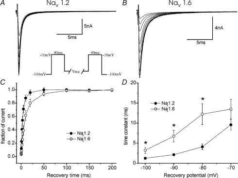
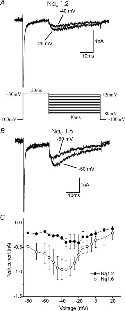
Sodium channels Na(v)1.2 and Na(v)1.6 are both normally expressed along premyelinated and myelinated axons at different stages of maturation and are also expressed in a subset of demyelinated axons, where coexpression of Na(v)1.6 together with the Na(+)/Ca(2+) exchanger is associated with axonal injury. It has been difficult to distinguish the currents produced by Na(v)1.2 and Na(v)1.6 in native neurones, and previous studies have not compared these channels within neuronal expression systems. In this study, we have characterized and directly compared Na(v)1.2 and Na(v)1.6 in a mammalian neuronal cell background and demonstrate differences in their properties that may affect neuronal behaviour. The Na(v)1.2 channel displays more depolarized activation and availability properties that may permit conduction of action potentials, even with depolarization. However, Na(v)1.2 channels show a greater accumulation of inactivation at higher frequencies of stimulation (20-100 Hz) than Na(v)1.6 and thus are likely to generate lower frequencies of firing. Na(v)1.6 channels produce a larger persistent current that may play a role in triggering reverse Na(+)/Ca(2+) exchange, which can injure demyelinated axons where Na(v)1.6 and the Na(+)/Ca(2+) exchanger are colocalized, while selective expression of Na(v)1.2 may support action potential electrogenesis, at least at lower frequencies, while producing a smaller persistent current.


Figures

Figure 1
Figure 1. Comparison of Nav1.2R and Nav1.6R current properties
Representative families of currents recorded from Nav1.8-null DRG neurones expressing Nav1.2R channels (A) or Nav1.6R channels (B) are shown. Currents were elicited by 40 ms depolarizations from a holding potential of −100 mV to a range of potentials between −65 mV and +60 mV (inset). C, average absolute current–voltage relationship for the two channels (Nav1.2, n = 25; Nav1.6, n = 27). D, activation and availability curves for Nav1.2R (n = 23/10) and Nav1.6R (n = 24/11). Activation was calculated from current–voltage experiments, as detailed in Methods. Availability of channels was estimated by measuring the peak current amplitude elicited by 40 ms test pulses to −10 mV following 500 ms prepulses to a range of voltages from −110 mV to 0 mV. Values were normalized to peak and plotted versus voltage. E and F, Inactivation time constants (E, fast; F, slow) for double exponential fits of the decay phase of currents elicited at the potentials shown for 40 ms. Relative amplitudes of the fits were 60–88% for τfast and 12–40% for τslow. CF show mean ± s.e.m.
Figure 2
Figure 2. Nav1.6R displays a larger persistent current than Nav1.2R in Nav1.8-null DRG neurones
Representative currents, on an expanded scale, demonstrating the larger persistent current, in response to a −10 mV stimulus from a holding potential of −100 mV displayed by Nav1.6r (B) (n = 12), compared to Nav1.2r (A) (n = 14). Currents were elicited from a holding potential of −100 mV to the voltages between −65 mV and +20 mV for 40 ms. The amplitude of the current was measured 30 ms into the voltage step, as the arrows indicate, and plotted for a range of voltages (C). *P < 0.05. Scale in B refers to panels A and B. C show mean ± s.e.m.
Figure 3
Figure 3. Repriming (recovery from inactivation) from a single depolarizing stimulus is faster for Nav1.2R than Nav1.6R channels, expressed in DRG neurones
Families of currents of DRG neurones expressing Nav1.2R (A) and Nav1.6R (B) elicited using a recovery from inactivation protocol (inset), using −80 mV as the recovery voltage (Vrec) voltage in these cases. For this set of experiments, cells were held at −100 mV, depolarized to −10 mV for 40 ms and then allowed to recover for increasing amounts of time, at a variety of voltages, before the test potential to −10 mV for 40 ms to measure the extent of recovery. Averaged data at a Vrec of −80 mV are plotted in C, demonstrating single exponential fits to the results. The fitted time constants at the range of voltages tested (−100 to −70 mV) are plotted in D, showing fast repriming for both channels and faster recovery from inactivation for Nav1.2R*P < 0.05, under these conditions (n = 6–8). C, D show mean ± s.e.m.
Figure 4
Figure 4. Development of inactivation is more rapid for Nav1.6R than Nav1.2R channels
Experiments designed to examine onset of inactivation were carried out using the protocol shown in the inset, and representative records are shown for Nav1.2R (A) and Nav1.6R (B). Cells were held at −100 mV, depolarized to a particular potential (Vdev) for increasing amounts of time, until a test depolarization to −10 mV for 40 ms was given to measure the extent of inactivation. Averaged data are presented at Vdev potentials of −80 mV (C) and −60 mV (D). Double exponential decay fits were used to compare the development of inactivation and the fast time constants are shown in E and the slow in F (n = 5–6; *P < 0.02). Relative amplitudes of the fits were 38–94% for τfast and 6–62% for τslow. Development of inactivation was rapid for both channels but was faster for Nav1.6R than Nav1.2R. CF show mean ± s.e.m.
Figure 5
Figure 5. Nav1.2R and Nav1.6R can produce resurgent current in Nav1.8-null DRG neurones
Representative resurgent currents are shown for Nav1.2R (A) and Nav1.6R (B) after activation by a protocol (inset) where cells were held at −100 mV, depolarized to +30 mV for 20 ms, followed by repolarizations to a range of voltages (−80 to +20 mV) to elicit resurgent current. In 8% (2 out of 25) of the cells transfected with Nav1.2R, a resurgent current could be clearly observed. Resurgent current could be found in 22% (6 out of 27) of cells where Nav1.6R was expressed. Averaged data from only those cells producing the current are shown in C. C show mean ± s.e.m.
Figure 6
Figure 6. Nav1.6R currents can follow high-frequency stimulation more faithfully than Nav1.2R currents
Experiments were performed to examine the behaviour of the two currents with high-frequency stimulation using two protocols (both insets). Cells were held at −80 mV and depolarized to −10 mV for 40 ms for 20 episodes at a variety of frequencies. (A) Representative first and last currents from such a train are shown at 0.5 Hz (top panels) or 20 Hz (bottom panels). Averaged data are summarized in C. B, representative first and last currents from a train using 5 ms episodes are shown at 0.5 Hz (top panels) or 100 Hz (bottom panels). Averaged data are summarized in D. These experiments were performed at a range of frequencies (0.5–20/100 Hz) and the fraction of current remaining over the 20 episodes is plotted versus frequency for 40 ms episodes (E) and for 5 ms episodes (F) (n = 6–11). Nav1.6R currents were able to maintain their amplitude more than Nav1.2R currents (*P < 0.05), suggesting that cells that express Nav1.6 may be better high-frequency followers than cells that express Nav1.2. CF show mean ± s.e.m.

Similar articles

Cited by

References

    1. Afshari FS, Ptak K, Khaliq ZM, Grieco TM, Slater NT, McCrimmon DR, Raman IM. Resurgent Na currents in four classes of neurons of the cerebellum. J Neurophysiol. 2004;92:2831–2843. 10.1152/jn.00261.2004. - DOI - PubMed
    1. Akopian AN, Souslova V, England S, Okuse K, Ogata N, Ure J, Smith A, Kerr BJ, McMahon SB, Boyce S, Hill R, Stanfa LC, Dickenson AH, Wood JN. The tetrodotoxin-resistant sodium channel SNS has a specialized function in pain pathways. Nat Neurosci. 1999;2:541–548. 10.1038/9195. - DOI - PubMed
    1. Baker MD, Bostock H. Low-threshold, persistent sodium current in rat large dorsal root ganglion neurons in culture. J Neurophysiol. 1997;77:1503–1513. - PubMed
    1. Black JA, Dib-Hajj S, McNabola K, Jeste S, Rizzo MA, Kocsis JD, Waxman SG. Spinal sensory neurons express multiple sodium channel alpha-subunit mRNAs. Mol Brain Res. 1996;43:117–131. - PubMed
    1. Black JA, Renganathan M, Waxman SG. Sodium channel Nav1.6 is expressed along nonmyelinated axons and it contributes to conduction. Mol Brain Res. 2002;105:19–28. - PubMed

Publication types

LinkOut - more resources