. 2005 Jun 1;565(Pt 2):449-61.
doi: 10.1113/jphysiol.2004.078097. Epub 2005 Mar 10.

Calcium oscillations in interstitial cells of the rabbit urethra

Affiliations

Calcium oscillations in interstitial cells of the rabbit urethra

L Johnston et al. J Physiol. .

Abstract

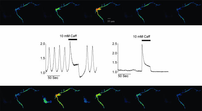
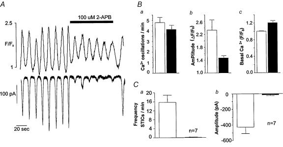
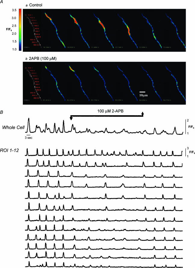
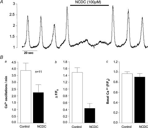
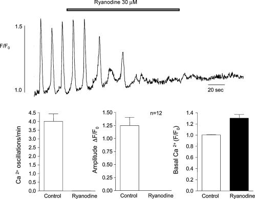
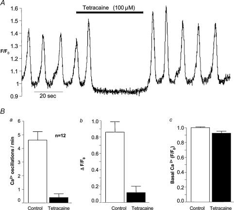
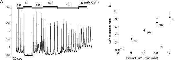
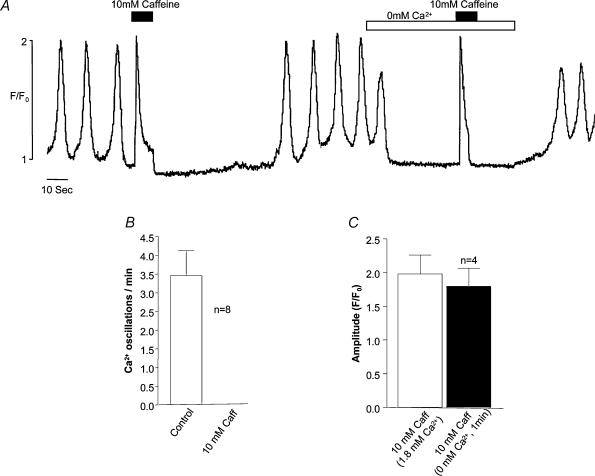
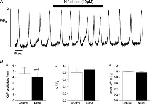
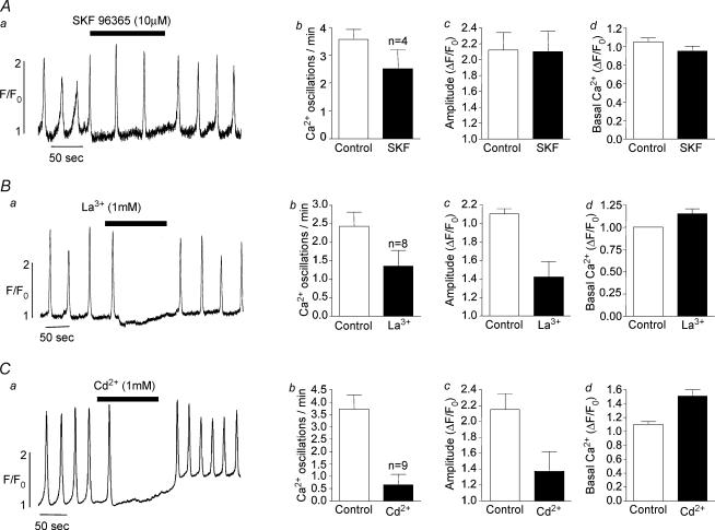
Measurements were made (using fast confocal microscopy) of intracellular Ca2+ levels in fluo-4 loaded interstitial cells isolated from the rabbit urethra. These cells exhibited regular Ca2+ oscillations which were associated with spontaneous transient inward currents recorded under voltage clamp. Interference with D-myo-inositol 1,4,5-trisphosphate (IP3) induced Ca2+ release using 100 microm 2-aminoethoxydiphenyl borate, and the phospholipase C (PLC) inhibitors 2-nitro-4-carboxyphenyl N,N-diphenylcarbamate and U73122 decreased the amplitude of spontaneous oscillations but did not abolish them. However, oscillations were abolished when ryanodine receptors were blocked with tetracaine or ryanodine. Oscillations ceased in the absence of external Ca2+, and frequency was directly proportional to the external Ca2+ concentration. Frequency of Ca2+ oscillation was reduced by SKF-96365, but not by nifedipine. Lanthanum and cadmium completely blocked oscillations. These results suggest that Ca2+ oscillations in isolated rabbit urethral interstitial cells are initiated by Ca2+ release from ryanodine-sensitive intracellular stores, that oscillation frequency is very sensitive to the external Ca2+ concentration and that conversion of the primary oscillation to a propagated Ca2+ wave depends upon IP3-induced Ca2+ release.


Figures

Figure 1
Figure 1. Ca2+ changes in interstitial and smooth muscle cells
Sequence of 10 frames before (top panel, taken at 4 s intervals) and during addition of 10 mm caffeine (bottom panel, at 3 s intervals). The highly branched cell on the right of each frame is a typical interstitial cell, while the darker spindle-shaped cell on the left, scarcely visible under control conditions, is a smooth muscle cell. Regular spontaneous increases in fluorescent intensity were apparent in the perinuclear region of the branched cell, while the smooth muscle cell remained dark. However, when 10 mm caffeine was added both cells lit up simultaneously (frame taken at 64 s). When the caffeine-induced transient was complete, the smooth muscle cell again became dark, and remained so for the rest of the experiment, while the interstitial cell, after a brief pause, resumed its oscillatory behaviour. This experiment also illustrated the different contractile properties of both cells. Thus, while the smooth muscle cell contracted vigorously in response to caffeine addition, the interstitial cell did not contract either in response to the spontaneous increases in intracellular Ca2+ or in response to the caffeine-induced Ca2+ transient. Scale bar, 10 μm.
Figure 2
Figure 2. The effect of 2-APB on Ca2+ oscillations and STICs
A, the upper record shows Ca2+ oscillations imaged in the perinuclear region of the cell, while the lower record shows the spontaneous transient inward currents (STICs) resulting from the intracellular increases in Ca2+. Addition of 100 μm 2-aminoethoxydiphenyl borate (2-APB) had the effect of almost abolishing the STICS, but the underlying Ca2+ oscillations, while reduced in amplitude, retained their normal rhythm and returned to control amplitude after washout of the drug. The summarized results (B and C) show essentially the same pattern as the original record in A.
Figure 3
Figure 3. Effect of 2-APB on wave propagation
Series of images of the initiation and spread of a Ca2+ wave under control conditions (A, upper panel) The wave arose at the top of the cell (region of interest (ROI) 1) and propagated without decrement to the other end of the cell. Occasionally the wave originated at ROI 9 and propagated in the opposite direction, but generally the global event was well coordinated throughout the cell (as is apparent from the plotted regions of interest in B). In the presence of 2-APB (A, lower panel) the pattern was quite different. Oscillations continued in all parts of the cell, but these were now propagated to only one or two of the neighbouring ROIs, with the result that activity in the cell as a whole was now very poorly coordinated. Upon washout of 2-APB, the well-coordinated global events were quickly restored. Scale bar, 10 μm.
Figure 4
Figure 4. The effect of NCDC on Ca2+ oscillations
A, before drug addition oscillations were occurring at a frequency of about 3 min−1. In the presence of 100 μmN,N-diphenylcarbamate (NCDC), the amplitude of oscillations was much reduced, but they continued but at the slightly slower frequency of 2 min−1. The summarized results (B) showed a decrease in both frequency and amplitude, but there was little effect on basal Ca2+ levels.
Figure 5
Figure 5. The effect of ryanodine
The effect of 30 mm ryanodine on Ca2+ oscillations is shown. Within 80 s of drug addition, oscillations had ceased completely, and this was accompanied by an increase in basal Ca2+ concentration within the cell. The effects of ryanodine were irreversible even on prolonged washout. In 12 such experiments this concentration of ryanodine invariably abolished spontaneous oscillations. The effect on basal Ca2+ level was also a consistent one with normalized level increasing from 1.0 ± 0.007 before drug addition to 1.30 ± 0.065 in the presence of 30 mm ryanodine.
Figure 6
Figure 6. The effect of tetracaine
A, tetracaine at 100 μm abolished oscillations within 10 s of drug addition, and these were just as quickly restored when the drug was washed out. The summarized results (B) show that both frequency and amplitude of oscillations were profoundly depressed by this concentration of tetracaine.
Figure 7
Figure 7. Effect of external Ca2+ concentration
Reduction of [Ca2+]o from the normal value of 1.8–0 mm (nominally Ca2+-free, since no chelator was present) immediately caused cessation of oscillations and a decrease in basal cytosolic Ca2+. Frequency and amplitude of oscillations were promptly restored on readmission of normal Ca2+. When this solution was changed to one containing 0.9 mm Ca2+, frequency of oscillation was markedly reduced. On the other hand increasing [Ca2+]o to 3.6 mm had the effect of increasing frequency of oscillation. B, the averaged results (numbers of experiments at each concentration are shown alongside each point) (± s.e.m.) at five different Ca2+ concentrations. It can be seen that there is a roughly linear relationship between oscillation frequency and external Ca2+ concentration.
Figure 8
Figure 8. Removing external Ca2+ does not rapidly deplete stores
When 10 mm caffeine was added in 1.8 mm Ca2+ solution, a maximal Ca2+ transient was induced followed by inhibition of spontaneous oscillations (eight such experiments are summarized in B). A second introduction of 10 mm caffeine a minute after exposure to 0 mm[Ca2+]o still evoked a maximal Ca2+ transient suggesting that the stores had not been depleted in the nominally Ca2+-free solution. In four such experiments there was no significant difference between the transient evoked in 1.8 mm Ca2+ as compared with that evoked in nominally Ca2+-free solution (C).
Figure 9
Figure 9. The effect of nifedipine
Nifedipine at 10 μm increased rather than decreased oscillation frequency while having little effect on amplitude (an effect that was consistent in four preparations as shown in the summarized data, B) suggesting that Ca2+ influx through L-type channels is not important for store refilling.
Figure 10
Figure 10. The effect of blockers of Ca2+ influx
SKF-96365 at 10 μm approximately halved the frequency of oscillation (Aa) and, although this was a greater reduction than the average of four such experiments (30% reduction, Ab), it would suggest that Ca2+ entry through store-operated channels does play a part in modulating oscillation frequency. The nonspecific blockers lanthanum and cadmium chloride had a more dramatic blocking effect confirming the importance of Ca2+ influx in the modulation of intracellular oscillations.

Similar articles

Cited by

References

    1. Aoyama M, Yamada A, Wang J, Ohya S, Furuzono S, Goto T, Hotta S, Ito Y, Matsubara T, Shimokata K, Chen SR, Imaizumi Y, Nakayama S. Requirement of ryanodine receptors for pacemaker Ca2+ activity in ICC and HEK293 cells. J Cell Sci. 2004;117:2813–2825. 10.1242/jcs.01136. - DOI - PubMed
    1. Bayguinov O, Hagen B, Bonev AD, Nelson MT, Sanders KM. Intracellular calcium events activated by ATP in murine colonic myocytes. Am J Physiol Cell Physiol. 2000;279:C126–135. - PubMed
    1. Berridge MJ. Elementary and global aspects of calcium signalling. J Physiol. 1997;499:291–306. - PMC - PubMed
    1. Boittin FX, Coussin F, Macrez N, Mironneau C, Mironneau J. Inositol 1,4,5-trisphosphate-and ryanodine-sensitive Ca2+ release channel-dependent Ca2+ signalling in rat portal vein myocytes. Cell Calcium. 1998;23:303–311. - PubMed
    1. Boittin FX, Coussin F, Morel JL, Halet G, Macrez N, Mironneau J. Ca2+ signals mediated by Ins(1,4,5)P3-gated channels in rat ureteric myocytes. Biochem J. 2000;349:323–332. - PMC - PubMed

Publication types

MeSH terms

LinkOut - more resources