. 2005 Apr;115(4):860-9.
doi: 10.1172/JCI23640. Epub 2005 Mar 3.

The IL-6-gp130-STAT3 pathway in hepatocytes triggers liver protection in T cell-mediated liver injury

Affiliations

The IL-6-gp130-STAT3 pathway in hepatocytes triggers liver protection in T cell-mediated liver injury

Christian Klein et al. J Clin Invest. 2005 Apr.

Abstract

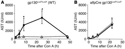
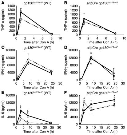
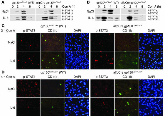
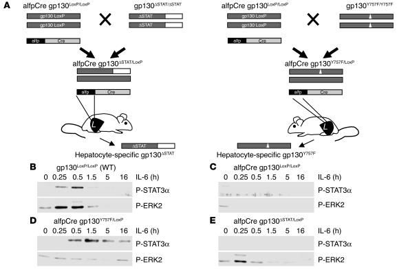
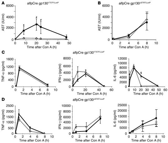
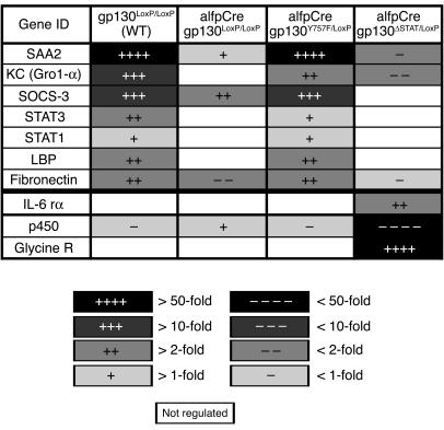
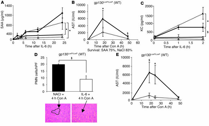
Increasing evidence demonstrates that IL-6 has a protective role during liver injury. IL-6 activates intracellular pathways via the gp130 receptor. In order to identify IL-6-gp130 pathways involved in mediating liver protection, we analyzed hepatocyte-specific gp130 knockout mice in a concanavalin A-induced (Con A-induced) model of immune-mediated hepatitis. We demonstrated that IL-6-gp130-dependent pathways in hepatocytes alone are sufficient for triggering protection in Con A-induced hepatitis. gp130-STAT3 signaling in hepatocytes mediates the IL-6-triggered protective effect. This was demonstrated by analysis of IL-6-induced protection in mice selectively deficient for gp130-dependent STAT1/3 or gp130-SHP2-RAS signaling in hepatocytes. To identify IL-6-gp130-STAT1/3 dependently expressed liver-protective factors, we performed gene array analysis of hepatic gene expression in hepatocyte-specific gp130(-/-) mice as well as in gp130-STAT1/3- and gp130-SHP2-RAS-MAPK-deficient mice. The mouse IL-8 ortholog KC (also known as Gro-alpha) and serum amyloid A2 (SAA2) was identified as differentially IL-6-gp130-STAT3-regulated genes. Hepatic expression of KC and SAA2 mediate the liver-protective potential of IL-6, since treatment with recombinant KC or serum SAA2 effectively reduced liver injury during Con A-induced hepatitis. In summary, this study defines IL-6-gp130-STAT3-dependent gene expression in hepatocytes that mediates IL-6-triggered protection in immune-mediated Con A-induced hepatitis. Additionally, we identified the IL-6-gp130-STAT3-dependent proteins KC and SAA2 as new candidates for therapeutic targets in liver diseases.


Figures

Figure 1
Figure 1
gp130 in hepatocytes is essential for IL-6–dependent protection in Con A–induced hepatitis. Con A hepatitis was induced by intravenous injection of 32.5 mg/kg Con A in wild-type (gp130LoxP/LoxP) and hepatocyte-specific gp130-null (alfpCre gp130LoxP/LoxP) mice. Mice were pretreated for 3 hours with NaCl (solid line) or IL-6 (200 μg/kg) (dotted line) before i.v. injection of Con A. Liver injury was quantified by detection of AST. (A) Time course of AST serum levels during Con A–induced hepatitis in wild-type mice pretreated with NaCl (solid line) or IL-6 (dotted line). #P < 0.01 and *P < 0.05 vs. corresponding IL-6–pretreated wild-type control group at the same time point. (B) Time course of AST serum levels during Con A–induced hepatitis in alfpCre gp130LoxP/LoxP mice pretreated with NaCl (solid line) or IL-6 (dotted line).
Figure 2
Figure 2
Cytokine secretion during Con A–induced hepatitis. Cytokine levels in the serum after Con A injection in wild-type (gp130LoxP/LoxP) and alfpCre gp130LoxP/LoxP mice. Three hours before Con A administration, animals were treated with NaCl (solid line) or IL-6 (200 μg/kg) (dotted line) by i.p. injection. (A and B) TNF-α levels of wild-type and alfpCre gp130LoxP/LoxP mice. #P < 0.01 vs. corresponding IL-6–pretreated wild-type control group at the same time point. (C and D) IFN-γ levels of wild-type and alfpCre gp130LoxP/LoxP mice. (E and F) IL-6 concentrations of wild-type and alfpCre gp130LoxP/LoxP mice. §P < 0.001 and #P < 0.01 vs. corresponding IL-6–pretreated wild-type or alfpCre gp130LoxP/LoxP mice at the same time point.
Figure 3
Figure 3
Changes in liver nuclear phosphorylation of STAT1 and STAT3 during Con A–induced hepatitis. (A) Phosphorylation of STAT1 in wild-type and alfpCre gp130LoxP/LoxP mice. Western blot analysis of STAT1 and STAT3 activation, as detected by phosphorylation of STAT1 and STAT3, in liver nuclear protein extracts of wild-type and alfpCre gp130LoxP/LoxP mice. Animals were pretreated 3 hours before Con A administration with NaCl or IL-6 (200 μg/kg) by i.p. injection as indicated. Representative results of 3 independent experiments are shown. (B) Phosphorylation of STAT3 in wild-type and alfpCre gp130LoxP/LoxP mice. (C) Double immunofluorescence of liver sections for P-STAT3 and CD11b expression. CD11b detects activated macrophages, NK cells, and monocytes. Mice were killed 2 hours after Con A injection. DAPI counterstaining was used to detect nuclei. (D) Double immunofluorescence of liver sections for P-STAT3 and CD11b expression. CD11b detects activated macrophages, NK cells, and monocytes. Mice were killed 4 hours after Con A injection. Representative areas are shown. Slides of 3 animals per group were analyzed. Magnification, ×400.
Figure 4
Figure 4
Generation and functional characterization of hepatocyte-specific gp130ΔSTAT/LoxP and gp130Y757F/LoxP mice. (A) alfpCre gp130LoxP/LoxP mice were generated by breeding alfpCre mice with animals expressing LoxP-flanked gp130 alleles (25). Hepatocyte-specific gp130ΔSTAT/LoxP animals (alfpCre gp130ΔSTAT/LoxP) were generated by crossing alfpCre gp130LoxP/LoxP with gp130ΔSTAT/ΔSTAT mice, which express a truncated gp130 knockin allele encoding a truncated gp130 protein that lacks the domains mediating STAT1 and STAT3 activation (26). Hepatocyte-specific gp130Y757F/LoxP animals (alfpCre gp130Y757F/LoxP) were bred by crossing alfpCre gp130LoxP/LoxP with gp130Y757F/Y757F mice, which express a gp130 allele encoding a phenylalanine substitution of the Y757 residue (32), thereby rendering gp130 incapable of recruiting SHP2 and activating the RAS-MAPK pathway. Animals were genotyped by PCR analysis for alfpCre, gp130LoxP, and gp130Y757F alleles. (B and E) The functional characterization of hepatocyte-specific gp130 mutant mice. Gp130 downstream-signaling pathways were analyzed by monitoring of phosphorylated STAT3 and phosphorylated ERK2 (p42) expression in whole cell extracts of primary hepatocytes isolated from wild-type (B), alfpCre gp130LoxP/LoxP (C), alfpCre gp130Y757F/LoxP (D), or alfp Cre gp130ΔSTAT/LoxP mice (E). Activation of phosphorylated STAT1 was not detected in any of the 4 mouse strains.
Figure 5
Figure 5
gp130-STAT3 signaling in hepatocytes is responsible for IL-6–induced liver protection. Con A–induced hepatitis was induced by intravenous injection of 32.5 mg/kg Con A in hepatocyte-specific gp130ΔSTAT (alfpCre gp130ΔSTAT/LoxP) and hepatocyte-specific gp130Y757F/LoxP mice (alfpCre gp130Y757F/LoxP). Mice were pretreated 3 hours before Con A injection with NaCl (solid line) or IL-6 (200 ng/g) (dotted line) by i.p. injection. (A and B) Liver damage was quantified by detection of ASTs. (A) AST levels of alfpCre gp130Y757F/LoxPmice in the time course of Con A–induced hepatitis, pretreated with NaCl (solid line) or IL-6 (dotted line). #P < 0.01 and *P < 0.05 vs. corresponding IL-6–pretreated control group at the same time point. (B) AST levels of alfpCre gp130ΔSTAT/LoxP in the time course of Con A–induced hepatitis, pretreated with NaCl (solid line) or IL-6 (dotted line). (C and D) TNF-α, IFN-γ, and IL-6 secretion in the serum during Con A–induced hepatitis in alfpCre gp130Y757F/LoxP (C) and alfpCre gp130ΔSTAT/LoxP mice (D) was determined by ELISA. §P < 0.001 vs. corresponding IL-6–pretreated alfpCre gp130Y757F/LoxP mice at the same time point.
Figure 6
Figure 6
IL-6–induced gene expression profile of wild-type, alfpCre gp130LoxP/LoxP, alfpCre gp130Y757F/LoxP, and alfpCre gp130ΔSTAT/LoxP mice. Affimetrix GeneChip analysis was used to detect gene expression of IL-6–stimulated gp130LoxP/LoxP, alfpCre gp130LoxP/LoxP, alfpCre gp130ΔSTAT/LoxP, and alfpCre gp130Y757F/LoxP mice. Three animals per group were treated in parallel by i.p. injection of recombinant IL-6 (IL-6 rα) (200 μg/kg) or NaCl. Liver RNA was isolated, and gene expression was compared. Most of the examples shown are differentially regulated by gp130-STAT signaling. Glycine receptor (Glycine R) and cytochrome p450 (p450) represent examples that are regulated by gp130-ras-MAPK signaling. The change of gene expression level is marked by increasing color intensity. LBP, LPS binding protein.
Figure 7
Figure 7
Acute-phase protein SAA2 and the chemokine KC are IL-6–gp130–STAT–induced, liver-protective proteins. (A) SAA2 serum levels after IL-6 (200 μg/kg) injection. Each time point represents the serum of 3–5 animals. Filled squares represent wild-type mice, open squares show alfpCre gp130LoxP/LoxP mice, filled triangles mark alfpCre gp130Y757F/LoxP mice, and open triangles represent alfpCre gp130ΔSTAT/LoxP. (B) Pretreatment with recombinant SAA (0.8 mg/kg) leads to a significant reduction of AST levels in the time course of Con A–induced hepatitis in wild-type mice. Either SAA (dotted line) or NaCl (solid line) was injected intravenously 1 hour before induction of Con A–induced hepatitis. (C) Serum KC levels after IL-6 injection. Filled squares represent wild-type mice, open squares show alfpCre gp130LoxP/LoxPmice, filled triangles mark alfpCre gp130Y757F/LoxP mice, and open triangles represent alfpCre gp130ΔSTAT/LoxP. (D) IL-6 treatment results in a significant reduction of PMN infiltration during Con A–induced hepatitis. Four hours after Con A injection, the influx of PMNs was counted on H&:E-stained paraffin sections by an experienced liver pathologist. The arrows mark liver infiltrating PMNs. hPF, high powered field. (E) KC (40 μg/kg) treatment leads to a significant reduction of AST levels during Con A–induced hepatitis. Eight mice per group were treated with either KC (dotted line) or NaCl (solid line) 1 hour before Con A injection. §P < 0.001 and *P < 0.05 vs. corresponding control groups at the same time point.

Similar articles

Cited by

References

    1. Mizuhara H, et al. T cell activation-associated hepatic injury: mediation by tumor necrosis factors and protection by interleukin 6. J. Exp. Med. 1994;179:1529–1537. - PMC - PubMed
    1. Kovalovich K, et al. Increased toxin-induced liver injury and fibrosis in interleukin-6-deficient mice. Hepatology. 2000;31:149–159. - PubMed
    1. Klein C, et al. ME3738 protects from concanavalin A-induced liver failure via an IL-6-dependent mechanism. Eur. J. Immunol. 2003;33:2251–2261. - PubMed
    1. Hong F, et al. Elevated interleukin-6 during ethanol consumption acts as a potential endogenous protective cytokine against ethanol-induced apoptosis in the liver: involvement of induction of Bcl-2 and Bcl-x(L) proteins. Oncogene. 2002;21:32–43. - PubMed
    1. Kovalovich K, et al. Interleukin-6 protects against Fas-mediated death by establishing a critical level of anti-apoptotic hepatic proteins FLIP, Bcl-2, and Bcl-xL. J. Biol. Chem. 2001;276:26605–26613. - PubMed

Publication types

MeSH terms