. 2005 Apr;115(4):1030-8.
doi: 10.1172/JCI23962. Epub 2005 Mar 10.

Prevention of obesity in mice by antisense oligonucleotide inhibitors of stearoyl-CoA desaturase-1

Affiliations

Prevention of obesity in mice by antisense oligonucleotide inhibitors of stearoyl-CoA desaturase-1

Guoqiang Jiang et al. J Clin Invest. 2005 Apr.

Erratum in

  • J Clin Invest. 2005 Aug;115(8):2297

Abstract

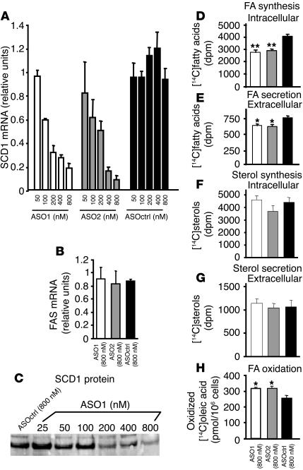
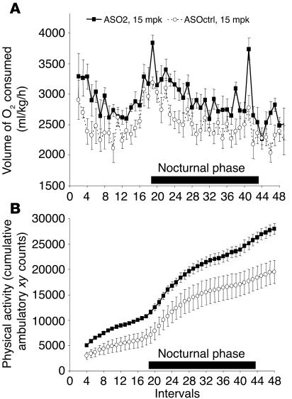
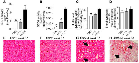
Effective therapies for the treatment of obesity, a key element of metabolic syndrome, are urgently needed but currently lacking. Stearoyl-CoA desaturase-1 (SCD1) is the rate-limiting enzyme catalyzing the conversion of saturated long-chain fatty acids into monounsaturated fatty acids, which are major components of triglycerides. In the current study, we tested the efficacy of pharmacological inhibition of SCD1 in controlling lipogenesis and body weight in mice. SCD1-specific antisense oligonucleotide inhibitors (ASOs) reduced SCD1 expression, reduced fatty acid synthesis and secretion, and increased fatty acid oxidization in primary mouse hepatocytes. Treatment of mice with SCD1 ASOs resulted in prevention of diet-induced obesity with concomitant reductions in SCD1 expression and the ratio of oleate to stearoyl-CoA in tissues and plasma. These changes correlated with reduced body adiposity, hepatomegaly and steatosis, and postprandial plasma insulin and glucose levels. Furthermore, SCD1 ASOs reduced de novo fatty acid synthesis, decreased expression of lipogenic genes, and increased expression of genes promoting energy expenditure in liver and adipose tissues. Thus, SCD1 inhibition represents a new target for the treatment of obesity and related metabolic disorders.


Figures

Figure 1
Figure 1
Effects of SCD1 ASOs on SCD1 mRNA and protein levels, fatty acid and sterol synthesis and secretion, and fatty acid oxidation in primary mouse hepatocytes in vitro. Primary hepatocytes isolated from C57/B6 mice were transfected with ASO1, ASO2 and ASOctrl. Shown are the relative abundances of SCD1 (A) and FAS (B) mRNA levels in the transfected hepatocytes as quantified by TaqMan real-time PCR. SCD1 protein levels were determined by Western blot analysis (C). Also shown are the levels of conversion of [14C]acetate into [14C]-labeled fatty acids in hepatocytes (intracellular; D), [14C]-labeled fatty acids secreted into the tissue culture medium (extracellular; E), [14C]sterols in hepatocytes (intracellular; F), [14C]sterols in the culture medium (extracellular; G), and conversion of [14C]oleic acid into soluble [14C]-labeled products in the cells (H). The experiments were repeated 3 times with similar results.
Figure 2
Figure 2
Effect of SCD1 ASOs on weight gain, body adiposity, and plasma metabolic parameters in C57/B6 mice on HFD. C57/B6 mice were given HFD and treated with ASO1, ASO2, or ASOctrl at 5 and 15 mpk twice a week for 9–10 weeks as described in Methods. Shown are the body weight (A) and food intake (B) of the animals measured weekly. Also shown are body composition (C), and postprandial plasma insulin (D) and glucose (E) measured at the end of the treatments. While ASO2 was only tested in the current experiment, similar results on ASO1 and ASOctrl were obtained in 2 experiments. *P < 0.05; **P < 0.01.
Figure 3
Figure 3
Effect of SCD1 ASO2 on oxygen consumption and physical activity of C57/B6 mice on HFD. Interval measurements of oxygen consumption and physical activity were done every 30 minutes for 24 hours on C57/B6 mice fed HFD and treated with ASO2 or ASOctrl at 15 mpk for 10 weeks as described in Methods. Shown are the volume of oxygen consumed (A) and the cumulative ambulatory xy counts (i.e., the number of times that light beams in the x and y axes were broken due to animal movement; B). Both oxygen consumption and physical activity are significantly higher in ASO2-treated mice than in ASOctrl-treated mice (P < 0.05).
Figure 4
Figure 4
Effect of SCD1 ASOs on SCD1 enzymatic activity, de novo fatty acid synthesis activity, and fat accumulation in the liver of treated C57/B6 mice on HFD. SCD1 enzymatic activity in the liver of mice treated with the various ASOs at 15 mpk for 4 weeks (A) and 10 weeks (B). The de novo fatty acid synthesis activity as indicated by the level of conversion of [3H]water into fatty acids (C) or sterols (D) in the liver tissues of mice treated with the various ASOs at 15 mpk for 10 weeks. (EG) Images of H&E staining of liver sections of mice treated with the ASO1 (E), ASO2 (F) and ASOctrl (G) at 15 mpk for 10 weeks. The clear vacuoles in the liver section are identified by arrows in G. (H) Images of oil red O staining of frozen liver section of a mouse treated with ASOctrl at 15 mpk for 10 weeks. The lipid drops stained by oil red O within the vacuoles are identified by arrows.
Figure 5
Figure 5
Effect of SCD1 ASOs on lipid desaturation index in liver and plasma of treated C57/B6 mice on HFD. Saturated fatty acids and MUFAs in liver and plasma were measured after mice were treated with the various ASOs at 15 mpk for 4 weeks as described in Methods. Shown are the ratios of the levels of MUFAs verses saturated fatty acids in liver (A and B) and in plasma (C and D).
Figure 6
Figure 6
Proposed molecular basis for SCD1 ASO-mediated metabolic effects in mice. SCD1 ASOs directly reduce SCD1 levels in liver. SCD1 ASOs also reduce SCD1 in WAT and BAT; however, whether the reduction is due to direct or indirect effects remains unknown and is indicated by question marks. In liver, the reduction of SCD1 leads to increased CPT1 levels first and decreased SREBP-1, FAS, ACC1, and ACC2 later. It is possible that the downregulation of FAS, ACC1, and ACC2 may be downstream of the transcription factor SREBP-1. In WAT, reduction of SCD1 leads to reduction of FAS. In BAT, reduction of SCD1 leads to increase in UCP2 first and then UCP1, UCP3, and β3-AR.

Comment in

References

    1. Kopelman PG. Obesity as a medical problem. Nature. 2000;404:635–643. - PubMed
    1. Abelson P, Kennedy D. The obesity epidemic. Science. 2004;304:1413. - PubMed
    1. Enoch HG, Catala A, Strittmatter P. Mechanism of rat liver microsomal stearyl-CoA desaturase. Studies of the substrate specificity, enzyme-substrate interactions, and the function of lipid. J. Biol. Chem. 1976;251:5095–5103. - PubMed
    1. Ntambi JM, Miyazaki M. Regulation of stearoyl-CoA desaturases and role in metabolism. Prog. Lipid Res. 2004;43:91–104. - PubMed
    1. Zheng Y, et al. Scd3--a novel gene of the stearoyl-CoA desaturase family with restricted expression in skin. Genomics. 2001;71:182–191. - PubMed

MeSH terms