

Tumor angiogenesis and dynamic CT in colorectal carcinoma: radiologic-pathologic correlation
- PMID: 15761965
- PMCID: PMC4250674
- DOI: 10.3748/wjg.v11.i9.1287
Tumor angiogenesis and dynamic CT in colorectal carcinoma: radiologic-pathologic correlation
Abstract
Aim: To investigate the correlation between microvessel density and spiral CT perfusion imaging in colorectal carcinoma.
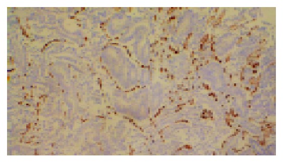
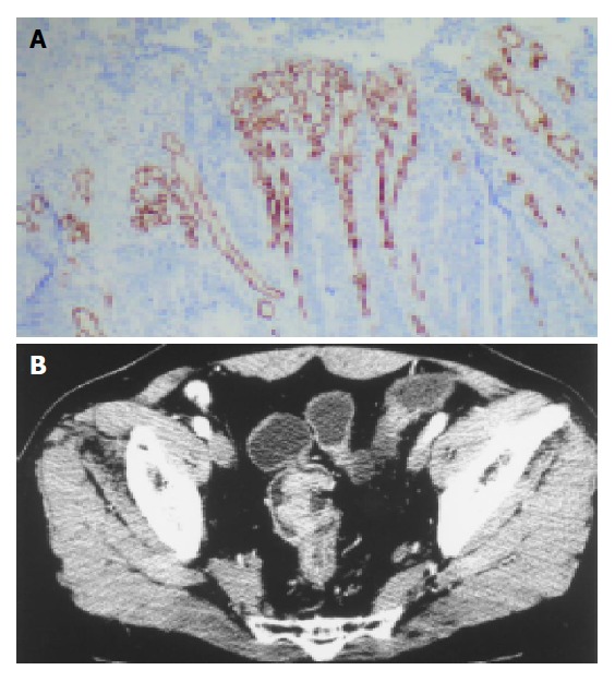
Methods: Thirty-seven patients, with histologically proven colorectal carcinoma, underwent water enema spiral CT scan. The largest axial surface of the primary tumor was searched on unenhanced spiral CT images. At this level, the enhanced dynamic scan series was acquired. Time-density curves (TDC) were created from the region of interest drawn over the tumor, target artery by Toshiba Xpress/SX spiral CT with perfusion functional software. Then the perfusion was calculated. Microvessel density (MVD) was evaluated using immunohistochemical staining of surgical specimens with anti-CD34, and then MVD was correlated with perfusion.
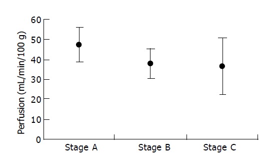
Results: MVD of colorectal carcinomas was 33.11-173.44, mean 87.28, and perfusion was 15.60-64.80 mL/min/100 g, mean 39.74 mL/min/100 g. MVD and perfusion were not associated with invasive depth, metastasis and disease stage, and they all decreased with increasing Dukes' stage, but no significant correlation was found between them (r = 0.18, P = 0.29).
Conclusion: There is no significant correlation between MVD and perfusion. Neovascularizaton and perfusion are highly presented in early colorectal carcinoma. CT perfusion imaging may be more suited for assessing tumorigenesis in colorectal carcinoma than histological MVD technique.
Figures
Similar articles
-
Evaluation of angiogenesis in colorectal carcinoma with multidetector-row CT multislice perfusion imaging.Eur J Radiol. 2010 Aug;75(2):191-6. doi: 10.1016/j.ejrad.2009.04.058. Epub 2009 May 29. Eur J Radiol. 2010. PMID: 19481397
-
[Evaluation of microvessel density and vascular endothelial growth factor in colorectal carcinoma with 64-multidetector-row CT perfusion imaging].Zhonghua Wei Chang Wai Ke Za Zhi. 2008 Nov;11(6):537-41. Zhonghua Wei Chang Wai Ke Za Zhi. 2008. PMID: 19031129 Chinese.
-
Assessment of tumor grade and angiogenesis in colorectal cancer: whole-volume perfusion CT.Acad Radiol. 2014 Jun;21(6):750-7. doi: 10.1016/j.acra.2014.02.011. Acad Radiol. 2014. PMID: 24809317
-
Diagnostic value of one-stop CT energy spectrum and perfusion for angiogenesis in colon and rectum cancer.BMC Med Imaging. 2024 May 21;24(1):116. doi: 10.1186/s12880-024-01291-8. BMC Med Imaging. 2024. PMID: 38773384 Free PMC article.
-
Perfusion CT imaging of colorectal cancer.Br J Radiol. 2014 Feb;87(1034):20130811. doi: 10.1259/bjr.20130811. Br J Radiol. 2014. PMID: 24434157 Free PMC article. Review.
Cited by
-
Assessment of blood flow in hepatocellular carcinoma: correlations of computed tomography perfusion imaging and circulating angiogenic factors.Int J Mol Sci. 2013 Aug 27;14(9):17536-52. doi: 10.3390/ijms140917536. Int J Mol Sci. 2013. PMID: 23985826 Free PMC article.
-
Does arterial spin-labeling MR imaging-measured tumor perfusion correlate with renal cell cancer response to antiangiogenic therapy in a mouse model?Radiology. 2009 Jun;251(3):731-42. doi: 10.1148/radiol.2521081059. Radiology. 2009. PMID: 19474376 Free PMC article.
-
Imaging of tumor angiogenesis: functional or targeted?AJR Am J Roentgenol. 2009 Aug;193(2):304-13. doi: 10.2214/AJR.09.2869. AJR Am J Roentgenol. 2009. PMID: 19620425 Free PMC article. Review.
-
Perfusion CT in solid body-tumours. Part II: Clinical applications and future development.Radiol Med. 2010 Sep;115(6):858-74. doi: 10.1007/s11547-010-0545-9. Epub 2010 Mar 9. Radiol Med. 2010. PMID: 20221706 English, Italian.
-
Protocol modifications for CT perfusion (CTp) examinations of abdomen-pelvic tumors: impact on radiation dose and data processing time.Eur Radiol. 2011 Jun;21(6):1293-300. doi: 10.1007/s00330-010-2048-4. Epub 2011 Jan 19. Eur Radiol. 2011. PMID: 21246200
References
-
- Hawighorst H, Knapstein PG, Knopp MV, Weikel W, Brix G, Zuna I, Schönberg SO, Essig M, Vaupel P, van Kaick G. Uterine cervical carcinoma: comparison of standard and pharmacokinetic analysis of time-intensity curves for assessment of tumor angiogenesis and patient survival. Cancer Res. 1998;58:3598–3602. - PubMed
-
- Bossi P, Viale G, Lee AK, Alfano R, Coggi G, Bosari S. Angiogenesis in colorectal tumors: microvessel quantitation in adenomas and carcinomas with clinicopathological correlations. Cancer Res. 1995;55:5049–5053. - PubMed
-
- Hermans R, Lambin P, Van der Goten A, Van den Bogaert W, Verbist B, Weltens C, Delaere PR. Tumoural perfusion as measured by dynamic computed tomography in head and neck carcinoma. Radiother Oncol. 1999;53:105–111. - PubMed
-
- Miles KA, Hayball MP, Dixon AK. Measurement of human pancreatic perfusion using dynamic computed tomography with perfusion imaging. Br J Radiol. 1995;68:471–475. - PubMed
Publication types
MeSH terms
LinkOut - more resources
Full Text Sources
Medical
