

In vivo demonstration of T lymphocyte migration and amelioration of ileitis in intestinal mucosa of SAMP1/Yit mice by the inhibition of MAdCAM-1
- PMID: 15762871
- PMCID: PMC1809333
- DOI: 10.1111/j.1365-2249.2005.02742.x
In vivo demonstration of T lymphocyte migration and amelioration of ileitis in intestinal mucosa of SAMP1/Yit mice by the inhibition of MAdCAM-1
Abstract
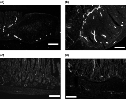
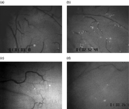
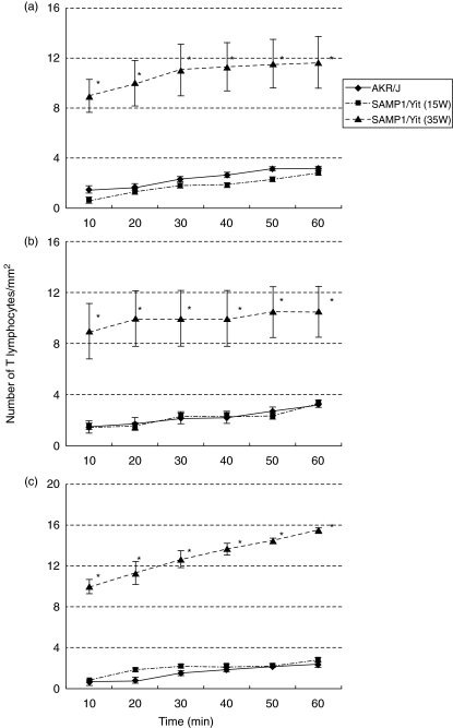
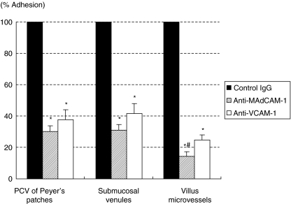
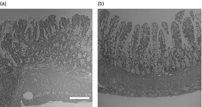
The aetiology of Crohn's disease (CD) remains unknown. Since SAMP1/Yit mice have been reported to develop CD-like spontaneous enteric inflammation, such mice have been studied as an animal model of CD. In this study, using this model we examined T lymphocyte migration in microvessels of intestinal mucosa in vivo and the expression of adhesion molecules by immunohistochemistry. Fluorescence-labelled T lymphocytes isolated from AKR/J (control) mice were injected into the tail veins of recipient mice, and T lymphocyte migration in the postcapillary venules of Peyer's patches, submucosal microvessels, and villus capillaries of the terminal ileum was monitored using an intravital microscope. Adhesion of T lymphocytes was significantly increased in 35 week old SAMP1/Yit mice compared with that in AKR/J or 15 week old SAMP1/Yit mice. Immunohistochemical study showed increased infiltration of CD4, CD8 and beta7-integrin-positive cells and increased expression of MAdCAM-1 and VCAM-1 in the terminal ileum of SAMP1/Yit mice. Antibodies against MAdCAM-1 and VCAM-1 significantly inhibited adhesion of T lymphocytes to microvessels of the terminal ileum, and anti-MAdCAM-1 antibody showed stronger suppressive effect than the anti-VCAM-1 antibody. Periodical administration of anti-MAdCAM-1 antibody twice a week for 7 weeks significantly ameliorated ileitis of SAMP1/Yit mice, but submucosal hypertrophy was not significantly suppressed. Anti-VCAM-1 antibody treatment failed to show significant resolution of ileitis. In addition, anti-MAdCAM-1 antibody treatment also attenuated established ileitis. The results demonstrate that, although MAdCAM-1 and VCAM-1 play an important role in T lymphocyte-endothelial cell interactions in SAMP1/Yit mice, MAdCAM-1 may be a more appropriate target for therapeutic modulation of chronic ileitis.
Figures
Similar articles
-
Blockade of PSGL-1 attenuates CD14+ monocytic cell recruitment in intestinal mucosa and ameliorates ileitis in SAMP1/Yit mice.J Leukoc Biol. 2005 Mar;77(3):287-95. doi: 10.1189/jlb.0204104. Epub 2004 Nov 29. J Leukoc Biol. 2005. PMID: 15569697
-
Lemon grass (Cymbopogon citratus) ameliorates murine spontaneous ileitis by decreasing lymphocyte recruitment to the inflamed intestine.Microcirculation. 2010 Jul;17(5):321-32. doi: 10.1111/j.1549-8719.2010.00032.x. Microcirculation. 2010. PMID: 20618690
-
Spatial heterogeneity of TNF-alpha-induced T cell migration to colonic mucosa is mediated by MAdCAM-1 and VCAM-1.Am J Physiol Gastrointest Liver Physiol. 2002 Dec;283(6):G1379-87. doi: 10.1152/ajpgi.00026.2002. Epub 2002 May 29. Am J Physiol Gastrointest Liver Physiol. 2002. PMID: 12388196
-
Pathogenesis of gastritis in ileitis-prone SAMP1/Yit mice.Keio J Med. 2011;60(2):65-8. doi: 10.2302/kjm.60.65. Keio J Med. 2011. PMID: 21720202 Review.
-
Effect of dietary fat on intestinal inflammatory diseases.J Gastroenterol Hepatol. 2013 Dec;28 Suppl 4:33-6. doi: 10.1111/jgh.12252. J Gastroenterol Hepatol. 2013. PMID: 24251701 Review.
Cited by
-
Blocking lymphocyte localization to the gastrointestinal mucosa as a therapeutic strategy for inflammatory bowel diseases.Gastroenterology. 2011 May;140(6):1776-84. doi: 10.1053/j.gastro.2011.02.015. Gastroenterology. 2011. PMID: 21530744 Free PMC article.
-
Shining a light on intestinal traffic.Clin Dev Immunol. 2012;2012:808157. doi: 10.1155/2012/808157. Epub 2011 Nov 22. Clin Dev Immunol. 2012. PMID: 22162719 Free PMC article. Review.
-
Rheostat regulation of integrin-mediated leukocyte adhesion.J Clin Invest. 2007 Sep;117(9):2391-5. doi: 10.1172/JCI33376. J Clin Invest. 2007. PMID: 17786236 Free PMC article.
-
MAdCAM-1-Mediated Intestinal Lymphocyte Homing Is Critical for the Development of Active Experimental Autoimmune Encephalomyelitis.Front Immunol. 2019 Apr 26;10:903. doi: 10.3389/fimmu.2019.00903. eCollection 2019. Front Immunol. 2019. PMID: 31114574 Free PMC article.
-
Tissue specific heterogeneity in effector immune cell response.Front Immunol. 2013 Aug 27;4:254. doi: 10.3389/fimmu.2013.00254. eCollection 2013. Front Immunol. 2013. PMID: 23986763 Free PMC article.
References
-
- Podolsky DK. Inflammatory bowel disease. N Engl J Med. 2002;347:417–29. - PubMed
-
- Marteau P. Inflammatory bowel disease. Endoscopy. 2000;32:131–7. - PubMed
-
- Satsangi J, Welsh KI, Bunce M, et al. Contribution of genes of the major histocompatibility complex to susceptibility and disease phenotype in inflammatory bowel disease. Lancet. 1996;347:1212–7. - PubMed
-
- Ogura Y, Bonen DK, Inohara N, et al. A frameshift mutation in NOD2 associated with susceptibility to Crohn's disease. Nature. 2001;411:603–6. - PubMed
-
- Hugot JP, Chamaillard M, Zouali H, et al. Association of NOD2 leucine-rich repeat variants with susceptibility to Crohn's disease. Nature. 2001;411:599–603. - PubMed
MeSH terms
Substances
LinkOut - more resources
Full Text Sources
Molecular Biology Databases
Research Materials
Miscellaneous
