. 2005 May;109(2):109-24.
doi: 10.1016/j.virusres.2004.11.014. Epub 2004 Dec 18.

Two cellular proteins that interact with a stem loop in the simian hemorrhagic fever virus 3'(+)NCR RNA

Affiliations

Two cellular proteins that interact with a stem loop in the simian hemorrhagic fever virus 3'(+)NCR RNA

Taronna R Maines et al. Virus Res. 2005 May.

Abstract

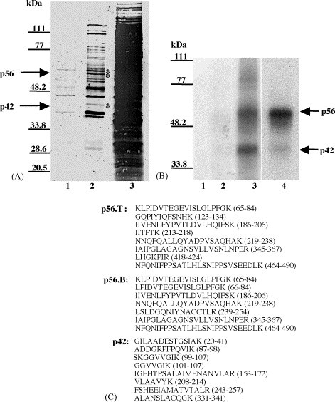
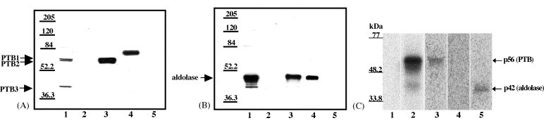
Both full-length and subgenomic negative-strand RNAs are initiated at the 3' terminus of the positive-strand genomic RNA of the arterivirus, simian hemorrhagic fever virus (SHFV). The SHFV 3'(+) non-coding region (NCR) is 76 nts in length and forms a stem loop (SL) structure that was confirmed by ribonuclease structure probing. Two cell proteins, p56 and p42, bound specifically to a probe consisting of the SHFV 3'(+)NCR RNA. The 3'(+)NCR RNAs of two additional members of the arterivirus genus specifically interacted with two cell proteins of the same size. p56 was identified as polypyrimidine tract-binding protein (PTB) and p42 was identified as fructose bisphosphate aldolase A. PTB binding sites were mapped to a terminal loop and to a bulged region of the SHFV 3'SL structure. Deletion of either of the PTB binding sites in the viral RNA significantly reduced PTB binding activity, suggesting that both sites are required for efficient binding of this protein. Changes in the top portion of the SHFV 3'SL structure eliminated aldolase binding, suggesting that the binding site for this protein is located near the top of the SL. These cell proteins may play roles in regulating the functions of the genomic 3' NCR.


Figures

Fig. 1
Fig. 1
Analysis of the interaction between proteins in MA104 cell extracts and 32P-labeled arterivirus 3′(+)NCR RNAs. (A) Gel mobility shift assay. Radiolabeled SHFV 3′(+)NCR RNA was incubated with an S100 cytoplasmic extract from either SHFV-infected or mock-infected MA104 cells. The RPCs were resolved on a 10% non-denaturing polyacrylamide gel and visualized by autoradiography. (Lane 1) free probe; (lanes 2–5) increasing amounts of mock-infected MA104 S100 cytoplasmic extract (100, 200, 300, and 400 ng); (lanes 6–10) increasing amounts of SHFV-infected MA104 S100 cytoplasmic extract (100, 200, 300, 400, and 500 ng). The locations of the RNA–protein complex and free probe are indicated by arrows. (B) Competition gel mobility shift assay. Different amounts of non-radiolabeled competitor RNAs were incubated with an MA104 S100 cytoplasmic extract before addition of the 32P-labeled SHFV 3′(+)NCR RNA. The RPCs were resolved on a 10% non-denaturing polyacrylamide gel and visualized by autoradiography. (Lane 1) free probe; (lane 2) no competitor; (lanes 3–6) increasing amounts of non-radiolabeled SHFV 3′(+)NCR RNA (5-, 10-, 20-, and 30-fold molar excess); (lane 7) 250-fold molar excess of yeast tRNA; (lane 8) 150-fold molar excess of WNV 3′(+)SL RNA; (lane 9) 250-fold molar excess of poly(I)–poly(C). The locations of the RNA–protein complex and free probe are indicated by arrows. (C) UV-induced cross-linking assay. MA104 S100 cytoplasmic extracts were incubated with radiolabeled SHFV 3′(+)NCR RNA and then were exposed to UV-irradiation. The unprotected RNA was digested with RNase A and the cross-linked proteins were resolved by 10% SDS–PAGE and visualized by autoradiography. (Lane 1) free probe; (lane 2) mock-infected MA104 S100 cytoplasmic extract (1 μg) and poly(I)–(C) (1 μg); (lane 3) SHFV-infected MA104 S100 cytoplasmic extract (1 μg) and poly(I)–(C) (1 μg); (lane 4) mock-infected MA104 S100 cytoplasmic extract (1 μg) and poly(I)–(C) (600 ng). Standard protein markers are indicated by lines and the positions of p56 and p42 are indicated by arrows. (D) UV-induced cross-linking assay. MA104 S100 cytoplasmic extracts and different arterivirus 32P-labeled RNA probes were cross-linked by UV-irradiation in the presence of 600 ng of poly(I)–(C). (Lane 1) free probe; (lane 2) SHFV 3′(+)NCR RNA; (lane 3) EAV 3′(+)NCR RNA; (lane 4) PRRSV 3′(+)NCR RNA. The gels shown in lanes 1 and 2 were analyzed by autoradiography and the gel shown in lanes 3 and 4 was analyzed using the FUJI Bio Imaging Analyzer. The positions of protein standard markers are indicated by lines on the right. The positions of the p56 and p42 bands are indicated by arrows. (E) Competition gel mobility shift assay. MA104 S100 cytoplasmic extracts were incubated with different amounts of non-radiolabeled arterivirus RNAs before addition of the 32P-labeled SHFV 3′(+)NCR RNA. (Lane 1) free probe; (lane 2) no competitor; (lanes 3–5) increasing amounts of unlabeled EAV 3′(+)NCR RNA (25-, 50-, and 75-fold molar excess); (lanes 6–8) increasing amounts of unlabeled PRRSV 3′(+)NCR RNA (25-, 50-, and 75-fold molar excess). The gels were analyzed using the FUJI Bio Imaging Analyzer. The locations of the RNA–protein complex and free probe are indicated by arrows.
Fig. 2
Fig. 2
Schematic representation of the truncated 32P-labeled SHFV 3′(+)NCR RNAs used in UV-induced cross-linking assays. Numbering was from the 3′ end. Three adenines of the poly(A) tract were included. The plus symbol (+) indicates wildtype binding observed with the full-length SHFV 3′(+)NCR. The symbol (✓) indicates various degrees of reduced binding and the minus symbol (–) indicates no detectable binding.
Fig. 3
Fig. 3
Ribonuclease probing of the SHFV 3′(+)NCR RNA. (A) Autoradiograph of primer extension products. SHFV 3′(+)NCR RNA was partially digested under native conditions with either RNase A (10−7 or 10−6 units), RNase T2 (10−2 or 10−1 units) or RNase T1 (10−3 or 10−2 units) and then used as a template for reverse transcription. cDNAs synthesized by primer extension were resolved by 9% denaturing-PAGE. Representative gels are shown. RNA sequencing reactions were performed as previously described (Nainan et al., 1991) and are shown on the left of the gel. FL (full length), 32P-labeled RNA template prior to treatment. (B) Summary of the ribonuclease probing data. Only strong cleavages that were consistently observed in multiple experiments are indicated.
Fig. 4
Fig. 4
Thermodynamically predicted secondary structures of the full length and deleted SHFV 3′(+)NCR RNAs and UV-induced cross-linking assays with these RNAs. (A) Thermodynamically predicted secondary structures of the indicated RNAs generated with Mfold (Mathews et al., 1999, Zuker et al., 1999). ΔG, the free energy values calculated for each structure. Locations of mutations within the RNAs are indicated by gray lines and arrows. (B) UV-induced cross-linking assays with an MA104 S100 cytoplasmic extract and a 32P-labeled SHFV RNA probe in the presence of 1 μg of poly(I)–(C). (Lane 1) free probe; (lane 2) full-length SHFV 3′(+)NCR RNA; (lane 3) RNA-E; (lane 4) RNA-F; (lane 5) RNA-G. The positions of the p56 and p42 bands are indicated by arrows.
Fig. 5
Fig. 5
Partial purification and identification of p56 and p42. (A) GoldBlot analysis of eluates of an RNA affinity column. (Lane 1) 0.2 M NaCl wash (4 μl of 300 μl); (lane 2) 3 M NaCl eluate (4 μl of 300 μl); (lane 3) original S100 extract (4 μl of 5 ml). The positions of the p56 and p42 bands are indicated by asterisks. (B) UV-induced cross-linking analysis of eluates of the RNA affinity column. (Lane 1) free probe; (lane 2) 0.2 M NaCl wash (1 μl of 300 μl); (lane 3) 3 M NaCl eluate (1 μl of 300 μl); (lane 4) original S100 extract (1 μl of 5 ml). The positions of the p56 and p42 bands are indicated by arrows. The positions of standard protein markers are indicated by lines on the left side of the gels. (C) Amino acid sequences of peptides derived by tryptic digestion of the purified proteins. p56.T and p56.B represent the top and bottom bands of the p56 doublet, respectively. The numbers to the right of each peptide denote its location within the full-length protein with which it matched.
Fig. 6
Fig. 6
Confirmation of the identity of p56 and p42. (A) Western blotting analysis of RNA affinity chromatography eluate fractions using anti-PTB antibody. (Lane 1) original S100 extract (10 μl of 5 ml); (lane 2) 0.2 M NaCl wash (10 μl of 300 μl); (lane 3) 3 M NaCl eluate (10 μl of 300 μl); (lane 4) recombinant His-PTB (36 ng); (lane 5) mock column 3 M NaCl eluate (10 μl of 300 μl). (B) Western blotting analysis of RNA affinity chromatography eluate fractions using anti-aldolase antibody. (Lane 1) original S100 extract (10 μl of 5 ml); (lane 2) 0.2 M NaCl wash (10 μl of 300 μl); (lane 3) 3 M NaCl eluate (10 μl of 300 μl); (lane 4) purified aldolase (33 ng); (lane 5) mock column 3 M NaCl eluate (10 μl of 300 μl). (C) Immunoprecipitation of proteins cross-linked to SHFV 3′(+)NCR RNA. Scaled up UV-induced cross-linking reactions (90 μl) using cytoplasmic extracts were subjected to immunoprecipitation with various antibodies. (Lane 1) free probe (only one-third of reaction volume was loaded onto the gel); (lane 2), UV-induced cross-linking reaction (only one-third of the reaction volume was loaded onto the gel); (lane 3) immunoprecipitation with anti-PTB antibody; (lane 4) immunoprecipitation with anti-hnRNP A1 antibody; (lane 5) immunoprecipitation with anti-aldolase antibody. The precipitate from the entire reaction volume was loaded onto the gel in lanes 3–5. Standard protein markers are indicated on the left.
Fig. 7
Fig. 7
Analysis of the binding specificity of recombinant His-PTB and aldolase and quantification of their interactions with the SHFV 3′(+)NCR RNA. (A) Competition gel mobility shift assay using constant amounts of recombinant His-PTB and 32P-labeled SHFV 3′(+)NCR RNA and various amounts of different competitor RNAs. (Lane 1) free probe; (lane 2) no competitor RNA; lanes 3–6, increasing amounts of non-radiolabeled SHFV 3′(+)NCR RNA (10-, 18-, 56-, and 94-fold molar excess). (Lane 7) 469-fold molar excess of poly(I)–poly(C); (lane 8) 200-fold molar excess of yeast tRNA; (lane 9) 500-fold molar excess of WNV 3′(+)SL RNA. (B) Gel mobility shift assay. Increasing amounts of His-PTB were incubated with a constant amount of 32P-labeled SHFV 3′(+)NCR RNA. (Lane 1) free probe; (lanes 2–9) various concentrations (2.5, 3.7, 5.5, 8.2, 12.5, 18.7, 28, and 42.3 nM) of His-PTB. The locations of the RPC and free probe bands are indicated by arrows. Asterisks indicate the locations of the RPC aggregates that were detected with increasing protein concentrations. (C) Theoretical saturation binding curve. The relative amounts of RPC formation and free probe were measured using a FUJIFILM Bio Imaging Analyzer and Image Reader 1.8 and Image Gauge 3.0 software. The entire free probe region and all of the RPC band regions, as indicated by brackets, were included in the calculation. The percent of SHFV 3′(+)NCR RNA bound was plotted against the concentration of His-PTB. The data were transformed and used to calculate the relative equilibrium dissociation constant and stoichiometry of the interaction. (D) Competition gel mobility shift assay using constant amounts of aldolase and 32P-labeled SHFV 3′(+)NCR RNA and various amounts of different competitor RNAs. (Lane 1) free probe; (lane 2) no competitor RNA; (lanes 3–6) increasing amounts of non-radiolabeled SHFV 3′(+)NCR RNA (10-, 18-, 56-, and 94-fold molar excess). (Lane 7) 469-fold molar excess of poly(I)–poly(C); (lane 8) 200-fold molar excess of yeast tRNA; (lane 9) 500-fold molar excess of WNV 3′(+)SL RNA. (E) Gel mobility shift assay. Increasing amounts of aldolase were incubated with a constant amount of 32P-labeled SHFV 3′(+)NCR RNA. (Lane 1) free probe; (lanes 2–6) various concentrations (24.8, 37.3, 55.9, 83.9, and 125.8 nM) of aldolase. The locations of the RPC and free probe bands are indicated by arrows. Asterisks indicate the locations of the RPC aggregates that were detected with increasing protein concentrations. (F) Theoretical saturation binding curve (generated as described above).

Similar articles

Cited by

References

    1. Ali N., Siddiqui A. The La antigen binds 5′ non-coding region of the hepatitis C virus RNA in the context of the initiator AUG codon and stimulates internal ribosome entry site-mediated translation. Proc. Natl. Acad. Sci. U.S.A. 1997;94:2249–2254. - PMC - PubMed
    1. Blackwell J.L., Brinton M.A. BHK cell proteins that bind to the 3′ stem-loop structure of the West Nile virus genome RNA. J. Virol. 1995;69:5650–5658. - PMC - PubMed
    1. Blackwell J.L., Brinton M.A. Translation elongation factor-1 alpha interacts with the 3′ stem-loop region of West Nile virus genomic RNA. J. Virol. 1997;71:6433–6444. - PMC - PubMed
    1. Blyn L.B., Swiderek K.M., Richards O., Stahl D.C., Semler B.L., Ehrenfeld E. Poly(rC) binding protein 2 binds to stem-loop IV of the poliovirus RNA 5′ non-coding region: identification by automated liquid chromatography-tandem mass spectrometry. Proc. Natl. Acad. Sci. U.S.A. 1996;93:11115–11120. - PMC - PubMed
    1. Blyn L.B., Towner J.S., Semler B.L., Ehrenfeld E. Requirement of poly(rC) binding protein 2 for translation of poliovirus DNA. J. Virol. 1997;71:6243–6246. - PMC - PubMed

Publication types

MeSH terms