. 2005 Apr;42(4):548-56.
doi: 10.1016/j.jhep.2004.11.043.

Interleukin-6 protects hepatocytes from CCl4-mediated necrosis and apoptosis in mice by reducing MMP-2 expression

Affiliations

Interleukin-6 protects hepatocytes from CCl4-mediated necrosis and apoptosis in mice by reducing MMP-2 expression

Meena B Bansal et al. J Hepatol. 2005 Apr.

Abstract

Background/aims: Interleukin-6 stimulates liver regeneration and promotes hepatoprotection following experimental liver injury, but underlying mechanisms have not been fully characterized. Because studies suggest matrix metalloproteinase-2 (MMP-2) may promote liver injury, we examined whether IL-6 exerted its protective effects via regulation of MMP-2.

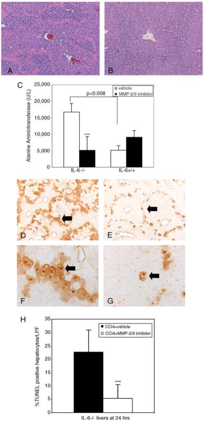
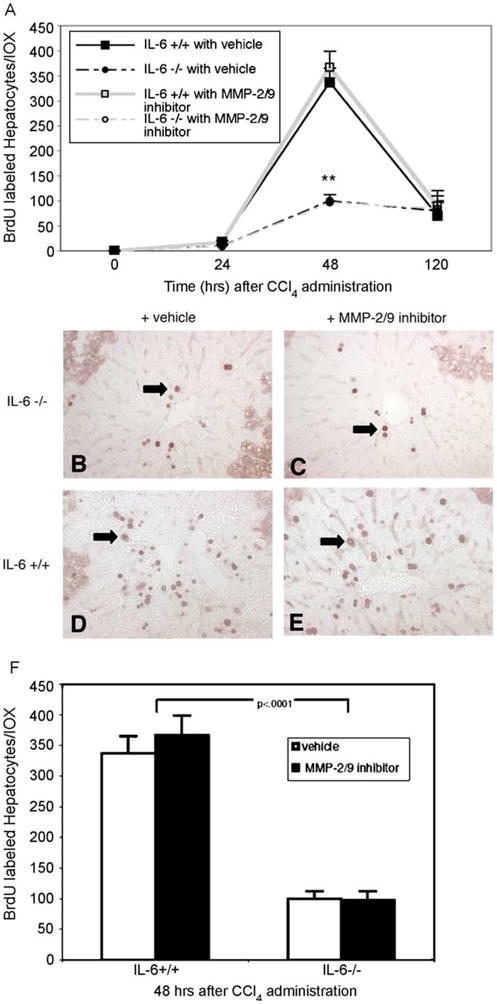
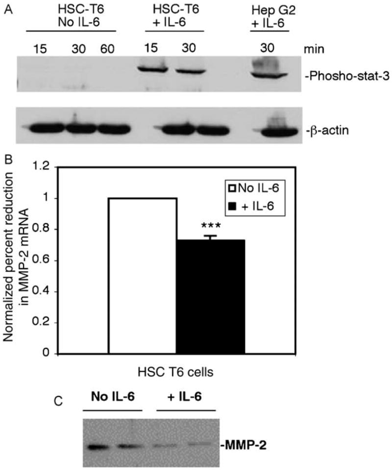
Methods: MMP-2 was analyzed in livers of IL-6-/- and IL-6+/+ mice following CCl(4) administration. IL-6-/- mice were pretreated with IL-6 and liver histology and MMP-2 expression were examined after liver injury. IL-6-/- mice were treated with an MMP-2 inhibitor and assessment of injury (histology and serum ALT levels), apoptosis by TUNEL assay, and hepatocyte proliferation by BRDU-labeling was performed. These studies were complemented by analysis of cultured stellate cells.

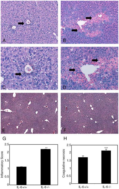
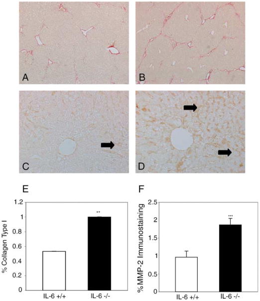
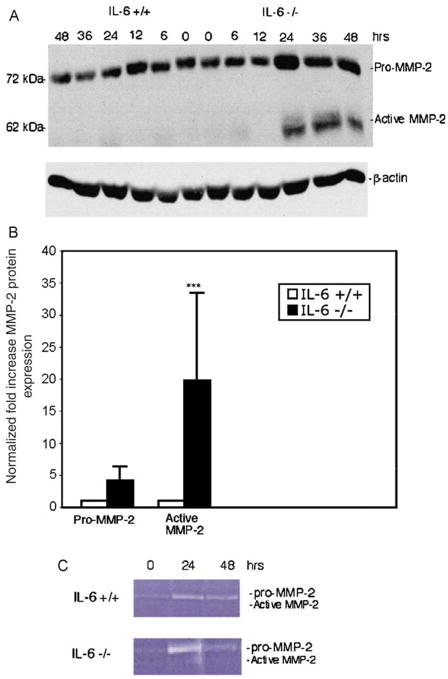
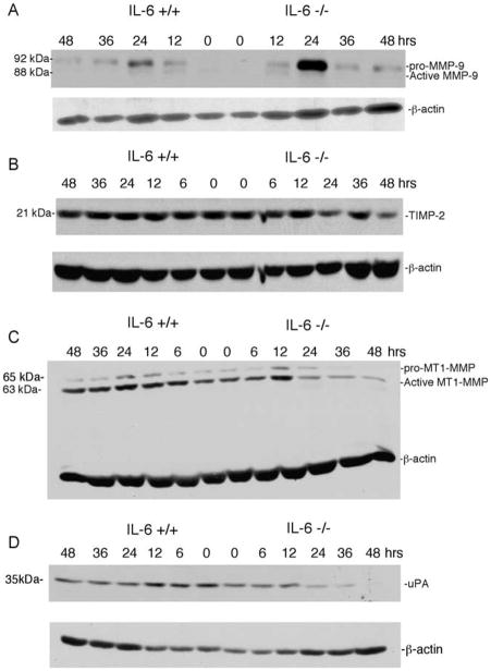
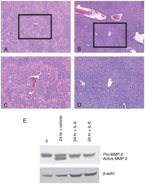
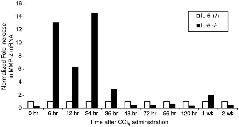
Results: MMP-2 mRNA, protein, and activity was increased in IL-6-/- livers. Restoration of IL-6 signaling in IL-6-/- mice rescued injury and restored MMP-2 expression to wild-type levels. Furthermore, pharmacologic inhibition of MMP-2 decreased hepatocellular injury and apoptosis in IL-6-/- mice. In cultured stellate cells, recombinant IL-6 suppressed endogenous MMP-2 mRNA and protein expression.

Conclusions: IL-6 may be hepatoprotective in acute injury through down-regulation of MMP-2. These findings suggest a role for MMP-2 in amplifying liver injury in vivo.


Figures

Fig. 1
Fig. 1
Protracted injury and inflammation following CCl4 administration in IL-6−/− livers. Increased mononuclear and lymphoid inflammatory infiltrate, coagulative necrosis, and hemorrhage in IL-6−/− livers 120 h after a single intraperitoneal dose of CCl4 (B) compared to IL-6+/+ livers (A). Higher magnification demonstrates increased inflammatory cells in bridging areas of necrosis in IL-6−/− livers (D) compared to IL-6+/+ livers (C). Arrows indicating inflammation and coagulative necrosis. Complete restoration of liver architecture is noted by 2 weeks in both IL-6−/− (F) and IL-6+/+ livers (E) (original magnification, 50× A, B; 100× C, D; 10× E, F). Inflammation and perivenular necrosis was graded on a 4-point scale (Table 1) by a blinded pathologist in 20 random high power fields per animal (n = 3 in each group) and represented graphically in panels G and H. Data represents mean±SEM; **P<0.0001; ***P<0.024.
Fig. 2
Fig. 2
Increased MMP-2 is associated with fibrosis in IL-6−/− livers. Sirius Red staining shows increased fibrosis in IL-6−/− livers (B) compared to IL-6+/+ livers (A) after 5 weeks of chronic administration of CCl4 which is associated with increased MMP-2 expression as assessed by immunostaining (D vs. C). Arrows denote sinusoidal staining for MMP-2. Bioquant analysis performed on 36 images per animal (n = 3) demonstrates 20% increase in collagen I (E; P<0.0001) and 49% increase in MMP-2 expression (F; P<0.001) in IL-6−/− fibrotic livers. Original magnification 5× (A, B) and 200× (C, D).
Fig. 3
Fig. 3
Increased expression and activity of MMP-2 in IL-6−/− livers after single dose of CCl4. (A) Representative immunoblot for MMP-2 on whole liver extracts harvested from IL-6−/− and IL-6+/+ mice after administration of CCl4 demonstrates a 20-fold increase (range 3.2–47; ***P<0.002) in expression of active MMP-2 in IL-6−/− vs. IL-6+/+ animals. (B) Quantitation of increases of both active and latent MMP-2 from three independent experiments. β-Actin expression was used as a loading control. (C) Gelatin zymography performed on liver extracts demonstrates 3-fold increase in MMP-2 activity at 24 h in the IL-6−/− livers. [This figure appears in colour on the web.]
Fig. 4
Fig. 4
Expression of active MMP-9, TIMP-2, MT1-MMP, or uPA are not increased in IL-6−/− mice following administration of CCl4. Immunoblot of whole liver extracts prepared from IL-6+/+ and IL-6−/− livers following a single dose of CCl4 were probed with antibodies to MMP-9, TIMP-2, MT1-MMP, and uPA. (A) An increase in latent but not active MMP-9 is apparent in IL-6−/− livers. (B) Decreased TIMP-2 expression in IL-6−/− livers is apparent, concomitant with increased MMP-2. (C) A transient peak of MT1-MMP expression at 12 h precedes activation of MMP-2 then declines rapidly in IL-6−/− animals at 36 and 48 h. (D) uPA expression declines in concert with MMP-2 activation in IL-6−/− livers. β-Actin protein expression was used as a loading control on each blot. Experiments were performed in duplicate.
Fig. 5
Fig. 5
Restoration of IL-6 signaling in IL-6−/− mice rescues CCl4-induced injury and restores MMP-2 expression to wild-type levels. Hematoxylin and eosin stained liver sections from IL-6−/− mice pretreated with either vehicle (A) or recombinant IL-6 (B) prior to CCl4 (original magnification 10×). Area outlined shown at higher magnification (100×; C, D). Those receiving recombinant IL-6 (B, D) had decreased injury at 24 h compared to control group (A, C). (E) Western blot of MMP-2 protein expression performed on extracts from IL-6−/− mice receiving vehicle (lane 2) compared with those receiving recombinant IL-6 (lanes 3 and 4). At 24 h, there is a significant increase in active MMP-2 expression in IL-6−/− animals, which is decreased in animals receiving recombinant IL-6. The blot probed for β-actin to confirm equal protein loading. Experiments were performed in triplicate.
Fig. 6
Fig. 6
Pharmacologic inhibition of MMP-2 rescues CCl4-induced liver injury and apoptosis in IL-6−/− mice but has no significant impact in IL-6+/+ mice. Liver sections 24 h after co-administration of MMP-2/9 inhibitor at a dose of 8 μl/g or vehicle control and CCl4 were stained with hematoxylin and eosin (100×). IL-6−/− mice receiving the inhibitor developed less injury (B) compared to control animals receiving vehicle control (A). (C) ALT levels measured in IL-6−/− and IL-6+/+ with and without MMP-2/9 inhibitor. IL-6+/+ mice had significantly less injury as measured by ALT levels (P<0.008). IL-6−/− mice receiving inhibitor had a ~75% reduction in serum ALT (***P<0.03). TUNEL staining was performed on liver sections from IL-6−/− mice 24 h after the administration of CCl4 with or without MMP-2/9 inhibitor. IL-6−/− mice receiving inhibitor (E) demonstrated significantly less apoptosis than vehicle control group (D). High power magnification (400×) demonstrates fewer TUNEL-positive nuclei in animals receiving inhibitor (G) compared to those receiving vehicle (F). Arrows denote TUNEL-positive cells. (H) Five (100× magnification) fields were randomly selected per slide and 100 hepatocytes counted per field. Mean percent of apoptotic hepatocytes was calculated and compared between different study groups. IL-6−/− mice receiving inhibitor demonstrated a 17.4% absolute reduction and 80% relative reduction in hepatocyte apoptosis (***P<0.001). Bars represent means±SEM from three mice in each group.
Fig. 7
Fig. 7
Administration of MMP-2 inhibitor does not restore hepatocyte proliferative response in IL-6−/− mice following CCl4 administration. (A) Peak proliferative defect in IL-6−/− livers notable at 48 h after single dose of CCl4. Data points represent means±SEM (**P<0.0001; IL-6−/− vehicle vs. IL-6+/+ vehicle). (B–E) Representative photomicrographs of BRDU immunohistochemistry at 48 h with or without MMP-2/9 inhibitor (original magnification, 100×). Arrows indicate large round positively stained hepatocyte nuclei. (F) Quantification of percent BRDU-labelled hepatocytes per 10× field at 48 h after CCl4 administration in IL-6+/+and IL-6−/− demonstrates no significant impact of inhibitor within either group. [This figure appears in colour on the web.]
Fig. 8
Fig. 8
IL-6 suppresses MMP-2 mRNA levels in vivo. Real time PCR using RNA extracted from IL-6−/− and IL-6+/+ livers following a single dose of CCl4 was analyzed using primers specific for MMP-2 mRNA and normalized to β2-microglobulin. There was a significant increase in MMP-2 mRNA in IL-6−/− livers at 6, 12, 24, and 36 h after a single dose of CCl4. This increase was most notable at 24 h where a 13.6-fold increase in MMP-2 mRNA was noted in the IL-6−/− livers.
Fig. 9
Fig. 9
IL-6 suppresses endogenous MMP-2 mRNA and protein in a stellate cell line. (A) HSC-T6, an immortalized rat stellate cell line, demonstrates phoshphorylated STAT-3 after incubation with recombinant IL-6 for 15 and 30 min compared to vehicle control. Hep G2 cells incubated with IL-6 for 30 min served as positive control. (B) RNA extracted from HSC-T6 cells incubated with IL-6 for 2 h demonstrated a mean 27% reduction in MMP-2 mRNA (range 18–58%, ***P<0.0012) in MMP-2 mRNA compared with control as assessed by real time PCR. Data from representative experiment shown here. (C) Immunoblot analysis of cell culture supernatant of HSC-T6 cells after 24 h incubation with IL-6 demonstrates an average 86% reduction (range 60–93%, P<0.0015) in MMP-2 protein expression compared to control. Quantification performed using Bioquant image analysis software.

References

    1. Sun Y, Tokushige K, Isano E, Yamauchi K, Ohata H. Elevated serum interleukin-6 levels in patients with acute hepatitis. J Clin Immunol. 1992;12:192–200. - PubMed
    1. Hill D, Marsano L, Cohen D, Allen J, Shedlofsky S, McClain C. Increased plasma interleukin-6 concentrations in alcoholic hepatitis. J Clin Immunol. 1992;119:547–552. - PubMed
    1. Taub R. Hepatoprotection via the IL-6/stat 3 pathway. J Clin Investig. 2003;112:978–980. - PMC - PubMed
    1. Cressman D, Greenbaum L, DeAngelis R, Cilberto G, Furth E, Poli V, et al. Liver failure and defective hepatocyte regeneration in interleukin-6 deficient mice. Science. 1996;274:1379–1383. - PubMed
    1. Kovalovich K, Li W, DeAngelis R, Greenbaum L, Cilberto G, Taub R. Interleukin-6 protects against Fas-mediated death by establishing a critical level of anti-apoptotic hepatic proteins FLIP, Bcl-2, and Bcl-XL. J Biol Chem. 2001;276:26605–26613. - PubMed

Publication types

MeSH terms