

Electrical inhibition of lens epithelial cell proliferation: an additional factor in secondary cataract?
- PMID: 15764648
- PMCID: PMC1459287
- DOI: 10.1096/fj.04-2733fje
Electrical inhibition of lens epithelial cell proliferation: an additional factor in secondary cataract?
Abstract
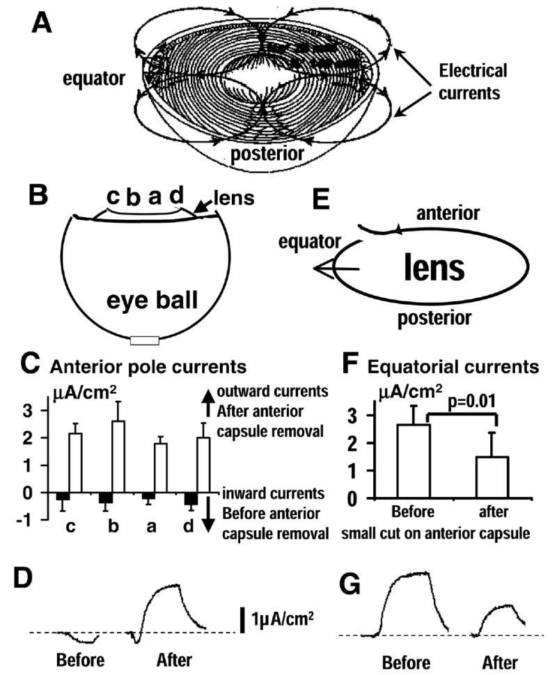
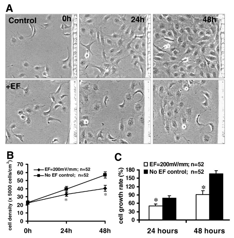
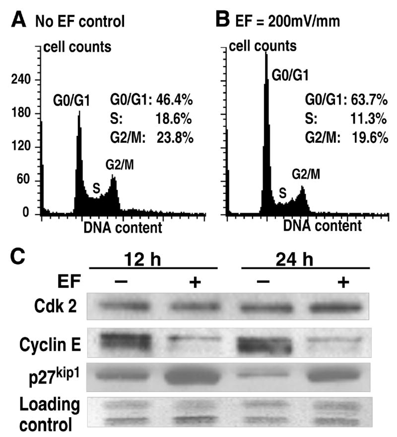
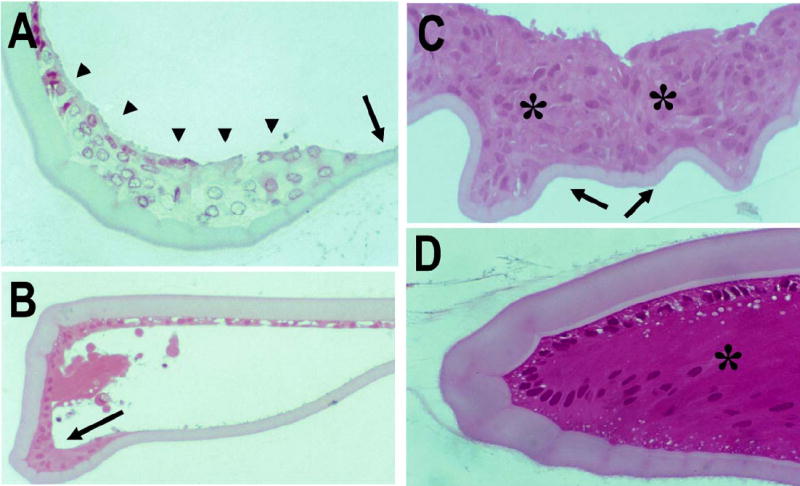
Cataract is the most common cause of blindness but is at least curable by surgery. Unfortunately, many patients gradually develop the complication of posterior capsule opacification (PCO) or secondary cataract. This arises from stimulated cell growth within the lens capsule and can greatly impair vision. It is not fully understood why residual lens epithelial cell growth occurs after surgery. We propose and show that cataract surgery might remove an important inhibitory factor for lens cell growth, namely electric fields. The lens generates a unique pattern of electric currents constantly flowing out from the equator and entering the anterior and posterior poles. We show here that cutting and removing part of the anterior capsule as in cataract surgery significantly decreases the equatorial outward electric currents. Application of electric fields in culture inhibits proliferation of human lens epithelial cells. This inhibitory effect is likely to be mediated through a cell cycle control mechanism that decreases entry of cells into S phase from G1 phase by decreasing the G1-specific cell cycle protein cyclin E and increasing the cyclin-Cdk complex inhibitor p27kip1. Capsulorrhexis in vivo, which reduced endogenous lens electric fields, significantly increased LEC growth. This, together with our previous findings that electric fields have significant effects on the direction of lens cell migration, points to a controlling mechanism for the aberrant cell growth in posterior capsule opacification. A novel approach to control growth of lens epithelial cells using electric fields combined with other controlling mechanisms may be more effective in the prevention and treatment of this common complication of cataract surgery.
Figures
Similar articles
-
Posterior capsule opacification. Part 1: Experimental investigations.J Cataract Refract Surg. 1999 Jan;25(1):106-17. doi: 10.1016/s0886-3350(99)80020-0. J Cataract Refract Surg. 1999. PMID: 9888086 Review.
-
[Secondary cataract: a problem resolved].Bull Mem Acad R Med Belg. 2006;161(7-9):469-78; discussion 478. Bull Mem Acad R Med Belg. 2006. PMID: 17304977 Review. French.
-
[The influence of the lens capsule mechanical polishing to the secondary cataract development].Cesk Slov Oftalmol. 2010 Feb;66(1):21-5. Cesk Slov Oftalmol. 2010. PMID: 20521506 Czech.
-
Posterior capsule opacification.Exp Eye Res. 2009 Feb;88(2):257-69. doi: 10.1016/j.exer.2008.10.016. Epub 2008 Oct 29. Exp Eye Res. 2009. PMID: 19013456 Review.
-
Posterior capsular opacification: a problem reduced but not yet eradicated.Arch Ophthalmol. 2009 Apr;127(4):555-62. doi: 10.1001/archophthalmol.2009.3. Arch Ophthalmol. 2009. PMID: 19365040 Review.
Cited by
-
Elucidating the Role of Injury-Induced Electric Fields (EFs) in Regulating the Astrocytic Response to Injury in the Mammalian Central Nervous System.PLoS One. 2015 Nov 12;10(11):e0142740. doi: 10.1371/journal.pone.0142740. eCollection 2015. PLoS One. 2015. PMID: 26562295 Free PMC article.
-
Electric field exposure promotes epithelial‑mesenchymal transition in human lens epithelial cells via integrin β1‑FAK signaling.Mol Med Rep. 2017 Oct;16(4):4008-4014. doi: 10.3892/mmr.2017.7086. Epub 2017 Jul 26. Mol Med Rep. 2017. PMID: 28765922 Free PMC article.
-
Electrical signaling in control of ocular cell behaviors.Prog Retin Eye Res. 2012 Jan;31(1):65-88. doi: 10.1016/j.preteyeres.2011.10.001. Epub 2011 Oct 17. Prog Retin Eye Res. 2012. PMID: 22020127 Free PMC article. Review.
-
Transmembrane voltage potential controls embryonic eye patterning in Xenopus laevis.Development. 2012 Jan;139(2):313-23. doi: 10.1242/dev.073759. Epub 2011 Dec 7. Development. 2012. PMID: 22159581 Free PMC article.
-
Lens Stretching Modulates Lens Epithelial Cell Proliferation via YAP Regulation.Invest Ophthalmol Vis Sci. 2019 Sep 3;60(12):3920-3929. doi: 10.1167/iovs.19-26893. Invest Ophthalmol Vis Sci. 2019. PMID: 31546253 Free PMC article.
References
-
- Ibaraki N. A brighter future for cataract surgery. Nat Med. 1997;3:958–960. - PubMed
-
- Apple DJ, Solomon KD, Tetz MR, Assia EI, Holland EY, Legler UFC, Tsai JC, Castaneda VE, Hoggatt JP, Kostick AMP. Posterior capsule opacification. Surv Ophthalmol. 1992;37:73–116. - PubMed
-
- Clark DS. Posterior capsule opacification. Curr Opin Ophthalmol. 2000;11:56–64. - PubMed
-
- Moisseiev J, Bartov E, Schochat A, Blumenthal M. Long-term study of the prevalence of capsular opacification following extracapsular cataract extraction. J Cataract Refract Surg. 1989;15:531–533. - PubMed
Publication types
MeSH terms
Grants and funding
LinkOut - more resources
Full Text Sources
Medical
