Review
. 2005 Mar;115(3):527-37.
doi: 10.1172/JCI24178.

Protein kinase cascades in the regulation of cardiac hypertrophy

Affiliations
Review

Protein kinase cascades in the regulation of cardiac hypertrophy

Gerald W Dorn 2nd et al. J Clin Invest. 2005 Mar.

Abstract

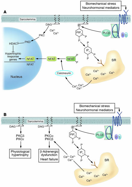
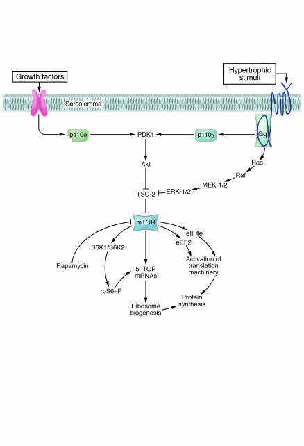
In broad terms, there are 3 types of cardiac hypertrophy: normal growth, growth induced by physical conditioning (i.e., physiologic hypertrophy), and growth induced by pathologic stimuli. Recent evidence suggests that normal and exercise-induced cardiac growth are regulated in large part by the growth hormone/IGF axis via signaling through the PI3K/Akt pathway. In contrast, pathological or reactive cardiac growth is triggered by autocrine and paracrine neurohormonal factors released during biomechanical stress that signal through the Gq/phospholipase C pathway, leading to an increase in cytosolic calcium and activation of PKC. Here we review recent developments in the area of these cardiotrophic kinases, highlighting the utility of animal models that are helping to identify molecular targets in the human condition.


Figures

Figure 1
Figure 1
Mechanisms of activation of PI3K/Akt signaling in adaptive versus maladaptive hypertrophy. In adaptive hypertrophy, binding of growth factors to their cognate receptors triggers translocation of the PI3K isoform p110α to the cell membrane, a process triggered by the interaction of the p85 subunit of PI3K with specific tyrosine phosphorylated residues in the growth factor receptor. p110α then phosphorylates phosphatidylinositols in the membrane at the 3′ position of the inositol ring. The pleckstrin homology (PH) domains of both Akt and its activator, PDK1, associate with the 3′ phosphorylated lipids, allowing PDK1 to activate Akt. Full activation of Akt requires phosphorylation by a second kinase, PDK2 (not shown), that may be the DNA-dependent protein kinase (DNA-PK). Activation of Akt then leads to activation of mTOR, a central regulator of protein synthesis, via its effects on both ribosome biogenesis and activation of the protein translation machinery. Akt also phosphorylates and inhibits a kinase, GSK-3 (of which there are 2 isoforms, α and β). Since GSK-3 inhibits a key component of the protein translation machinery, as well as a number of transcription factors believed to play roles in the induction of the hypertrophic program of gene expression, inhibition of GSK-3 promotes both protein synthesis and gene transcription. Maladaptive hypertrophy, triggered by neurohormonal mediators and biomechanical stress, also activates Akt, but the mechanism involves activation of heterotrimeric G-protein–coupled receptors coupled to the G-protein family Gq/G11. The PI3K isoform p110γ associates with the βγ subunits of Gq and phosphorylates membrane phosphatidylinositols, which leads to the recruitment of PDK1 and Akt. Maladaptive hypertrophy also recruits alternative pathways to activation of mTOR and Akt. Also shown is the phosphatase, PTEN, which, by dephosphorylating the 3′ position of phosphatidylinositol trisphosphate (PIP3), shuts off signaling down the pathway.
Figure 2
Figure 2
Gq/11-activated pathways in maladaptive hypertrophy. (A) Calcineurin/NFAT pathway. Hypertrophic stimuli, acting via the α subunit of Gq or G11, recruit PLCβ to the membrane, where it hydrolyses phosphatidylinositol 4,5 bisphosphate (PIP2), releasing inositol 1,4,5-triphosphate (IP3) and diacylglycerol (DAG). IP3 binds to receptors in the sarcoplasmic reticulum (SR), releasing calcium. The increase in cytosolic [Ca2+], together with calmodulin, activates the protein phosphatase calcineurin. Calcineurin dephosphorylates several residues in the amino-terminal region of the transcription factor NFAT, allowing it to translocate to the nucleus and activate transcription of hypertrophic response genes. (B) PKCs. Activation of PKC isoforms is accomplished by the IP3-mediated release of calcium from the SR together with DAG (classical isoforms), whereas the so-called novel isoforms are activated by DAG alone. See text for details of the roles of the various PKC isoforms in hypertrophy. One PKC-regulated pathway not discussed is that leading to the inhibition of a subset of histone deacetylases (HDACs 5 and 9) that appear to specifically regulate cellular hypertrophy. In this pathway, one or more PKC isoforms activate another protein kinase, PKD, that then phosphorylates the HDAC, leading to its export from the nucleus and, thus, inactivation. This pathway is the subject of a review in this series (109).
Figure 3
Figure 3
GSK-3 as a convergence point in hypertrophic signaling. Inhibition of GSK-3 appears to be a key element in both adaptive and maladaptive hypertrophy. Growth factors, acting via Akt; neurohormonal mediators, acting via both Akt and PKCs (particularly PKCα); β-adrenergic agonists, acting via PKA; and biomechanical stress, acting via several mechanisms, possibly involving the integrin-linked kinase (ILK) or an ILK-associated protein, all lead to the inactivation of GSK-3. Therefore, GSK-3 appears to serve as a convergence point, integrating inputs from many prohypertrophic signals. Inhibition of GSK-3 releases a number of transcription factors from tonic inhibition, and also releases eIF2B, allowing activation of the protein synthetic machinery. Thus GSK-3 affects both key components of the response, reprogramming of gene expression and activation of protein synthesis. Additional negative regulators of GSK-3 not shown include the serum and glucocorticoid–induced kinase (SGK) and, possibly, the ERK pathway target p90 ribosomal S6 kinase (RSK1).
Figure 4
Figure 4
Regulation of protein translation in adaptive versus maladaptive hypertrophy. It is likely that all hypertrophic stimuli must activate mTOR and the general protein translational machinery in order to allow the full expression of the phenotype. This is mediated via the inhibition of the tuberous sclerosis gene product, tuberin (TSC2), mutations of which lead to benign hamartomas in various tissues including the heart. TSC2 can be phosphorylated and inhibited by Akt and, in some instances, via ERK-1/2 or an ERK target. The latter may be an Akt-independent mechanism of activation of mTOR that may be particularly relevant to pathologic stress–induced growth. Shown are the pathways to ribosome biogenesis as well as the regulators of the translational machinery (initiation factors [IF] and elongation factors [EF]) regulated by mTOR in the heart. As noted in the text, recent surprising findings related to this pathway have included the limited role for S6K1 and S6K2 in both adaptive and, particularly, maladaptive hypertrophy and the identification of Akt1 as a possible antihypertrophic factor in pathologic hypertrophy but a prohypertrophic factor in physiologic hypertrophy.

Similar articles

Cited by

References

    1. Simpson P. Norepinephrine-stimulated hypertrophy of cultured rat myocardial cells is an α 1 adrenergic response. J. Clin. Invest. 1983;72:732–738. - PMC - PubMed
    1. Sadoshima J, Izumo S. Molecular characterization of angiotensin II–induced hypertrophy of cardiac myocytes and hyperplasia of cardiac fibroblasts. Critical role of the AT1 receptor subtype. Circ. Res. 1993;73:413–423. - PubMed
    1. Shubeita HE, et al. Endothelin induction of inositol phospholipid hydrolysis, sarcomere assembly, and cardiac gene expression in ventricular myocytes. A paracrine mechanism for myocardial cell hypertrophy. J. Biol. Chem. 1990;265:20555–20562. - PubMed
    1. Bristow MR. beta-adrenergic receptor blockade in chronic heart failure. Circulation. 2000;101:558–569. - PubMed
    1. Mehra MR, Uber PA, Francis GS. Heart failure therapy at a crossroad: are there limits to the neurohormonal model? J. Am. Coll. Cardiol. 2003;41:1606–1610. - PubMed

Publication types

MeSH terms