Review
. 2005 Mar;115(3):538-46.
doi: 10.1172/JCI24144.

Toward transcriptional therapies for the failing heart: chemical screens to modulate genes

Affiliations
Review

Toward transcriptional therapies for the failing heart: chemical screens to modulate genes

Timothy A McKinsey et al. J Clin Invest. 2005 Mar.

Abstract

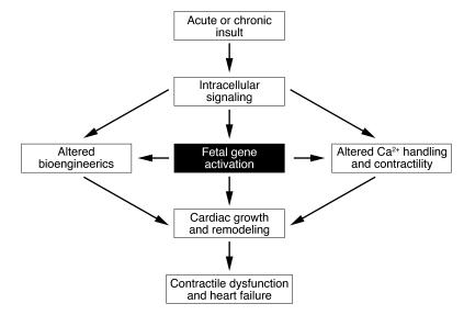
In response to acute and chronic stresses, the heart frequently undergoes a remodeling process that is accompanied by myocyte hypertrophy, impaired contractility, and pump failure, often culminating in sudden death. The existence of redundant signaling pathways that trigger heart failure poses challenges for therapeutic intervention. Cardiac remodeling is associated with the activation of a pathological gene program that weakens cardiac performance. Thus, targeting the disease process at the level of gene expression represents a potentially powerful therapeutic approach. In this review, we describe strategies for normalizing gene expression in the failing heart with small molecules that control signal transduction pathways directed at transcription factors and associated chromatin-modifying enzymes.


Figures

Figure 1
Figure 1
Abnormalities associated with cardiac remodeling during pathological hypertrophy and heart failure.
Figure 2
Figure 2
Model of HDAC function and hypersensitivity of HDAC9-knockout mice to cardiac stress. (A) Schematic of chromatin structure and the actions of HATs and HDACs. HATs acetylate (Ac) histones, causing relaxation of nucleosomal structure and transcriptional activation. HDACs oppose the actions of HATs by deacetylating histones, causing chromatin condensation and transcriptional repression. (B) Histological sections of adult mouse hearts of the indicated genotypes are shown. HDAC9-null mice do not display a cardiac phenotype at early age. The cardiac calcineurin transgene (Calcineurin-Tg) induces dramatic cardiac growth, which is exacerbated in the absence of HDAC9. Adapted with permission from Cell (26).
Figure 3
Figure 3
Agonist-dependent nuclear export of HDAC5 correlates with cardiomyocyte hypertrophy. Stimulation of cardiomyocytes with neurohumoral agonists evokes a hypertrophic response characterized by fetal gene activation, sarcomere assembly, and hypertrophy. The upper panels show the subcellular distribution of HDAC5 fused to GFP. In unstressed myocytes, HDAC5-GFP is localized to the nucleus, whereas stimulation with a hypertrophic agonist causes it to redistribute to the cytoplasm. A small molecular inhibitor of the signaling pathway from the cell surface to HDAC5 prevents nuclear export of HDACs and blocks hypertrophy, as detected by staining for sarcomeric α-actinin.
Figure 4
Figure 4
Signal-dependent modulation of cardiac genes and hypertrophy by class II HDACs. MEF2 recruits class II HDACs to target genes, which results in transcriptional repression due to chromatin condensation. Stimulation of cardiomyocytes with neurohumoral agonists acting through G-protein coupled receptors (GPCRs) activates kinase pathways that culminate with the phosphorylation of class II HDACs and their export to the cytoplasm as a complex with 14-3-3 proteins. The nuclear export protein CRM1 is required for HDAC nuclear export. The release of class II HDACs from MEF2 allows for the association of HATs with MEF2 and consequentially chromatin relaxation and transcriptional activation of fetal cardiac genes.
Figure 5
Figure 5
Signal-dependent modulation of cardiac genes and hypertrophy by calcineurin/NFAT signaling. Stimulation of cardiomyocytes with neuro-humoral agonists acting through GPCRs activates calcineurin, which dephosphorylates NFAT, allowing its entry into the nucleus, where it acts with other transcription factors, such as GATA4, to activate stress-response genes. Glycogen synthase kinase–3 (GSK3) phosphorylates the serine residues dephosphorylated by calcineurin, driving NFAT back to the cytoplasm and terminating the growth signal. The MCIP1 gene is a target of calcineurin/NFAT signaling, and its protein product associates with calcineurin to restrain its activity, thereby creating a negative feedback loop to govern cardiac growth.
Figure 6
Figure 6
Modulation of cardiac calcineurin signaling by MCIP expression. The upper set of panels shows the effect of calcineurin signaling in MCIP1-knockout mice. In the absence of MCIP, the heart is sensitized to calcineurin signaling and rapidly undergoes fatal hypertrophy. Because of the heightened sensitivity of MCIP1-knockout mice to calcineurin, only a weak calcineurin transgene can be expressed in the absence of MCIP1. A stronger calcineurin transgene, as used in the lower panels, results in heart failure and death within the first few days after birth. The lower set of panels shows the effect of calcineurin signaling in mice that overexpress MCIP1 in the heart. Overexpression of MCIP1 prevents pathological hypertrophy. Adapted with permission from Proceedings of the National Academy of Sciences of the United States of America (58, 59).
Figure 7
Figure 7
Structures of serotonin, PAMH, and A-PAMH.
Figure 8
Figure 8
Potential roles of HDACs in cardiac myocytes. Class II HDACs repress hypertrophy. Their activity is repressed by signal-dependent phosphorylation. Class I HDACs appear to be prohypertrophic based on the ability of HDAC inhibitors to prevent hypertrophy. Class I HDACs could act by inhibiting prohypertrophic signaling pathways, by inhibiting the expression of growth inhibitory genes or by activating the expression of pro-growth genes.

Similar articles

Cited by

References

    1. Linseman JV, Bristow MR. Drug therapy and heart failure prevention. Circulation. 2003;107:1234–1236. - PubMed
    1. Vakili BA, Okin PM, Devereux RB. Prognostic implications of left ventricular hypertrophy. Am. Heart J. 2001;141:334–341. - PubMed
    1. Okin PM, et al. for the LIFE Study Investigators. Regression of electrocardiographic left ventricular hypertrophy during antihypertensive treatment and the prediction of major cardiovascular events. JAMA. 2004;292:2343–2349. - PubMed
    1. Devereux RB, et al. Prognostic significance of left ventricular mass change during treatment of hypertension. JAMA. 2004;292:2350–2356. - PubMed
    1. Gardin JM, Lauer MS. Left ventricular hypertrophy: the next treatable, silent killer? JAMA. 2004;292:2396–2398. - PubMed

Publication types

MeSH terms