. 2005 Mar;115(3):653-63.
doi: 10.1172/JCI22655.

Neurotrophins promote revascularization by local recruitment of TrkB+ endothelial cells and systemic mobilization of hematopoietic progenitors

Affiliations

Neurotrophins promote revascularization by local recruitment of TrkB+ endothelial cells and systemic mobilization of hematopoietic progenitors

Pouneh Kermani et al. J Clin Invest. 2005 Mar.

Abstract

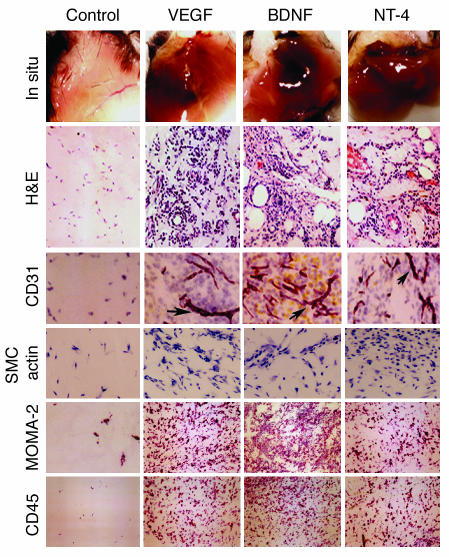
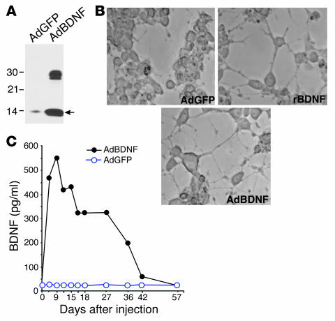
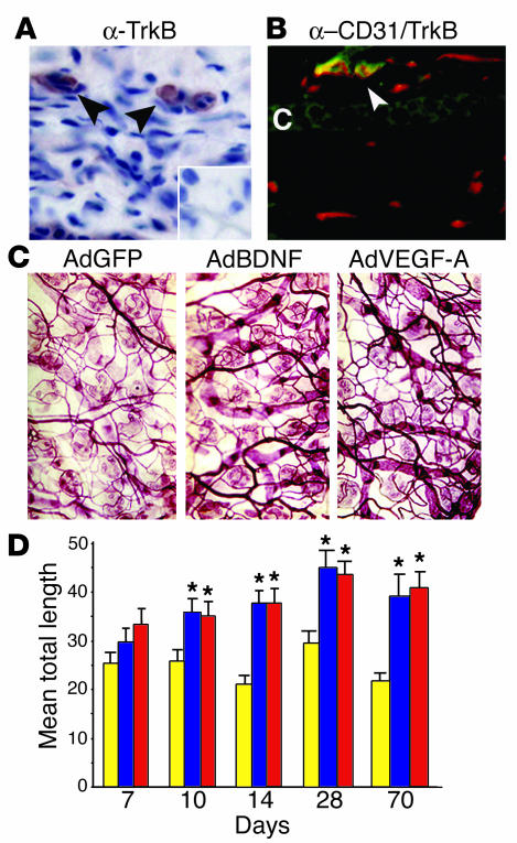
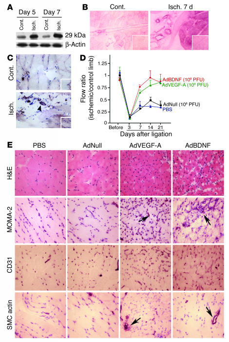
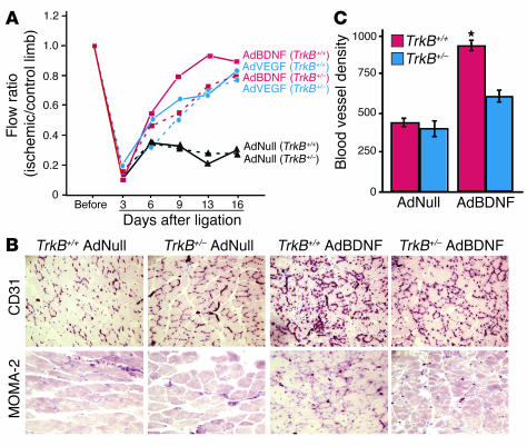
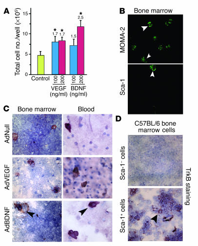
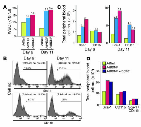
The neurotrophin brain-derived neurotrophic factor (BDNF) is required for the maintenance of cardiac vessel wall stability during embryonic development through direct angiogenic actions on endothelial cells expressing the tropomysin receptor kinase B (TrkB). However, the role of BDNF and a related neurotrophin ligand, neurotrophin-4 (NT-4), in the regulation of revascularization of the adult tissues is unknown. To study the potential angiogenic capacity of BDNF in mediating the neovascularization of ischemic and non-ischemic adult mouse tissues, we utilized a hindlimb ischemia and a subcutaneous Matrigel model. Recruitment of endothelial cells and promotion of channel formation within the Matrigel plug by BDNF and NT-4 was comparable to that induced by VEGF-A. The introduction of BDNF into non-ischemic ears or ischemic limbs induced neoangiogenesis, with a 2-fold increase in the capillary density. Remarkably, treatment with BDNF progressively increased blood flow in the ischemic limb over 21 days, similar to treatment with VEGF-A. The mechanism by which BDNF enhances capillary formation is mediated in part through local activation of the TrkB receptor and also by recruitment of Sca-1+CD11b+ pro-angiogenic hematopoietic cells. BDNF induces a potent direct chemokinetic action on subsets of marrow-derived Sca-1+ hematopoietic cells co-expressing TrkB. These studies suggest that local regional delivery of BDNF may provide a novel mechanism for inducing neoangiogenesis through both direct actions on local TrkB-expressing endothelial cells in skeletal muscle and recruitment of specific subsets of TrkB+ bone marrow-derived hematopoietic cells to provide peri-endothelial support for the newly formed vessels.


Figures

Figure 1
Figure 1
BDNF induces neoangiogenesis in an in vivo Matrigel assay. Young adult female mice were injected subcutaneously with 0.3 ml of growth factor–depleted Matrigel containing 64 U/ml heparin and recombinant human VEGF165 (30–50 ng/ml), recombinant human BDNF (50–100 ng/ml), NT-4 (50–100 ng/ml), or no growth factor addition (Control). After 14 days, the animals were sacrificed and the Matrigel plug was photographed (In situ) and harvested. Serial sections for each Matrigel plug were analyzed by investigators “blinded” to sample identity, and the degree of cellularity was quantified in central regions of the Matrigel (H&E). Immunoreactivity for CD31, smooth muscle α-actin (SMC actin), MOMA-2, and CD45 was assessed, followed by hematoxylin counterstaining. Matrigel plugs containing BDNF, NT-4, or VEGF-A showed an increase in the number of infiltrating endothelial-like cells arborizing (arrowheads) throughout the tissue compared with the control samples. Magnification, ×40 (CD31) or ×20 (H&E, smooth muscle α-actin, MOMA-2 and CD45).
Figure 2
Figure 2
Expression of biologically active BDNF protein after adenovirus-mediated gene delivery. (A) 293 cells were infected with AdBDNF or AdGFP at an MOI of 100, and media was harvested 48 hours after infection. Secretion of BDNF was confirmed by Western blot analysis after protein separation by SDS-PAGE. The 13-kDa immunoreactive band (arrow) is consistent with mature BDNF, and the 29-kDa and 17-kDa species correspond to incompletely processed proforms that are released when the gene is expressed at high levels. (B) The biological activity of BDNF expressed by adenovirus vector was evaluated using PC12 cells stably expressing TrkB. Media from 293 cells infected with AdBDNF, AdGFP, or rBDNF were added to cells, and neuritogenesis (the presence of neurite processes greater than one cell body in length) was assessed 24 hours after treatment using Normasky imaging; neurite outgrowths were visualized with a 20× objective. (C) In vivo expression of BDNF following injection of AdBDNF in the tail veins was measured by ELISA using a commercially available kit and a recombinant BDNF standard (BDNF Emax ImmunoAssay System). The BDNF Emax kit has a minimum sensitivity of 7.8 pg/ml. The ELISA for BDNF was carried out on plasma samples of SCID mice injected in the tail vein with 1 × 10⁹ PFU of AdBDNF. Blood was collected every 2–3 days after adenovirus vector administration. Absorbance was measured at 450 nm using a microplate reader and the BDNF concentration was normalized to that of recombinant protein.
Figure 3
Figure 3
AdBDNF promotes neoangiogenesis in the mouse ear model. (A) Immunohistochemical analysis of expression of TrkB in a section from the base of the ear pinna of a wild-type mouse. The black arrowheads show blood vessels positive for TrkB. α-, anti-. Magnification, ×40. (B) Section from the base of the ear pinna of an untreated mouse was examined for expression of TrkB and CD31 using double-immunofluorescence microscopy. Section was incubated with biotinylated anti-CD31, followed by rhodamine-avidin, and with TrkB antisera, detected with fluorescein-conjugated secondary antibody. The white arrowhead shows the colocalization of CD31 and TrkB. C, cartilage. Magnification, ×40. (C) Whole-mount immunostaining with anti-CD31 of ear skin of mice treated with AdGFP, AdVEGF-A, or AdBDNF (n = 3/group). One week to 10 weeks after injection, ears were removed and the skin was separated from cartilage. Skin was permeablized with Triton and was incubated with antisera against CD31 for whole-mount immunohistochemistry. VIP-based immunodetection yielded a red reaction product. Magnification, ×10. (D) Quantitative analysis of the total vessel length from 4 peripheral fields of tissues obtained at 7, 10, and 14 days and 4 and 10 weeks after injection was performed by investigators, who were “blinded” to sample identity, using NIH Image. *P < 0.001. Yellow, AdGFP; blue, AdVEGF-A; red, AdBDNF.
Figure 4
Figure 4
BDNF accelerates the revascularization of the ischemic limbs. (A and B) Western blot analysis of tissue lysates at 5 and 7 days after ligation using polyclonal anti-BDNF (Santa Cruz Biotechnology) (A), followed by a chemiluminescence-based detection, ECL and immunohistochemical detection of tissue sections to assess BDNF expression (B) in ischemic (Isch.) or non-ischemic (Cont.) limb at day 7 after ligation. BDNF expression was detected using frozen sections, anti-BDNF, and a VIP substrate, resulting in a red-purple reaction product. Magnification, ×10. (C) Immunohistochemical detection of phosphorylated TrkB in mouse muscle sections, in ischemic (Isch.) or non-ischemic (Cont.) limb, 21 days after vessel ligation. The boxes in the lower right corners of B and C represent the negative controls. (D) Hindlimb blood flow monitored serially for 3–21 days after ligation in mice receiving 1 × 10⁸ PFU AdBDNF (red), AdVEGF-A (green), AdNull (black), or PBS (blue). Blood flow is calculated as the ratio of flow in the ischemic limb to that in the non-ischemic limb. Values are expressed as the mean ± SEM for 5 animals per condition. (E) Immunohistochemical analysis, 21 days after ligation, of quadriceps muscle sections from animals receiving the indicated treatment (above columns). Endothelial cells, smooth muscle cells, and monocytes/macrophages were identified using antibodies against CD31, smooth muscle cell α-actin (arrows), and MOMA-2 (arrows), respectively. Magnification, ×40. Sections were analyzed and photographed by investigators “blinded” to sample identity and are representative of tissue obtained from 5 animals evaluated per group.
Figure 5
Figure 5
Assessment of the potential role of TrkB receptor in mediating the angiogenic effects of BDNF in the ischemic hindlimb model. (A) Hindlimb blood flow monitored serially in TrkB+/+ (solid line) and TrkB+/– (dashed line) mice receiving 1 × 10⁸ PFU of AdBDNF, AdVEGF-A, or AdNull. Hindlimb blood flow was monitored serially before and after ligation for 3–16 days. An increase of blood flow is noted in wild-type animals treated with AdBDNF or AdVEGF-A. However, a delay in blood flow recovery is detected in TrkB+/– mice treated with AdBDNF compared with that of the respective wild-type mice. (B) Immunohistochemical analysis, 21 days after ligation, of quadriceps muscle sections from animals receiving the indicated treatment (above columns). Endothelial cells and monocytes/macrophages were identified using antibodies against CD31 and or MOMA-2, respectively. Magnification, ×40. Sections were analyzed and photographed by investigators “blinded” to sample identity and are representative of tissue obtained from 3 animals evaluated per group. (C) Quantitative morphometric analysis of the total blood vessel density from 6 sections/condition from tissues obtained at 21 days after ligation, by investigators “blinded” to sample identity, disclosed 2-fold increase in CD31+ vessels.
Figure 6
Figure 6
Chemotactic effect of BDNF on bone marrow–derived cells. (A) Bone marrow was harvested from femurs and chemotactic assays were performed using Transwell plates. After 6 hours of exposure to 100 or 200 ng/ml of recombinant VEGF-A or BDNF, cells were counted using Trypan Blue exclusion by an observer “blinded” to experimental conditions. *P < 0.001. The numbers above the bars indicate the fold increase in migrated cells. This experiment is representative of 2 experiments, with 4 different animals for each condition. (B) Immunofluorescence microscopy was performed on migrating cells in the presence of BDNF to detect MOMA-2 and Sca-1 immunoreactivity using fluoroscein-avidin detection. MOMA-2+ and Sca-1+ cells are indicated by white arrowheads. Magnification, ×100. (C) Immunohistochemical analysis of TrkB expression by nucleated cells from the bone marrow (left) or peripheral blood (right) of C57BL/6 mice injected in the tail vein with AdNull, AdVEGF-A, or AdBDNF (arrowheads show TrkB+ cells). (D) Immunohistochemical analysis of TrkB expression in a Sca-1+ population isolated from the bone marrow of wild-type C57BL/6 mice. Sca-1+TrkB+ cells are indicated by the black arrowhead. Magnification, ×40.
Figure 7
Figure 7
AdBDNF induces mobilization of hematopoietic cells. (A) Total white blood cells (WBCs) in peripheral blood were quantified at 6 or 11 days after the injection of AdNull (yellow), AdVEGF-A (blue), or AdBDNF (red) into the tail veins of C57BL/6 mice. (B) Flow cytometry analysis of peripheral blood from animals treated with AdBDNF. CD11b+ and Sca-1+ cells are mobilized by AdBDNF. Percentages indicate percentage of positive cells detected by flow cytometry. (C) Flow cytometry analysis detected a 3.1-fold increase for Sca-1 at day 6 and 3.7 and 3.1-fold increases for Sca-1 and CD11b, respectively, 11 days after injection, in animals treated with AdBDNF. The bar graph illustrates the calculated cell number obtained by multiplying the percentage of positive cells, as measured by flow cytometry, by total WBC count. This experiment was done with 2 animals in each group treated. The numbers above the bars indicate the fold increase in WBC count. (D) Flow cytometry analysis of the peripheral blood in animals injected with AdNull (yellow), AdBDNF (red), or AdBDNF in combination with DC101 antibody (blue), 6 days after injection. Treatment with the DC101 antibody had no effect on the mobilization of Sca-1+ cells in animals treated with AdBDNF. The bar graph illustrates the calculated cell number obtained by multiplying the percentage of positive cells, as measured by flow cytometry, by total WBC count. This experiment was performed with 3 animals in each group treated.

Comment in

Similar articles

Cited by

References

    1. Dor Y, et al. Conditional switching of VEGF provides new insights into adult neovascularization and pro-angiogenic therapy. EMBO J. 2002;21:1939–1947. - PMC - PubMed
    1. Carmeliet P. Mechanisms of angiogenesis and arteriogenesis. Nat. Med. 2000;6:389–395. - PubMed
    1. Yancopoulos GD, et al. Vascular-specific growth factors and blood vessel formation. Nature. 2000;407:242–248. - PubMed
    1. Cao R, et al. Angiogenic synergism, vascular stability and improvement of hind-limb ischemia by a combination of PDGF-BB and FGF-2. Nat. Med. 2003;9:604–613. - PubMed
    1. Aicher A, et al. Essential role of endothelial nitric oxide synthase for mobilization of stem and progenitor cells. Nat. Med. 2003;9:1370–1376. - PubMed

Publication types

MeSH terms