

beta-Arrestin2 regulates the differential response of cortical and trabecular bone to intermittent PTH in female mice
- PMID: 15765183
- PMCID: PMC1586119
- DOI: 10.1359/JBMR.041204
beta-Arrestin2 regulates the differential response of cortical and trabecular bone to intermittent PTH in female mice
Abstract
Cytoplasmic arrestins regulate PTH signaling in vitro. We show that female beta-arrestin2(-/-) mice have decreased bone mass and altered bone architecture. The effects of intermittent PTH administration on bone microarchitecture differed in beta-arrestin2(-/-) and wildtype mice. These data indicate that arrestin-mediated regulation of intracellular signaling contributes to the differential effects of PTH at endosteal and periosteal bone surfaces.
Introduction: The effects of PTH differ at endosteal and periosteal surfaces, suggesting that PTH activity in these compartments may depend on some yet unidentified mechanism(s) of regulation. The action of PTH in bone is mediated primarily by intracellular cAMP, and the cytoplasmic molecule beta-arrestin2 plays a central role in this signaling regulation. Thus, we hypothesized that arrestins would modulate the effects of PTH on bone in vivo.
Materials and methods: We used pDXA, muCT, histomorphometry, and serum markers of bone turnover to assess the skeletal response to intermittent PTH (0, 20, 40, or 80 mug/kg/day) in adult female mice null for beta-arrestin2 (beta-arr2(-/-)) and wildtype (WT) littermates (7-11/group).
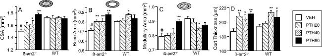
Results and conclusions: beta-arr2(-/-) mice had significantly lower total body BMD, trabecular bone volume fraction (BV/TV), and femoral cross-sectional area compared with WT. In WT females, PTH increased total body BMD, trabecular bone parameters, and cortical thickness, with a trend toward decreased midfemoral medullary area. In beta-arr2(-/-) mice, PTH not only improved total body BMD, trabecular bone architecture, and cortical thickness, but also dose-dependently increased femoral cross-sectional area and medullary area. Histomorphometry showed that PTH-stimulated periosteal bone formation was 2-fold higher in beta-arr2(-/-) compared with WT. Osteocalcin levels were significantly lower in beta-arr2(-/-) mice, but increased dose-dependently with PTH in both beta-arr2(-/-) and WT. In contrast, whereas the resorption marker TRACP5B increased dose-dependently in WT, 20-80 mug/kg/day of PTH was equipotent with regard to stimulation of TRACP5B in beta-arr2(-/-). In summary, beta-arrestin2 plays an important role in bone mass acquisition and remodeling. In estrogen-replete female mice, the ability of intermittent PTH to stimulate periosteal bone apposition and endosteal resorption is inhibited by arrestins. We therefore infer that arrestin-mediated regulation of intracellular signaling contributes to the differential effects of PTH on cancellous and cortical bone.
Figures


References
-
- Neer RM, Arnaud CD, Zanchetta JR, Prince R, Gaich GA, Reginster JY, Hodsman AB, Eriksen EF, Ish-Shalom S, Genant HK, Wang O, Mitlak BH. Effect of parathyroid hormone (1-34) on fractures and bone mineral density in postmenopausal women with osteoporosis. N Engl J Med. 2001;344:1434–1441. - PubMed
-
- Dempster DW, Cosman F, Kurland ES, Zhou H, Nieves J, Woelfert L, Shane E, Plavetic K, Muller R, Bilezikian J, Lindsay R. Effects of daily treatment with parathyroid hormone on bone microarchitecture and turnover in patients with osteoporosis: A paired biopsy study. J Bone Miner Res. 2001;16:1846–1853. - PubMed
-
- Jiang Y, Zhao JJ, Mitlak BH, Wang O, Genant HK, Eriksen EF. Recombinant human parathyroid hormone (1-34) [teriparatide] improves both cortical and cancellous bone structure. J Bone Miner Res. 2003;18:1932–1941. - PubMed
-
- Horwitz M, Stewart A, Greenspan SL. Sequential parathyroid hormone/alendronate therapy for osteoporosis-robbing Peter to pay Paul? J Clin Endocrinol Metab. 2000;85:2127–2128. - PubMed
-
- Burr DB, Hirano T, Turner CH, Hotchkiss C, Brommage R, Hock JM. Intermittently administered human parathyroid hormone(1-34) treatment increases intracortical bone turnover and porosity without reducing bone strength in the humerus of ovariectomized cynomolgus monkeys. J Bone Miner Res. 2001;16:157–165. - PubMed
Publication types
MeSH terms
Substances
Grants and funding
LinkOut - more resources
Full Text Sources
Molecular Biology Databases
