. 2005 Mar 14:6:6.
doi: 10.1186/1471-2199-6-6.

Recognition and binding of mismatch repair proteins at an oncogenic hot spot

Affiliations

Recognition and binding of mismatch repair proteins at an oncogenic hot spot

Michael Edelbrock et al. BMC Mol Biol. .

Abstract

Background: The current investigation was undertaken to determine key steps differentiating G:T and G:A repair at the H-ras oncogenic hot spot within the nuclear environment because of the large difference in repair efficiency of these two mismatches.

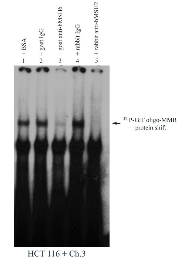
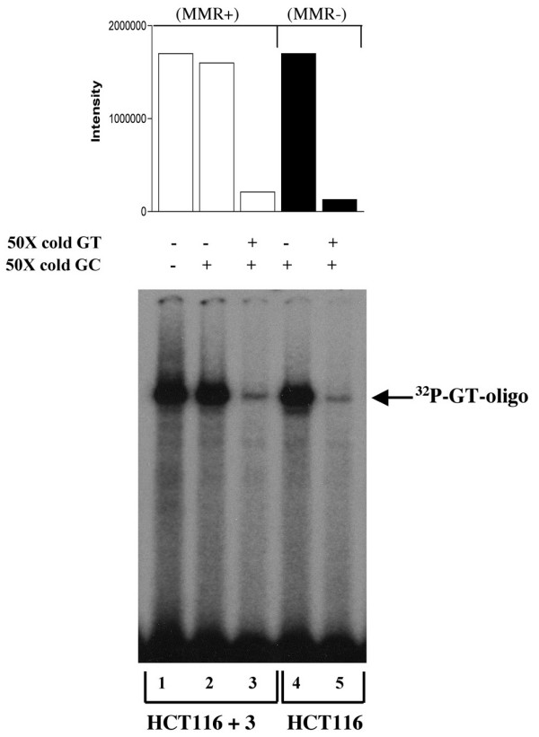
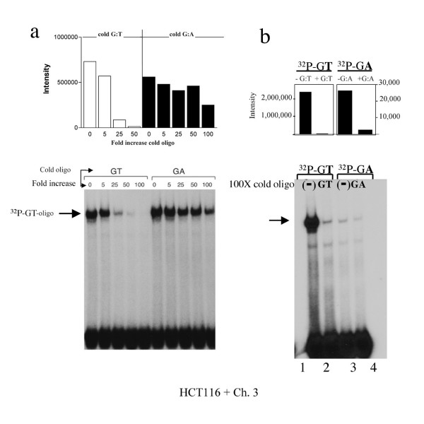
Results: Electrophoretic mobility shift (gel shift) experiments demonstrate that DNA containing mismatched bases are recognized and bound equally efficiently by hMutSalpha in both MMR proficient and MMR deficient (hMLH1-/-) nuclear extracts. Competition experiments demonstrate that while hMutSalpha predictably binds the G:T mismatch to a much greater extent than G:A, hMutSalpha demonstrates a surprisingly equal ratio of competitive inhibition for both G:T and G:A mismatch binding reactions at the H-ras hot spot of mutation. Further, mismatch repair assays reveal almost 2-fold higher efficiency of overall G:A repair (5'-nick directed correct MMR to G:C and incorrect repair to T:A), as compared to G:T overall repair. Conversely, correct MMR of G:T --> G:C is significantly higher (96%) than that of G:A --> G:C (60%).

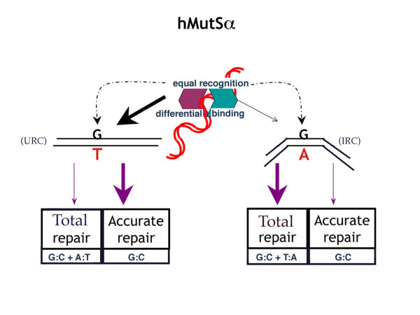
Conclusion: Combined, these results suggest that initiation of correct MMR requires the contribution of two separate steps; initial recognition by hMutSalpha followed by subsequent binding. The 'avidity' of the binding step determines the extent of MMR pathway activation, or the activation of a different cellular pathway. Thus, initial recognition by hMutSalpha in combination with subsequent decreased binding to the G:A mismatch (as compared to G:T) may contribute to the observed increased frequency of incorrect repair of G:A, resulting in the predominant GGC --> GTC (Gly --> Val) ras-activating mutation found in a high percentage of human tumors.


Figures

Figure 1
Figure 1
Specific interaction of hMSH6 and hMSH2 with site-specific mismatched DNA at H-ras codon 12. Protein-DNA binding reactions and gel shifts were performed using nuclear extracts from HCT 116 + Ch. 3 and equal cpm of [32P]-G:T-oligo (69-mer) in the presence of 100X molar excess of cold (unlabeled) homoduplex (G:C). BSA, goat-, or rabbit-nonspecific IgG (lanes 1, 2, 4 respectively), goat anti-hMSH6 (lane 3), and rabbit anti-hMSH2 (lane 5) were also included in the binding reactions as indicated. The lower gel shift band in each lane is due to biotin end-labeling of the probe.
Figure 2
Figure 2
Comparison of binding avidity of G:T-containing oligomer by MMR proficient (HCT 116 + Ch. 3) and MMR deficient (HCT 116; hMLH1-/-) nuclear extracts. Equal aliquots of nuclear extracts from HCT 116 + Ch. 3 (lanes 1–3) or HCT 116 (lanes 4–5) were incubated with equal cpm of [32P]-G:T-oligo (69-mer) and 50X molar excess cold (unlabeled) oligo as indicated.
Figure 3
Figure 3
Binding avidity of hMutSα to G:T versus G:A at H-ras codon 12 (a) Equal cpm of [32P]-G:T-oligo (69 mer) were incubated with equal concentrations of nuclear extract (HCT116 + Ch. 3) in the presence of increasing concentrations of unlabeled (cold) G:T- or G:A-containing oligo, and 100X cold G:C oligo. (b) Equal aliquots of nuclear extract were incubated for 2.5 hours (versus 30 minutes) with equal cpm of each [32P]-G:T- or [32P]-G:A-oligo alone (lanes 1 and 3), or also with respective 100X molar excess cold G:T or G:A oligo (lanes 2 and 4). Bar graphs are densitometry results of corresponding radioactive band intensities.
Figure 4
Figure 4
Repair model of hMutSα MMR. Model of hMutSα initial recognition (equal) of G:T and G:A, followed by repair (differential) of DNA containing a G:T or a G:A mismatch at the H-ras codon 12 location. hMutSα recognizes and forms an initial recognition complex (IRC) with both mismatches equally, and then binds more strongly to G:T, perhaps by undergoing an additional conformational step to the ultimate recognition complex (URC), which does not occur with G:A [40]. This results in more accurate repair of G:T, but more frequent total repair of G:A. See text for further discussion.

References

    1. Jiricny J. Replication errors: cha(lle)nging the genome. EMBO J. 1998;17:6427–6436. doi: 10.1093/emboj/17.22.6427. - DOI - PMC - PubMed
    1. Wood RD, Mitchell M, Sgouros J, Lindahl T. Human DNA repair genes. Science. 2001;291:1284–1289. doi: 10.1126/science.1056154. - DOI - PubMed
    1. Jiricny J. Mismatch repair and cancer. Cancer Surv. 1996;28:47–68. - PubMed
    1. Marra G, Boland CR. Hereditary nonpolyposis colorectal cancer: the syndrome, the genes, and the historical perspectives. J Natl Cancer Inst. 1995;87:1114–1125. - PubMed
    1. Orth K, Hung J, Gazdar A, Bowcock A, Mathis JM, Sambrook J. Genetic instability in human ovarian cancer cells. Proc Natl Acad Sci USA. 1994;91:9495–9499. - PMC - PubMed

Publication types