

Review
doi: 10.1016/j.str.2005.01.005.
Combining X-ray crystallography and electron microscopy
Affiliations
- PMID: 15766536
- PMCID: PMC7173138
- DOI: 10.1016/j.str.2005.01.005
Item in Clipboard
Review
Combining X-ray crystallography and electron microscopy
Structure.
2005 Mar.
Abstract
The combination of cryo-electron microscopy to study large biological assemblies at low resolution with crystallography to determine near atomic structures of assembly fragments is quickly expanding the horizon of structural biology. This technique can be used to advantage in the study of large structures that cannot be crystallized, to follow dynamic processes, and to "purify" samples by visual selection of particles. Factors affecting the quality of cryo-electron microscopy maps and limits of accuracy in fitting known structural fragments are discussed.
Figures
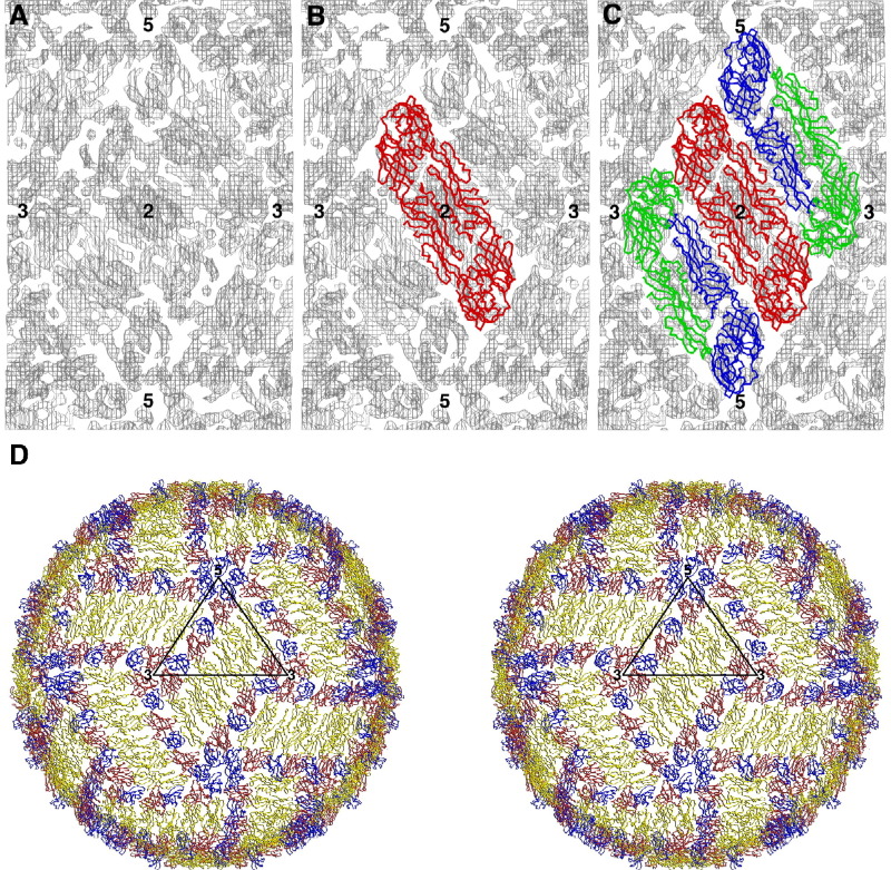
Fitting the Icosahedrally Averaged, Cryo-EM Density of Dengue Virus at 9 Å Resolution with the Crystal Structure of the Envelope (E) Ectodomain Protein Dimer Cα Backbone (A) The cryo-EM density (Kuhn et al., 2002; Zhang et al., 2003) and the position of icosahedral 5-fold, 3-fold, and 2-fold symmetry axes surrounding one asymmetric unit. (B) One E dimer (Modis et al., 2004; Rey et al., 1995; Zhang et al., 2004) fitted with its 2-fold axis coincident with an icosahedral 2-fold axis. (C) A second E dimer fitted into the remaining density. (D) The icosahedral symmetry has been used to generate the whole of the top surface of the virus shown as a stereo diagram. Each E monomer is colored red (domain I), yellow (domain II), and blue (domain III).

Diagrammatic Representation of Bacteriophage T4 The dsDNA genome is protected by the head capsid. The head is attached to the tail, a highly specialized and extremely efficient phage component required for infecting the E. coli host. The hexagonally shaped baseplate is situated at the distal end of the tail. The baseplate coordinates the movement of the six long tail fibers that initially sense the presence of the host, the short tail fibers that unfold from underneath the baseplate to firmly anchor on the E. coli surface, and the tail sheath surrounding the tail tube that contracts, thereby ejecting DNA into the host. The numbers identify the gene products of the various proteins that are in the assembled virion. It would be difficult to place this complex virus into a well-packed crystal lattice both because of its shape and because of the variably angled fibers (Eiserling and Black, 1994; Leiman et al., 2003).
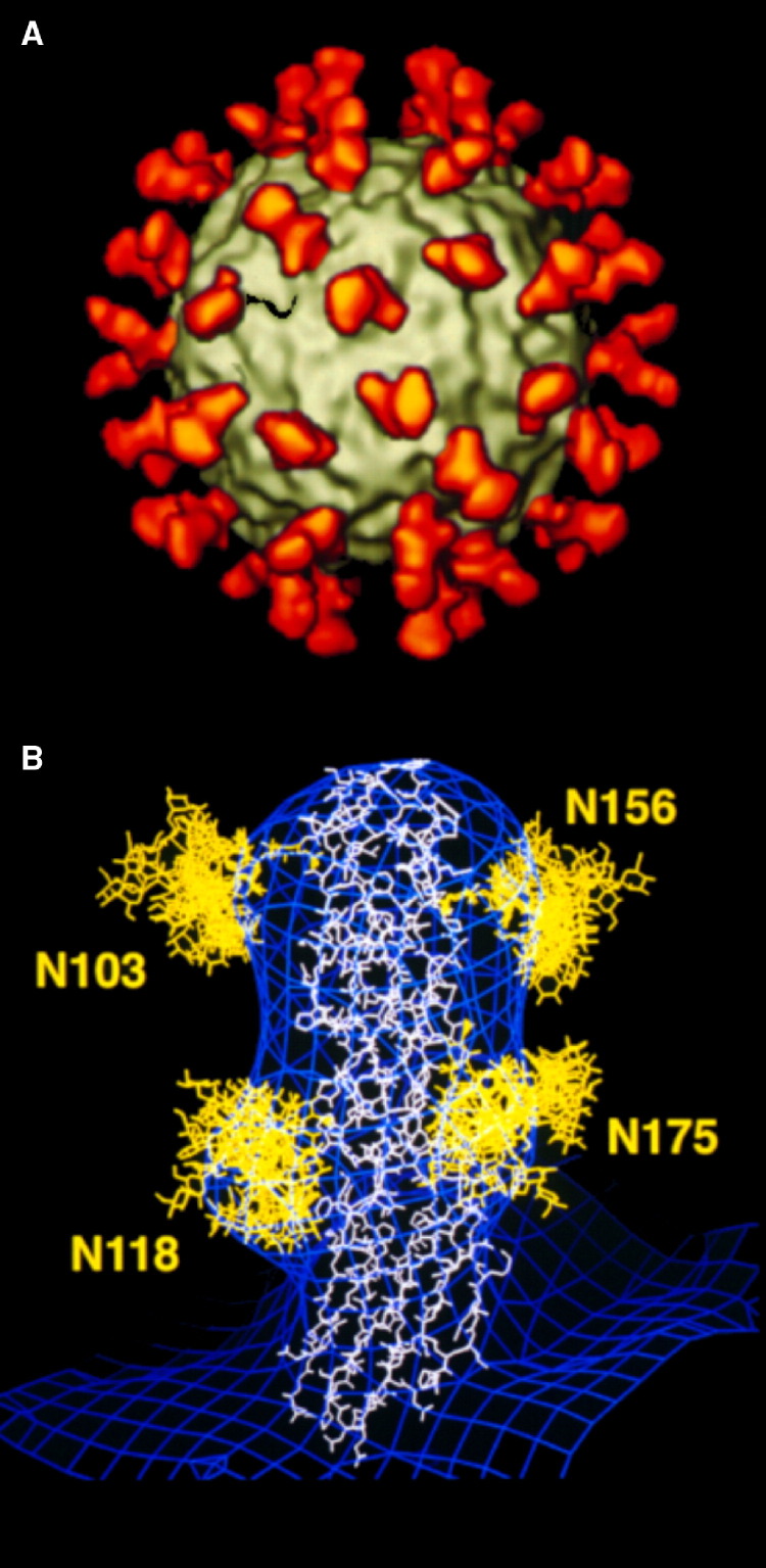
The Structure of Human Rhinovirus 14 Complexed with Its Cellular Receptor Molecule, Intercellular Adhesion Molecule 1, at 26 Å Resolution (A) The icosahedral virus (gray) complexed with the two amino-terminal domains of intercellular adhesion molecule 1 (ICAM1) (red) at all icosahedrally equivalent positions. (B) The cryo-EM density of the complex (blue), fitted with the X-ray crystal structure of the two amino-terminal domains decorated with multiple conformations of the carbohydrates at the four potential N-glycosylation sites. The Asn residues at positions 103, 118, and 156, but not 175, had to be mutated in order to form viable crystals. However, note the bulges of the blue density indicating the presence of the carbohydrate moieties in the wild-type ICAM1 used to form the complex. (Modified and reprinted by permission from Kolatkar et al., 1999, Macmillan Publishers Ltd.)

The Membrane Structure of Dengue Virus (A) A central cross-section through the cryo-EM density at 9.5 Å resolution showing the E glycoprotein ectodomain, the lipid bilayer, and the internal nucleocapsid. (B) Radial density section at a radius of 185 Å, showing higher density blacker than lower density, with the superimposed envelopes of the fitted E ectodomain. Note the four blacker regions associated similarly with each monomer corresponding to four transmembrane helices per monomer. (C) Diagrammatic side view of the E protein (domains I, II, and III). Domain III connects with the EH1 and EH2 helices of the stem region in the outer lipid leaflet, and ET1 and ET2 antiparallel transmembrane helices. Also shown are the two antiparallel transmembrane helices MH1 and MH2 of the membrane protein. (Reprinted with permission from Zhang et al., 2003, Nature Publishing Group.)

Cryo-EM Micrograph of Mature Dengue Virus Note the many broken particles, indicated by arrows, that can be neglected for an image reconstruction, but that are likely to inhibit crystallization attempts.
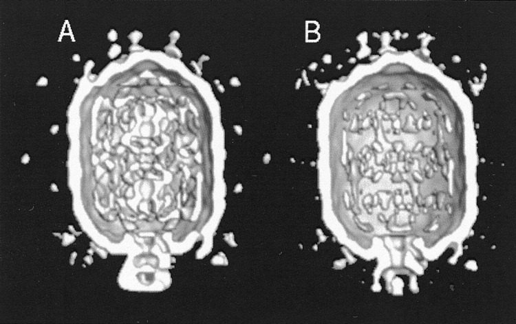
Cryo-EM Reconstruction of ϕ29 Prohead The packaging reaction was stopped by freezing 2 minutes after initiation. Particles partially packaged with genomic DNA (A) and empty (B) were selected by eye for each reconstruction from the same micrographs. Note the additional density, around the special pentagonal entry vertex, representing the ATPase (gp16) required to hydrolyze ATP for DNA packaging (Morais et al., 2001; Tao et al., 1998).

The Structure of the ϕ29 DNA-Packaging Machine (A) The crystal structure of the dodecameric connector (blue with one monomer picked out in red). (B) Fit of the Cα trace of the connector (yellow) into the cryo-EM density of the prohead. Shown also is the fit of the pRNA derived from a difference map between prohead and RNAase-treated connector (Morais et al., 2001; Tao et al., 1998). (Reprinted with permission from Simpson et al., 2000, Nature Publishing Group.)
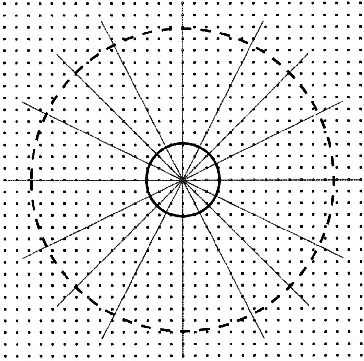
A Representation of Reciprocal Space with Lines Showing the Random Position of the Projection Planes of Randomly Selected Particle Projections When relatively few particle images are included in a reconstruction, only a low-resolution region of reciprocal space has moderately complete sampling (inside solid circle). Many more particle orientations are required for a fuller sampling at higher resolution (inside dashed circle).

Reconstruction of the T4 Head Capsid Using 5-Fold Symmetry Different proteins are identified by different colors: the major capsid protein, gp23, is blue, the vertex protein is magenta, the highly antigenic outer capsid proteins (hoc) are colored yellow, and the small outer capsid proteins (soc) are colored white. The scale bar represents 100 Å. Note the blurring of the tail, which has 6-fold symmetry (adapted from Fokine et al., 2004).
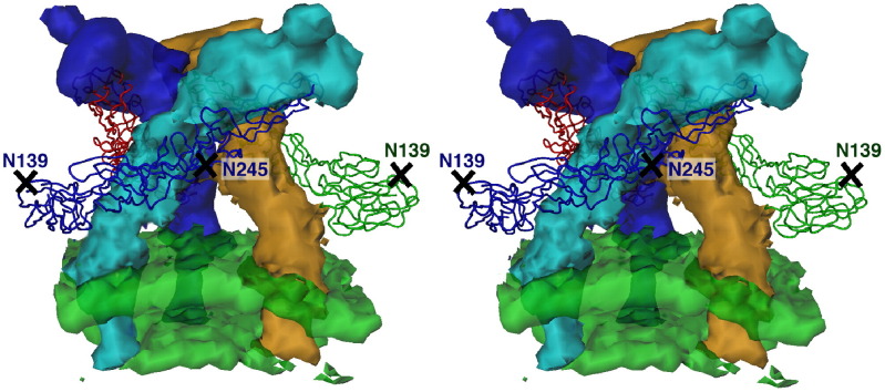
The Trimeric (E1E2)3 Spike of Sindbis Virus The known crystal structure of E1 was fitted into the cryo-EM density, assuming 3-fold symmetry, aided by the glycosylation sites at residues Asn139 and Asn245. The carbohydrate positions (crosses) had been determined from difference maps between wild-type and deglycosylated virus. After the E1 molecules had been fitted, the density at all grid points in the map that were within 4 Å of each atom in E1 was set to zero, leaving the density of E2. Hence, the three E2 molecules (cyan, brown, and blue) were shown to be long and thin (Zhang et al., 2002).
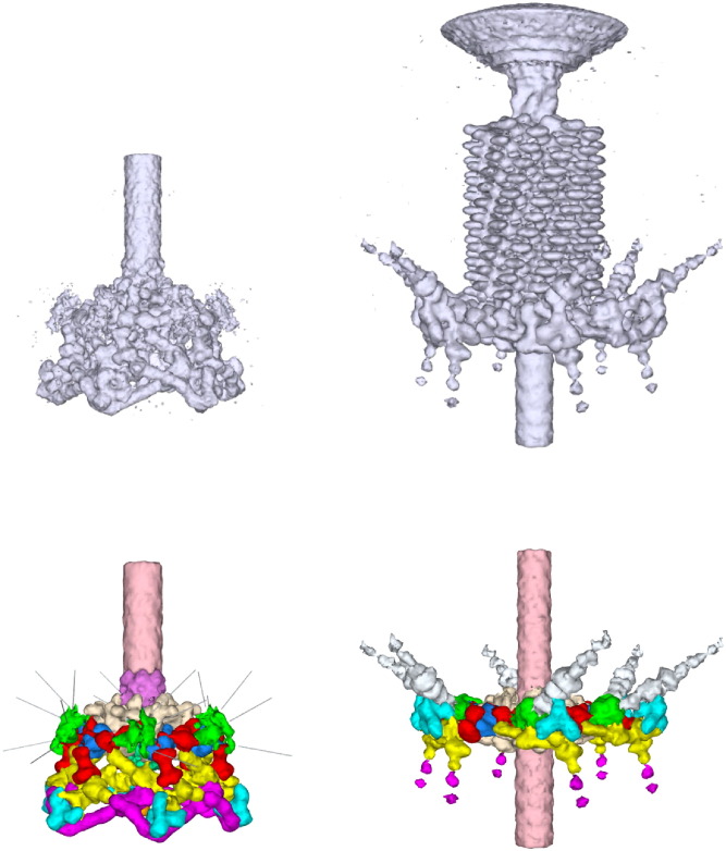
The Cryo-EM Densities (Top) of the Hexagonal (Left) and Star-Shaped (Right) Bacteriophage T4 Baseplate These densities were interpreted (bottom) by fitting the crystal structures of gene products (gp) 12 (magenta), 11 (blue), 8 (dark blue), 9 (green), 5 (mostly obscured), and 27 (obscured). The position and shape of other proteins (gp 6, 7, 10, 25, 48, 53, and 54) could then be interpreted from biochemical information (Kostyuchenko et al., 2003; Leiman et al., 2004).
References
-
- Baumeister W., Steven A.C. Macromolecular electron microscopy in the era of structural genomics. Trends Biochem. Sci. 2000;25:624–631. - PubMed
-
- Dubochet J., Adrian M., Chang J.J., Homo J.C., Lepault J., McDowall A.W., Schultz P. Cryo-electron microscopy of vitrified specimens. Q. Rev. Biophys. 1988;21:129–228. - PubMed
-
- Eiserling F.A., Black L.W. In: Molecular Biology of Bacteriophage T4. Karam J.D., editor. American Society for Microbiology; Washington, DC: 1994. Pathways in T4 morphogenesis; pp. 209–212.
Publication types
MeSH terms
Substances
LinkOut - more resources
Full Text Sources
