. 2005 Mar 14;33(5):1513-23.
doi: 10.1093/nar/gki284. Print 2005.

Identification of the 'NORE' (N-Oct-3 responsive element), a novel structural motif and composite element

Affiliations

Identification of the 'NORE' (N-Oct-3 responsive element), a novel structural motif and composite element

Robert Alazard et al. Nucleic Acids Res. .

Abstract

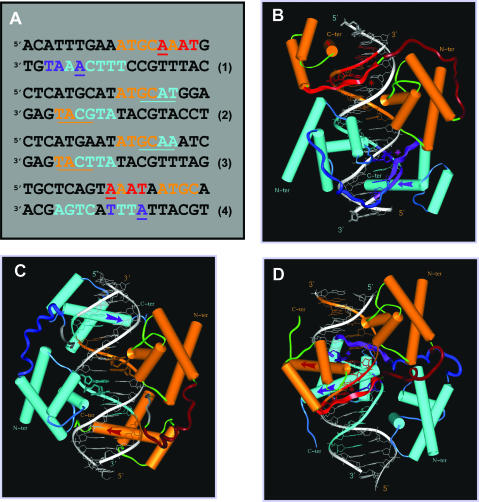
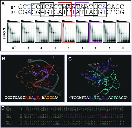
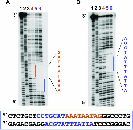
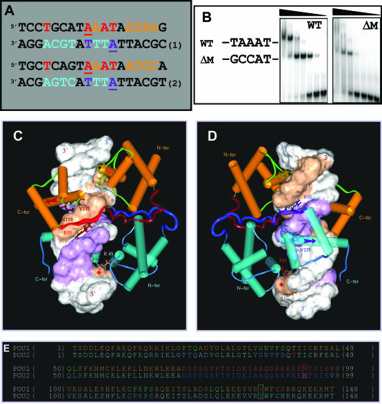
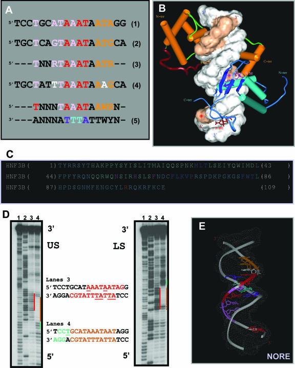
N-Oct-3 is a neuronal transcription factor widely expressed in the developing mammalian central nervous system, and necessary to maintain neural cell differentiation. The key role of N-Oct-3 in the transcriptional regulation of a multiplicity of genes is primarily due to the structural plasticity of its so-called 'POU' (acronym of Pit, Oct, Unc) DNA-binding domain. We have recently reported about the unusual dual neuro-specific transcriptional regulation displayed by N-Oct-3 [Blaud,M., Vossen,C., Joseph,G., Alazard,R., Erard,M. and Nieto,L. (2004) J. Mol. Biol., 339, 1049-1058]. To elucidate the underlying molecular mechanisms, we have now made use of molecular modeling, DNA footprinting and electrophoretic mobility shift assay techniques. This combined approach has allowed us to uncover a novel mode of homodimerization adopted by the N-Oct-3 POU domain bound to the neuronal aromatic amino acids de-carboxylase and corticotropin-releasing hormone gene promoters and to demonstrate that this pattern is induced by a structural motif that we have termed 'NORE' (N-Oct-3 responsive element), comprising the 14 bp sequence element TNNRTAAATAATRN. In addition, we have been able to explain how the same structural motif can also induce the formation of a heterodimer in association with hepatocyte nuclear factor 3beta(/Forkhead box a2). Finally, we discuss the possible role of the NORE motif in relation to neuroendocrine lung tumor formation, and in particular the development of small cell lung cancer.


Figures

Figure 1
Figure 1
The three generic modes of N-Oct-3 POU domain homodimerization. (A) Eighteen base pair DNA sequences of: (1) the osteopontin gene enhancer fragment defining the PORE motif, (2) the prototypic MORE motif, (3) the MORE-type IgG VH gene promoter canonical fragment and (4) the AADC gene promoter fragment defining the new NORE motif. In all the cases, the first and second N-Oct-3 POU domain binding sites are coded in brown and turquoise, respectively. In the PORE (1) and NORE (4) motifs, critical nucleotides of the first and second POUh tetrameric sub-sites are coded in red and indigo respectively. In the MORE motifs (2 and 3), the first and second POUh tetrameric sub-sites are underlined in brown and turquoise, respectively, to compensate for the overlap with the POUs binding sub-sites. (B–D) Modeled structures of the homodimeric complexes between the N-Oct-3 DBD and (B) a PORE motif (sequence 1), (C) a MORE motif (sequence 3) and (D) a NORE motif (sequence 4). In all the cases, the respective display-codes for the first and second N-Oct-3 POU domains are as follows: brown- and turquoise-colored cylinders for the α-helices, red- and indigo-colored arrows for the POUh recognition helices, dark brown- and blue-colored coils for the linkers. In the PORE (B) and NORE (D) types of complexes, the first and second POUh N-terminal extensions are displayed as red- and indigo-colored ribbons, respectively, their insertion into the DNA minor groove being indicated by asterisks of the identical color.
Figure 2
Figure 2
Structural determinants of the N-Oct-3 POU homodimerization on the AADC gene promoter (A) Mutation scanning of the 18 bp core region of the AADC fragment (the first and last base pairs are colored in blue) was carried out by selecting in turn 8 overlapping nucleotide triplets, in which A and G were substituted by C, and C and T substituted by G. The interactions between the N-Oct-3 DBD and the mutated AADC fragments were analyzed by EMSA and compared with the interaction with the non-mutated sequence (WT). The two most critical triplet mutations and their corresponding EMSA patterns have been highlighted. (B) Canonical interaction between the first monomer POUh and the optimal AAAT sub-site: anchoring into the DNA minor groove via the Arg 93/Ade 9 contact (red asterisk) and insertion of the recognition helix into the major groove via the Asn 139/Ade 11 and Val 135/Thy 12 contacts (pink arrow). (C) Interaction between the second monomer POUh and the ATTT sub-site: anchoring into the DNA minor groove via the Arg 93/Ade 7B contact (indigo asterisk) and insertion of the recognition helix into the major groove via the hydrophobic contacts between Val 135 and both Thy 9B and Thy 10B (turquoise arrow). (D) Location of Arg 93 (in red box), Val 135 (in turquoise box) and several important residues within the secondary structure elements of the first (POU1) and second (POU2) DBDs. Respective color coding is as follows: α-helices in brown and turquoise; POUh N-terminal extensions in red and indigo; linkers in dark brown and blue. POUs1 Gln 40, Thr41 and Arg 45 are colored in red, and POUs2 Thr 41 and Arg 45 in white.
Figure 3
Figure 3
DNAse I footprinting analysis of N-Oct-3 POU homodimerization on the CRH gene promoter. Autoradiograms of 12% polyacrylamide denaturing gels showing the DNAse I footprints on the upper (A) and lower (B) strands of the CRH promoter fragment. Lanes 1 and 2, Maxam–Gilbert chemical sequencing references (cleavage after pyrimidine and purine residues, respectively). Lane 3, free DNA cleavage products. Lane 4, first DBD footprint (brown color coding). Lanes 5 and 6, POU homodimer footprint (blue color coding).
Figure 4
Figure 4
Structural determinants of the N-Oct-3 POU homodimerization on the CRH gene promoter. (A) Alignment of the 18 bp core regions of the CRH (sequence 1) and AADC (sequence 2) promoter fragments based on the homology between the respective high-affinity binding sites for N-Oct-3 DBD. Same color coding as in Figure 1A. (B) Conversion of the TAA triplet (in boldface) of the 24 bp CRH promoter fragment (‘WT’) GCTCCTGCATAAATAATAGGGCCC to GCC (mutant ‘ΔM’) and EMSA analysis of the differential binding pattern of the N-Oct-3 DBD to the two oligonucleotides. (C and D) Modeled structure of the homodimeric complex between the N-Oct-3 DBD and the 18 bp CRH promoter fragment (sequence 1). Front views focusing on the first (C) and second (D) POU domain interactions with DNA. See the text for a detailed description. Same color coding as in Figure 1B–D. The ‘footprints’ on both strands of the first and second DBDs are shown as brown and purple-colored Connolly surfaces, respectively. (E) Same comment and color coding as in Figure 2D, except for POUs1 Gln 40, Thr41 and Arg 45 colored in yellow, and POUs2 Thr 41 and Arg 45 in red and white, respectively.
Figure 5
Figure 5
Identification of the NORE as a structural motif and dual transcription factor binding site. (A) Structural homology between the HNF-3β binding sites on the CRH (sequence 1) and AADC (sequence 2) gene promoters (pink-coded nucleotides) and overlap with the N-Oct-3 high-affinity binding sites. Sequence 3, optimal NORE motif (N: any nucleotide and R: purine residues). Sequence 4, 18 bp core of the aldolase C gene promoter fragment. Sequence 5, extended consensus NORE motif and its dual binding capacity specifying N-Oct-3 DBD homo or heterodimerization (W: A or T). (B) Modeled structure of the heterodimeric complex between the N-Oct-3 and HNF-3β DBDs and the 18 bp CRH promoter. Note the two major components of HNF-3β binding and structural similarities with the second POUh binding in the homodimeric complex. See the text for a detailed description. Display-code for the DNA and N-Oct-3 DBD as in Figure 4C and D, and for the HNF-3β DBD as follows: α-helices are turquoise-colored cylinders, β-strands are dark blue ribbons, and ‘wings’ are blue coils. (C) Location of the important residues within the secondary structure elements of HNF-3β DBD (‘HNF3B’); color coding as in (B). (D) Autoradiograms of 12% polyacrylamide denaturing gels showing the OP2-Cu cleavage products of the upper (‘US’) and lower (‘LS’) strands of the CRH promoter fragment. Lanes 1 and 2, Maxam–Gilbert chemical sequencing references (cleavage after pyrimidine and purine residues, respectively). Lanes 3, free DNA; cleavage enhancement sites highlighted in red. Lanes 4, DNA in the 1/1 high-affinity complex with the N-Oct-3 DBD; footprint and flanking cleavage enhancement sites highlighted in brown and green, respectively. (E) Model of the bound-form of the NORE motif [same color coding as in (A), sequence 5].

Similar articles

Cited by

References

    1. Veeramachaneni V., Makalowski W., Galdzicki M., Sood R., Makalowska I. Mammalian overlapping genes: the comparative perspective. Genome Res. 2004;14:280–286. - PMC - PubMed
    1. Warren A.J. Eukaryotic transcription factors. Curr. Opin. Struct. Biol. 2002;12:107–114. - PubMed
    1. Hahn S. Structure and mechanism of the RNA polymerase II transcription machinery. Nature Struct. Mol. Biol. 2004;11:394–403. - PMC - PubMed
    1. Johnson W., Jameson J.L. Transcriptional control of gene expression. In: Jameson J.L., editor. Principles of Molecular Medicine. Towota, NJ: Humana Press Inc.; 1998.
    1. Latchman D.S. Transcription factors as potential targets for therapeutic drugs. Curr. Pharm. Biotechnol. 2000;1:57–61. - PubMed

Publication types

MeSH terms