. 2005 Mar 14;33(5):1524-31.
doi: 10.1093/nar/gki289. Print 2005.

Dimeric configuration of SeqA protein bound to a pair of hemi-methylated GATC sequences

Affiliations

Dimeric configuration of SeqA protein bound to a pair of hemi-methylated GATC sequences

Sukhyun Kang et al. Nucleic Acids Res. .

Abstract

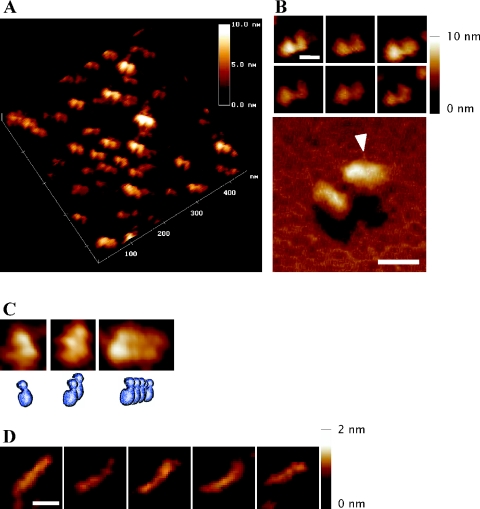
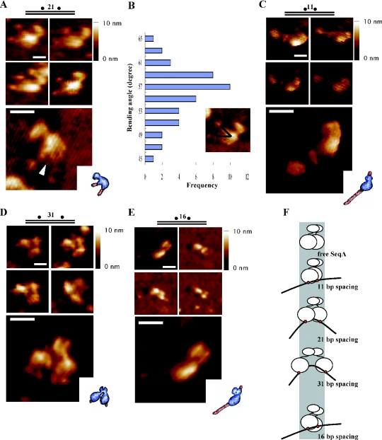
The binding of SeqA protein to hemi-methylated GATC sequences (hemi-sites) regulates chromosome initiation and the segregation of replicated chromosome in Escherichia coli. We have used atomic force microscopy to examine the architecture of SeqA and the mode of binding of one molecule of SeqA to a pair of hemi-sites in aqueous solution. SeqA has a bipartite structure composed of a large and a small lobe. Upon binding of a SeqA molecule to a pair of hemi-sites, the larger lobe becomes visibly separated into two DNA binding domains, each of which binds to one hemi-site. The two DNA binding domains are held together by association between the two multimerization domains that make up the smaller lobe. The binding of each DNA binding domain to a hemi-site leads to bending of the bound DNA inwards toward the bound protein. In this way, SeqA adopts a dimeric configuration when bound to a pair of hemi-sites. Mutational analysis of the multimerization domain indicates that, in addition to multimerization of SeqA polypeptides, this domain contributes to the ability of SeqA to bind to a pair of hemi-sites and to its cooperative behavior.


Figures

Figure 1
Figure 1
Atomic force microscopy of SeqA and 75 bp hemi-methylated DNA. (A) An aliquot of 10 μl of purified SeqA (50 μg/ml in buffer A containing 0.1 M KCl and no glycerol) was deposited on freshly cleaved mica and incubated for 30 min under ambient humidity to adsorb to the mica surface. Surface scanning of a 450 × 450 nm field was performed in height and amplitude mode. (B) Representative images of SeqA in computerized three dimensions (3D) (upper panel), and an enlarged 3D view (lower panel). The arrowhead points to the groove in the large lobe of SeqA. (C) Representative images of multimeric forms of SeqA molecules, with schematic pictures. A 10 μl of purified SeqA (200 μg/ml in buffer A containing 0.1 M KCl and no glycerol) was deposited on freshly cleaved mica and the surface was scanned as in (A). (D) A 75 bp DNA containing two hemi-sites with a spacing of 21 bp (Table 1) was deposited on a Ni-treated mica surface, and images were recorded in the tapping mode in aqueous solution. The horizontal scale bars represent 20 nm, and the upright scale bars, height of the particles from baseline to the top.
Figure 2
Figure 2
Dimeric configuration of SeqA bound to a pair of hemi-sites. To observe the complexes of SeqA with hemi-methylated DNA, 10 μl of a reaction mixture in buffer A containing SeqA (0.2–20 ng), the indicated DNA (50 fmol) and no glycerol was incubated for 15 min at 32°C, and the mixture was directly deposited on freshly cleaved mica. After further incubation for 30 min at room temperature, the specimen was gently washed with buffer A, fixed with 0.5% glutaraldehyde for 2–3 min, and washed several times again with buffer A. Images were recorded in the tapping mode in aqueous solution. (A) Representative images of complexes formed by SeqA and the 21 bp spacing DNA (upper panel), and a computerized 3D view of the complexes (lower panel) with a schematic picture (inner box). The arrowhead points to the portion of SeqA opened up as a result of binding to the DNA. (B) Relative frequency of the DNA bending angles in the complexes of SeqA with 21 bp spacing DNA. The degree of DNA bending was measured by drawing lines from the ends of the DNA strand to the apparent bending point of the bound DNA. (C) Representative images of complexes formed by SeqA and the 11 bp spacing DNA, and a computerized 3D view of the complexes. (D) Representative images of complexes formed by SeqA and 31 bp spacing DNA, and a computerized 3D view of the complexes. (E) Representative images of complexes formed by SeqA and 16 bp spacing DNA, and a computerized 3D view of the complexes. All the horizontal scale bars represent 20 nm, and the upright scale bars represent height of the particles from the baseline to the top. (F) Cartoon of the complexes formed between SeqA and a pair of hemi-sites spaced increasing distances apart.
Figure 3
Figure 3
The Thr-18 of SeqA contributes to cooperative interaction between SeqA molecules. The binding behavior of purified wild-type SeqA and SeqA(T18G) was analyzed by gel shift experiments: 10, 20 and 40 ng of wild-type SeqA (designated as WT) and SeqA(T18G) were incubated with P32-labeled DNA containing a pair of hemi-sites, which were separated by 16 bp (A), and two pairs of hemi-sites in which the pairs were separated by 16 bp (B), 19 bp (C) and 41 bp (D). Complexes of SeqA and DNA were separated on 5% polyacrylamide gels, dried and visualized by autoradiography.
Figure 4
Figure 4
N-terminal SeqA mutant proteins are defective in binding to a pair of hemi-sites, but interact cooperatively. Fractions containing mutant SeqA proteins were incubated with DNA containing a pair of hemi-sites separated by 21 bp (A), with DNA containing four hemi-sites with a 16 bp spacing between the pairs of hemi-sites (B) and DNA containing four hemi-sites with 32 bp spacing (C). In each experiment, 33 and 100 ng of wild-type SeqA (designated as WT) and SeqA(E5K), (D7K), (E8K), (E9K) and (E5,9K) partially purified fractions were added to the reaction. The amount of SeqA protein in each case was similar in western blot (data not shown).
Figure 5
Figure 5
Binding of a SeqA to a pair of hemi-methylated GATC sequences. The binding of one of the DNA binding domains of a SeqA molecule to a hemi-site leads to conformational change of the molecule that allows the other DNA binding domain to bind to an adjacent hemi-site within 32 bp. Binding is stabilized by bending the bound DNA.

Similar articles

Cited by

References

    1. Sarraf S.A., Stancheva I. Methyl-CpG binding protein MBD1 couples histone H3 methylation at lysine 9 by SETDB1 to DNA replication and chromatin assembly. Mol. Cell. 2004;15:595–605. - PubMed
    1. Wade P.A. Methyl CpG-binding proteins and transcriptional repression. Bioessays. 2001;23:1131–1137. - PubMed
    1. Reik W., Dean W., Walter J. Epigenetic reprogramming in mammalian development. Science. 2001;293:1089–1093. - PubMed
    1. Geier G.E., Modrich P. Recognition sequence of the dam methylase of Escherichia coli K12 and mode of cleavage of Dpn I endonuclease. J. Biol. Chem. 1979;254:1408–1413. - PubMed
    1. Campbell J.L., Kleckner N. E.coli oriC and the dnaA gene promoter are sequestered from dam methyltransferase following the passage of the chromosomal replication fork. Cell. 1990;62:967–979. - PubMed

Publication types

MeSH terms