. 2005 Apr;79(7):3949-61.
doi: 10.1128/JVI.79.7.3949-3961.2005.

ICP27 recruits Aly/REF but not TAP/NXF1 to herpes simplex virus type 1 transcription sites although TAP/NXF1 is required for ICP27 export

Affiliations

ICP27 recruits Aly/REF but not TAP/NXF1 to herpes simplex virus type 1 transcription sites although TAP/NXF1 is required for ICP27 export

I-Hsiung Brandon Chen et al. J Virol. 2005 Apr.

Abstract

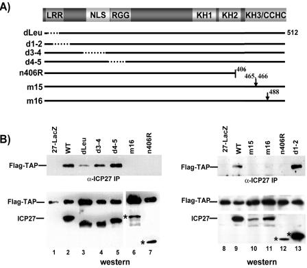
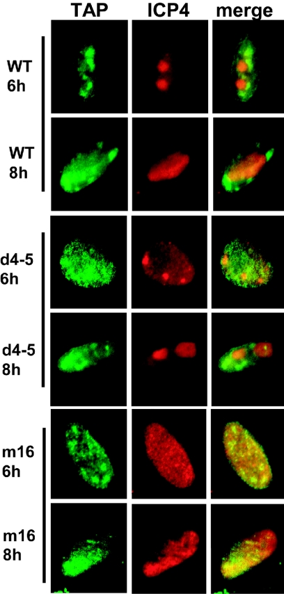
Herpes simplex virus type 1 (HSV-1) protein ICP27 interacts with the cellular export adaptor protein Aly/REF, which is part of the exon junction complex implicated in cellular mRNA export. We previously reported that Aly/REF was no longer associated with splicing factor SC35 sites during infection but instead colocalized with ICP27 in distinct structures. Here we show that these structures colocalize with ICP4 and are sites of HSV-1 transcription. ICP27 mutants with lesions in the region required for the interaction with Aly/REF failed to recruit Aly/REF to viral transcription sites; however, ICP27 export to the cytoplasm was unimpaired, indicating that the interaction of ICP27 with Aly/REF is not required for ICP27 shuttling. ICP27 has also been shown to interact with the cellular mRNA export receptor TAP/NXF1. We report that ICP27 interacts directly with TAP/NXF1 and does not require Aly/REF to bridge the interaction. The C terminus of ICP27 is required; however, the N-terminal leucine-rich region also contributes to the interaction of ICP27 with TAP/NXF1. In contrast to the results found for Aly/REF, mutants that failed to interact with TAP/NXF1 were not exported to the cytoplasm, and TAP/NXF1 was not recruited to sites of HSV-1 transcription. Therefore, the interaction of ICP27 with TAP/NXF1 occurs after ICP27 leaves viral transcription sites. We conclude that ICP27 and the viral RNAs to which it binds are exported via the TAP/NXF1 export receptor.


Figures

FIG. 1.
FIG. 1.
ICP27 interacts with Aly/REF, which is relocalized to centers of ICP4 staining and away from SC35 splice sites during infection. RSFs were transfected with EGFP-Aly/REF and 20 h later were infected with wild-type HSV-1 KOS. At the indicated times, cells were fixed and then stained with antibodies directed to the indicated proteins. Immunofluorescence staining was performed with anti-SC35 hybridoma supernatant (11), anti-ICP27 antibody H1119, or anti-ICP4 antibody H1114. EGFP expression was detected by direct fluorescence. WT, wild type.
FIG. 2.
FIG. 2.
ICP27 deletion, truncation, and substitution mutants. (A) A schematic representation of the ICP27 512-amino-acid protein shows the leucine-rich region (LRR), the NLS, the RGG box RNA binding domain, three predicted KH motifs, and a zinc finger-like domain (CCHC). The positions of the mutations are shown. (B) The residues that are deleted or altered are indicated. The ability of the mutant viruses to replicate on noncomplementing cells is indicated relative to that of the wild-type virus, with ++++ indicating nearly wild-type levels and − indicating that these mutants require an ICP27-complementing cell line to replicate. tsLG4 fails to replicate at a nonpermissive temperature. Ref., reference.
FIG. 3.
FIG. 3.
ICP27 recruits Aly/REF to ICP4 transcription sites. RSFs were transfected with EGFP-Aly/REF and then infected with the indicated ICP27 viral mutants. At 4 and 8 h after infection, cells were fixed and stained with anti-ICP27, anti-ICP4, and anti-SC35 antibodies.
FIG. 4.
FIG. 4.
In vivo RNA binding by wild-type (WT) ICP27 and ICP27 mutants at two RNA binding motifs. RSFs were infected with HSV-1 KOS, tsLG4, and d4-5 at 39.5°C for 6 h, at which time infected cell monolayers were UV irradiated as described previously (58). Nuclear extracts were prepared, and RNA bound to ICP27 was isolated by immunoprecipitation with anti-ICP27 antibody H1119. (A) ICP27-bound and unbound RNA fractions were hybridized to antisense RNA probes for ICP27, gB, and gD transcripts as described previously (58). (B) ICP27-bound RNA fractions were fractionated on SDS-polyacrylamide gels and transferred to nitrocellulose. The blots were probed with anti-ICP27 antibody.
FIG. 5.
FIG. 5.
ICP27 export to the cytoplasm is impaired in ICP27 N-terminal and C-terminal mutant-infected cells. RSFs were infected with HSV-1 KOS and the indicated viral mutants. In vitro export assays were performed at the indicated times after infection. The assays were stopped, and nuclear proteins were harvested at the indicated times. Western blots were scanned and quantified as described previously (4). All export assays were performed three or more times, and representative data are shown.
FIG. 6.
FIG. 6.
ICP27 interacts with TAP/NXF1 in vitro, and Aly/REF is not required to bridge the interaction. (A) A schematic diagram of ICP27 shows the leucine-rich region (LRR), the NLS, the RGG box RNA binding domain, three predicted KH domains, and a zinc finger-like motif (CCHC). The positions of the deletion mutations that were used in the in vitro binding assays are shown. (B) GST binding assays were performed with GST-TAP and in vitro-translated wild-type (WT) ICP27 and ICP27 mutants ΔLRR, D2ΔS5, S5, R1, ΔNLS, H17, and ΔC. Aly/Ref was included as a positive control, and luciferase (Luc) was included as a negative control. The in vitro binding assays were performed in the presence of RNase to eliminate the possibility of RNA bridging. (C) Input 35S-labeled proteins. (D) GST binding assays were performed with in vitro-translated WT ICP27, GST-TAP, and TAP truncations. A schematic diagram of the TAP/NXF1 protein is shown.
FIG. 7.
FIG. 7.
Mapping the regions of ICP27 that are involved in the interaction with TAP/NXF1 in infected cells. (A) Diagram of the ICP27 coding region, along with the positions of the mutations in the ICP27 viral mutants. See the legends to Fig. 2 and 6 for definitions. (B) RSFs were transfected with Flag-TAP and then infected with HSV-1 KOS and the indicated ICP27 viral mutants. At 6 h after infection, nuclear extracts were prepared, and immunoprecipitation (IP) was performed with anti-ICP27 antibody. Immune complexes were fractionated and transferred to nitrocellulose. Blots shown in the upper panels were probed with anti-Flag antibody. The middle and lower panels show Western blots of nuclear extracts before immunoprecipitation. The blots in the middle panels were probed with anti-Flag antibody, and the blots in the lower panels were probed with anti-ICP27 antibody. Asterisks mark the positions of ICP27 mutant proteins. WT, wild type.
FIG. 8.
FIG. 8.
ICP27 mutants that do not interact with TAP/NXF1 are confined to the nucleus. RSFs were transfected with EGFP-TAP and then infected with HSV-1 KOS or the indicated viral mutants. At 4 and 8 h after infection, cells were fixed and stained with anti-ICP27 antibody. EGFP expression was detected by direct fluorescence. WT, wild type.
FIG. 9.
FIG. 9.
TAP/NXF1 does not move to centers of ICP4 staining. RSFs were transfected with EGFP-TAP and then infected with HSV-1 KOS or mutant d4-5 or m16. At 6 and 8 h after infection, cells were stained with anti-ICP4 antibody. WT, wild type.
FIG. 10.
FIG. 10.
HSV-1 infection does not affect the export of cellular shuttling proteins. HeLa cells were either mock infected or infected with HSV-1 KOS. At the indicated times, in vitro export assays were performed. The amount of protein remaining in the nucleus at each time point after the start of the assay was quantified by Western blot analysis with anti-ICP27 (A), anti-TAP/NXF1 (B), anti-CRM1 (C), anti-Smad2 (D), anti-Aly/REF (E), and anti-hnRNP A1 (F) antibodies.

References

    1. Bachi, A., I. C. Braun, J. P. Rodrigues, N. Pante, K. Ribbeck, C. von Kobbe, U. Kutay, M. Wilm, D. Gorlich, M. Carmo-Fonseca, and E. Izaurralde. 2000. The C-terminal domain of TAP interacts with the nuclear pore complex and promotes export of specific CTE-bearing RNA substrates. RNA 6:136-158. - PMC - PubMed
    1. Braun, I. C., A. Herold, M. Rode, E. Conti, and E. Izaurralde. 2001. Overexpression of TAP/p15 heterodimers bypasses nuclear retention and stimulates nuclear mRNA export. J. Biol. Chem. 276:20536-20543. - PubMed
    1. Bryant, H. E., S. Wadd, A. I. Lamond, S. J. Silverstein, and J. B. Clements. 2001. Herpes simplex virus IE63 (ICP27) protein interacts with spliceosome-associated protein 145 and inhibits splicing prior to the first catalytic step. J. Virol. 75:4376-4385. - PMC - PubMed
    1. Chen, I. B., K. S. Sciabica, and R. M. Sandri-Goldin. 2002. ICP27 interacts with the export factor Aly/REF to direct herpes simplex virus 1 intronless RNAs to the TAP export pathway. J. Virol. 76:12877-12889. - PMC - PubMed
    1. Cullen, B. R. 2003. Nuclear RNA export. J. Cell Sci. 116:587-597. - PubMed

Publication types

MeSH terms

LinkOut - more resources