

The protein encoded by the US3 orthologue of Marek's disease virus is required for efficient de-envelopment of perinuclear virions and involved in actin stress fiber breakdown
- PMID: 15767401
- PMCID: PMC1061555
- DOI: 10.1128/JVI.79.7.3987-3997.2005
The protein encoded by the US3 orthologue of Marek's disease virus is required for efficient de-envelopment of perinuclear virions and involved in actin stress fiber breakdown
Abstract
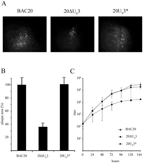
Marek's disease virus (MDV) encodes a protein exhibiting high amino acid similarity to the US3 protein of herpes simplex virus type 1 and the gene 66 product of varicella-zoster virus. The MDV US3 orthologue was replaced with a kanamycin resistance gene in the infectious bacterial artificial chromosome clone BAC20. After transfection of US3-negative BAC20 DNA (20DeltaUS3), the resulting recombinant 20DeltaUS3 virus exhibited markedly reduced growth kinetics. Virus titers on chicken embryo cells were reduced by approximately 10-fold, and plaque sizes were significantly smaller (65% reduction) compared to parental BAC20 virus. The defect of the US3-negative MDV was completely restored in a revertant virus (20US3*) expressing a US3 protein with a carboxy-terminal FLAG tag. Electron microscopical studies revealed that the defect of the 20DeltaUS3 mutant to efficiently spread from cell to cell was concomitant with an accumulation in the perinuclear space of primarily enveloped virions in characteristic vesicles containing several virus particles, which resulted in reduced numbers of particles in the cytoplasm. The formation of these vesicles was not observed in cells infected with either parental BAC20 virus or the 20US3* revertant virus. The role of the MDV US3 protein in actin stress fiber breakdown was investigated by visualizing actin with phalloidin-Alexa 488 after infection or transfection of a US3 expression plasmid. Addition of the actin-depolymerizing drug cytochalasin D to cells transfected or infected with BAC20 resulted in complete inhibition of plaque formation with as little as 50 nM of the drug, while concentrations of nocodazole as high as 50 microM only had a relatively minor effect on MDV plaque formation. The results indicated that the MDV US3 serine-threonine protein kinase is transiently involved in MDV-mediated stress fiber breakdown and that polymerization of actin, but not microtubules, plays an important role in MDV cell-to-cell spread.
Figures


Similar articles
-
Morphogenesis of a highly replicative EGFPVP22 recombinant Marek's disease virus in cell culture.J Virol. 2007 Nov;81(22):12348-59. doi: 10.1128/JVI.01177-07. Epub 2007 Sep 12. J Virol. 2007. PMID: 17855520 Free PMC article.
-
Enzymatically inactive U(S)3 protein kinase of Marek's disease virus (MDV) is capable of depolymerizing F-actin but results in accumulation of virions in perinuclear invaginations and reduced virus growth.Virology. 2008 May 25;375(1):37-47. doi: 10.1016/j.virol.2008.01.026. Epub 2008 Mar 4. Virology. 2008. PMID: 18304599 Free PMC article.
-
Role of Marek's Disease Virus (MDV)-Encoded US3 Serine/Threonine Protein Kinase in Regulating MDV Meq and Cellular CREB Phosphorylation.J Virol. 2020 Aug 17;94(17):e00892-20. doi: 10.1128/JVI.00892-20. Print 2020 Aug 17. J Virol. 2020. PMID: 32581093 Free PMC article.
-
Us3, a multifunctional protein kinase encoded by herpes simplex virus 1: how does it function in vivo?Cornea. 2013 Nov;32 Suppl 1:S22-7. doi: 10.1097/ICO.0b013e3182a0a320. Cornea. 2013. PMID: 24104928 Review.
-
Marek's disease virus latency.Curr Top Microbiol Immunol. 2001;255:223-43. Curr Top Microbiol Immunol. 2001. PMID: 11217424 Review.
Cited by
-
Manipulation of Promyelocytic Leukemia Protein Nuclear Bodies by Marek's Disease Virus Encoded US3 Protein Kinase.Microorganisms. 2021 Mar 26;9(4):685. doi: 10.3390/microorganisms9040685. Microorganisms. 2021. PMID: 33810320 Free PMC article.
-
Alphaherpesviral US3 kinase induces cofilin dephosphorylation to reorganize the actin cytoskeleton.J Virol. 2013 Apr;87(7):4121-6. doi: 10.1128/JVI.03107-12. Epub 2013 Jan 30. J Virol. 2013. PMID: 23365433 Free PMC article.
-
Phosphorylation of the varicella-zoster virus (VZV) major transcriptional regulatory protein IE62 by the VZV open reading frame 66 protein kinase.J Virol. 2006 Feb;80(4):1710-23. doi: 10.1128/JVI.80.4.1710-1723.2006. J Virol. 2006. PMID: 16439528 Free PMC article.
-
Morphogenesis of a highly replicative EGFPVP22 recombinant Marek's disease virus in cell culture.J Virol. 2007 Nov;81(22):12348-59. doi: 10.1128/JVI.01177-07. Epub 2007 Sep 12. J Virol. 2007. PMID: 17855520 Free PMC article.
-
Role of group A p21-activated kinases in the anti-apoptotic activity of the pseudorabies virus US3 protein kinase.Virus Res. 2011 Jan;155(1):376-80. doi: 10.1016/j.virusres.2010.11.003. Epub 2010 Nov 18. Virus Res. 2011. PMID: 21093504 Free PMC article.
References
-
- Cudmore, S., P. Cossart, G. Griffiths, and M. Way. 1995. Actin-based motility of vaccinia virus. Nature 378:636-638. - PubMed
MeSH terms
Substances
LinkOut - more resources
Full Text Sources
