. 2005 Apr;79(7):3998-4011.
doi: 10.1128/JVI.79.7.3998-4011.2005.

Human papillomavirus type 16 E1 E4-induced G2 arrest is associated with cytoplasmic retention of active Cdk1/cyclin B1 complexes

Affiliations

Human papillomavirus type 16 E1 E4-induced G2 arrest is associated with cytoplasmic retention of active Cdk1/cyclin B1 complexes

Clare E Davy et al. J Virol. 2005 Apr.

Abstract

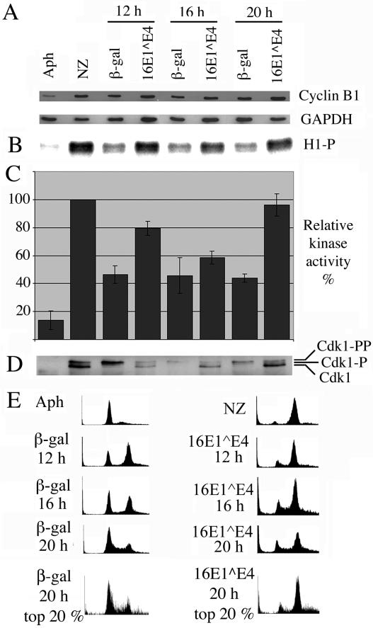
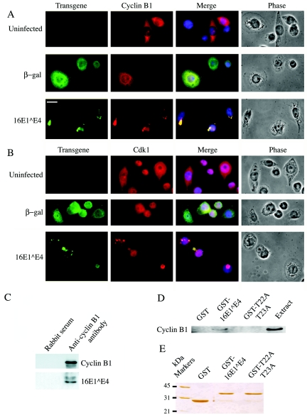
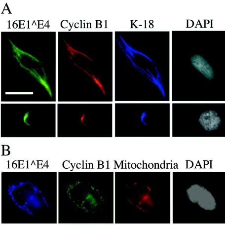
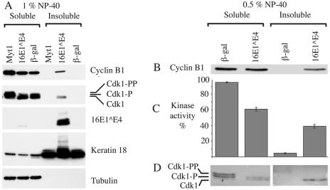
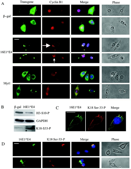
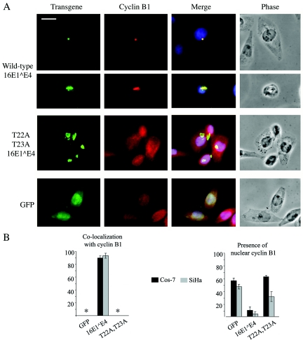
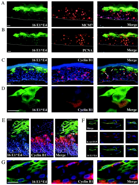
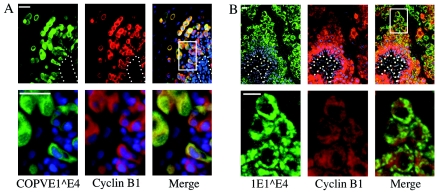
Human papillomavirus type 16 (HPV16) can cause cervical cancer. Expression of the viral E1 E4 protein is lost during malignant progression, but in premalignant lesions, E1 E4 is abundant in cells supporting viral DNA amplification. Expression of 16E1 E4 in cell culture causes G2 cell cycle arrest. Here we show that unlike many other G2 arrest mechanisms, 16E1 E4 does not inhibit the kinase activity of the Cdk1/cyclin B1 complex. Instead, 16E1 E4 uses a novel mechanism in which it sequesters Cdk1/cyclin B1 onto the cytokeratin network. This prevents the accumulation of active Cdk1/cyclin B1 complexes in the nucleus and hence prevents mitosis. A mutant 16E1 E4 (T22A, T23A) which does not bind cyclin B1 or alter its intracellular location fails to induce G2 arrest. The significance of these results is highlighted by the observation that in lesions induced by HPV16, there is evidence for Cdk1/cyclin B1 activity on the keratins of 16E1 E4-expressing cells. We hypothesize that E1 E4-induced G2 arrest may play a role in creating an environment optimal for viral DNA replication and that loss of E1 E4 expression may contribute to malignant progression.


Figures

FIG. 1.
FIG. 1.
16E1E4 expression does not inhibit Cdk1/cyclin B kinase activity. Control G1/S-synchronized SiHa cells were either harvested without releasing the G1/S block (Aph) or released from the block for 6 h and then treated for 12 h with nocodazole prior to harvesting (NZ). G1/S-synchronized SiHa cells infected with recombinant adenoviruses expressing β-galactosidase or 16E1E4 were harvested at 12, 16, and 20 h post-block release. (A) Cell extracts were Western blotted for cyclin B1 and glyceraldehyde-3-phosphate dehydrogenase (GAPDH). (B) Cdk1/cyclin B1 was immunoprecipitated from cell extracts and used in an in vitro kinase assay to phosphorylate histone H1. Protein labeled with 32P was detected with a phosphorimager. (C) Levels of histone H1 phosphorylation were quantitated with ImageQuant software and expressed as a percentage of that obtained for the nocodazole-treated samples. The results show the means of four experiments ± standard error of the mean. (D) Immunoprecipitated complexes were Western blotted for Cdk1. (E) Cells were stained with propidium iodide, immunostained for β-galactosidase or 16E1E4, and analyzed by flow cytometry. Plots show cell number versus DNA content. The final row shows plots in which the top 20% of high-β-galactosidase- or 16E1E4-expressing cells were selected for analysis of DNA content.
FIG. 2.
FIG. 2.
16E1E4 associates with Cdk1/cyclin B1. G1/S-synchronized SiHa cells expressing β-galactosidase or 16E1E4 were fixed at 16 h post-block release. Cells were double stained for β-galactosidase or 16E1E4 (green) and cyclin B1 (A) or Cdk1 (B) (red). A DAPI nuclear stain was included (blue). Bar, 10 μm. (C) Cell extracts were immunoprecipitated with anti-cyclin B1 rabbit polyclonal antibody or normal rabbit serum and Western blotted for 16E1E4 and cyclin B1. (D) Cell extracts from nocodazole-treated SiHa cells were used in pull-down experiments with GST, GST-16E1E4, or GST-T22A,T23A-16E1E4 and Western blotted along with the cell extract for cyclin B1. The levels of cyclin B1 pulled down are compared to 20% of the input amount of cyclin B1. (E) The input of GST, GST-16E1E4, and GST-T22A,T23A-16E1E4 into the pull-down experiments is shown by silver staining.
FIG. 3.
FIG. 3.
Cyclin B1 associated with 16E1E4 is found on the keratins or mitochondria. (A) G1/S-synchronized SiHa cells expressing 16E1E4 were fixed at 16 h post-block release. Cells were triple stained for 16E1E4 (green), cyclin B1 (red), and keratin 18 (K-18; blue). A DAPI nuclear stain was included (white). The upper panels show a cell prior to keratin collapse, while the lower panels show a cell with collapsed keratin. (B) Saos-2 cells infected with recombinant adenoviruses that express 16E1E4 were triple stained for 16E1E4 (blue), cyclin B1 (green), and mitochondria (red). A DAPI nuclear stain was included (white). Bar, 10 μm.
FIG. 4.
FIG. 4.
16E1E4 decreases the solubility of active Cdk1/cyclin B1. (A) G1/S-synchronized SiHa cells expressing Myt1, 16E1E4, or β-galactosidase were harvested at 20 h post-block release and fractionated with 1% NP-40 (NP-40-soluble fraction) and then with 1% SDS (NP-40-insoluble fraction). Fractions were Western blotted for 16E1E4, cyclin B1, Cdk1, keratin 18, and, as a control for fractionation, tubulin. (B) G1/S-synchronized SiHa cells expressing β-galactosidase or 16E1E4 were harvested at 20 h post-block release and fractionated first with 0.5% NP-40 (NP-40-soluble fraction) and then with 0.1% SDS (NP-40-insoluble fraction). The fractions were Western blotted for cyclin B1. (C) Cdk1/cyclin B1 was immunoprecipitated from fractions with anti-cyclin B1 monoclonal antibody. Immunoprecipitated complexes were used in an in vitro kinase assay to phosphorylate histone H1. 32P-labeled protein was detected with a phosphorimager and quantitated with ImageQuant. Kinase activity is expressed as a percentage of total activity in β-galactosidase- or 16E1E4-expressing cells. The results show the mean values of three experiments ± standard error of the mean. (D) Immunoprecipitated fractions were Western blotted for Cdk1.
FIG. 5.
FIG. 5.
16E1E4 expression reduces the ability of cyclin B1 to accumulate in the nucleus. G1/S-synchronized SiHa cells expressing β-galactosidase, 16E1E4, or Myt1 were treated with leptomycin B (LMB), beginning 14 h postrelease, and then harvested 2.5 h later. (A) The cells were double stained for β-galactosidase, 16E1E4, or Myt1 (green) and cyclin B1 (red). A DAPI nuclear stain was included (blue). The large white arrow indicates a cell with predominantly cytoplasmic cyclin B1, and the small white arrow indicates a cell in which some of the cyclin B1 has entered the nucleus. (B) Total cell extracts were Western blotted for phosphohistone H3 or phosphokeratin 18 serine 33 and compared to a glyceraldehyde-3-phosphate dehydrogenase (GAPDH) control. (C and D) G1/S-synchronized SiHa cells expressing 16E1E4 at 24 h post-block release were immunostained for 16E1E4 (green) and phosphokeratin 18 serine 33. A DAPI nuclear stain was included (blue). Bars, 10 μm.
FIG. 6.
FIG. 6.
Mutant unable to induce G2 arrest fails to colocalize with cyclin B1 and prevent its nuclear entry. Wild-type 16E1E4, mutant T22A,T23A-16E1E4, and GFP were expressed in SiHa and Cos-7 cells by transfection, and the localization of cyclin B1 was determined by immunofluorescence microscopy. (A) SiHa cells were double stained for 16E1E4 (green) and cyclin B1 (red). A DAPI nuclear stain was included (blue). Bar, 10 μm. (B) The percentages of cells showing cyclin B1 colocalized with 16E1E4 and showing some nuclear cyclin B1 were determined. The graphs show the mean values of three replicate experiments ± standard error of the mean.
FIG. 7.
FIG. 7.
Expression of 16E1E4 in cyclin B1-expressing cells during epithelial cell differentiation. HPV16-induced raft cultures were double stained for the presence of 16E1E4 (green) and (A) MCM7, (B) PCNA, or (C) cyclin B1 (red) to show that 16E1E4 levels begin to rise in cells that are expressing cell cycle markers. A subset of the cells that express moderate levels of 16E1E4 also express cyclin B1 and are arrowed in C. (D) An enlarged image of the cyclin B1/16 E1E4 double-positive cell that is marked with an asterisk in C. (E) HPV16-induced low-grade lesion double stained for the presence of 16E1E4 (green) and cyclin B1 (red). Coexpression of cyclin B1 and 16 E1E4 is limited to the region wherethe levels of 16E1E4 protein are beginning to rise during epithelial differentiation. Panel G shows a high-power image of cells from this region (indicated by an asterisk in E) that contain both cyclin B1 and 16 E1E4. Panel F shows that 16E1E4-expressing cells (green) in the upper layers of low-grade cervical lesions contain the keratin phosphoserine 33 epitope (red) that is also present following the expression of 16E1E4 in vitro (see Fig. 5). Merged images are shown in the uppermost panels within F. Where appropriate, the position of the basal layer is indicated by dotted white lines. A DAPI nuclear stain (blue) was included. Bars equal 20 μm except in panels D and G, where the bars equal 10 μm.
FIG. 8.
FIG. 8.
Coexpression of E4 and cyclin B1 in productive lesions caused by other papillomavirus types. (A) Canine oral papillomavirus (COPV)-induced lesion double stained for the presence of canine oral papillomavirus E1E4 (green) and cyclin B1 (red). The boxed region in the rightmost top panel is shown enlarged in the lower panels. Expression of E1E4 is apparent in a subset of cyclin B1-positive cells in the basal layer and in all the cyclin B1-expressing cells in the parabasal and superficial layers. (B) HPV1-induced lesion double stained for the presence of 1E1E4 (green) and cyclin B1 (red). The boxed region in the rightmost top panel is shown enlarged in the lower panels. HPV1 E1E4 expression begins in cyclin B1-positive cells in the parabasal layer. The intensity of cyclin B1 staining decreases as the cells migrate towards the epithelial surface. Bars, 20 μm.

Similar articles

Cited by

References

    1. Advani, S. J., R. R. Weichselbaum, and B. Roizman. 2000. The role of cdc2 in the expression of herpes simplex virus genes. Proc. Natl. Acad. Sci. USA 97:10996-11001. - PMC - PubMed
    1. Baker, C. C., W. C. Phelps, V. Lindgren, M. J. Braun, M. A. Gonda, and P. M. Howley. 1987. Structural and transcriptional analysis of human papillomavirus type 16 sequences in cervical carcinoma cell lines. J. Virol. 61:962-971. - PMC - PubMed
    1. Barnes, E. A., M. Kong, V. Ollendorff, and D. J. Donoghue. 2001. Patched1 interacts with cyclin B1 to regulate cell cycle progression. EMBO J. 20:2214-2223. - PMC - PubMed
    1. Bryan, J. T., and D. R. Brown. 2000. Association of the human papillomavirus type 11 E1()E4 protein with cornified cell envelopes derived from infected genital epithelium. Virology 277:262-269. - PubMed
    1. Chan, T. A., H. Hermeking, C. Lengauer, K. W. Kinzler, and B. Vogelstein. 1999. 14-3-3Sigma is required to prevent mitotic catastrophe after DNA damage. Nature 401:616-620. - PubMed

Publication types

MeSH terms

LinkOut - more resources