. 2005 Apr;79(7):4012-24.
doi: 10.1128/JVI.79.7.4012-4024.2005.

Feline calicivirus VP2 is essential for the production of infectious virions

Affiliations

Feline calicivirus VP2 is essential for the production of infectious virions

Stanislav V Sosnovtsev et al. J Virol. 2005 Apr.

Abstract

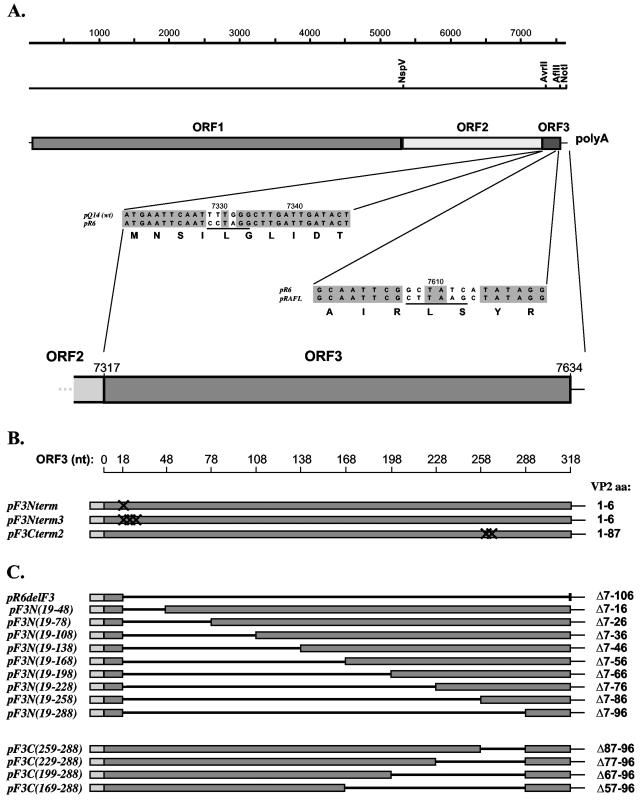
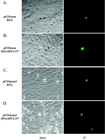
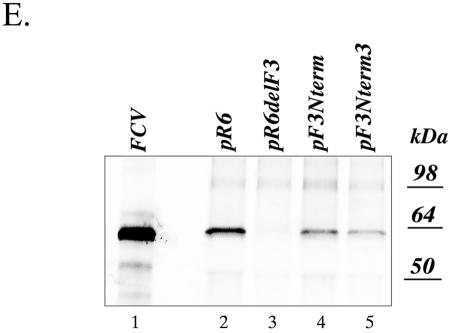
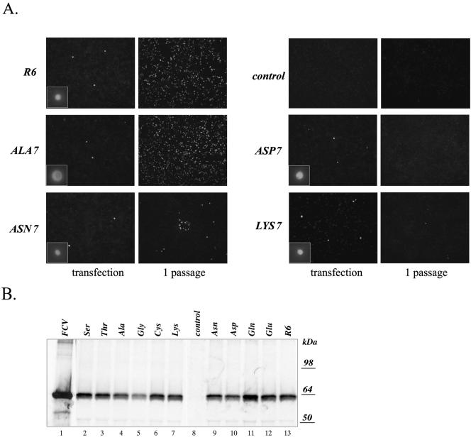
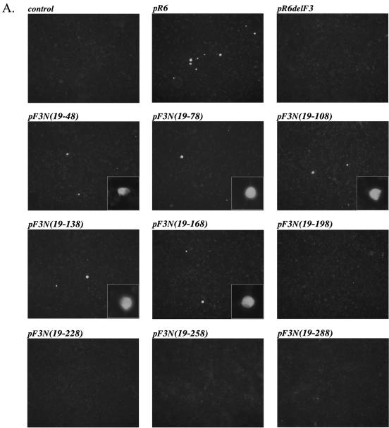
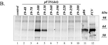
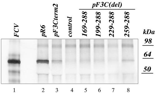
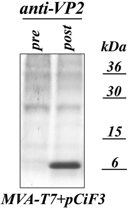
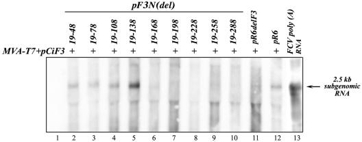
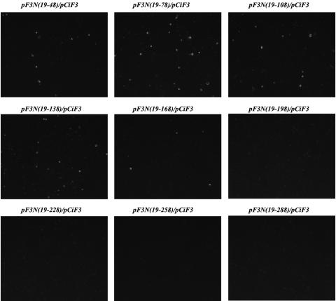
The third open reading frame (ORF3) located at the 3' end of the genomic RNA of feline calicivirus (FCV) encodes a small (12.2-kDa) minor structural protein of 106 amino acids designated VP2. Point mutations and deletions were introduced into an infectious FCV cDNA clone in order to evaluate the functional importance of ORF3 and its encoded protein, VP2. Deletion of the entire ORF3 sequence was lethal for the virus, and evidence was found for strong selective pressure to produce the VP2 protein. Extended deletions in the 5' end and small deletions in the 3' end of ORF3, as well as the introduction of stop codons into the ORF3 sequence, were tolerated by the viral replication machinery, but infectious virus could not be recovered. Infectious virus particles could be rescued from a full-length FCV cDNA clone encoding a nonfunctional VP2 when VP2 was provided in trans from a eukaryotic expression plasmid. Our data indicate that VP2, a protein apparently unique to the caliciviruses, is essential for productive replication that results in the synthesis and maturation of infectious virions and that the ORF3 nucleotide sequence itself overlaps a cis-acting RNA signal at the genomic 3' end.


Figures

FIG. 1.
FIG. 1.
Cloning cassette vectors and plasmid constructions. (A) Schematic diagram showing the FCV genome organization and locations of silent nucleotide changes introduced into the FCV FL cDNA clone (pQ14) to generate a unique AvrII restriction site (underlined) in the beginning of the ORF3 sequence or a unique AflII restriction site (underlined) in the end of the ORF3 sequence to generate cloning cassette vectors pR6 and pRAFL, respectively. (B) Plasmid constructions containing engineered stop codons in the ORF3 sequence of FL clones: pF3Nterm (introduction of terminator codon, TAG, at nucleotide positions 7335 to 7337 [nt 19 to 21 of ORF3], pF3Nterm3 (introduction of three terminatorcodons, TAGTAATGA, at the nucleotide positions 7335 to 7343 [nt 19 to 27 of ORF3], and pF3Cterm2 (introduction of two terminator codons, TAATAG, at the nucleotide positions 7578 to 7583 [nt 261 to 267 of ORF3]). (C) Plasmid constructions containing engineered deletions in the ORF3 sequence: pR6delF3 (with nearly all of ORF3 deleted), the pF3N series (with sequentially larger N-terminal deletions), and the pF3C series (with sequentially larger C-terminal deletions). The plasmids were engineered as described in the text. The borders (nucleotide positions) of introduced deletions in ORF3 are given on the scale above the plasmid diagrams, and the corresponding amino acid deletions (Δ) in VP2 are indicated on the right.
FIG. 2.
FIG. 2.
Analysis of replication of the ORF3 knockout mutants. (A to D) Immunofluorescent detection of expression of the FCV capsid protein in CRFK cells transfected with capped genomic RNA synthesized in vitro from pF3Nterm (A) and pF3Nterm3 (C) clones and the MVA-T7-infected CRFK cells transfected with their plasmid DNAs (B and D). CRFK cells were stained 20 h posttransfection with hyperimmune serum raised against purified FCV virions, and the binding of antibody was detected with a fluorescein isothiocyanate-conjugated anti-guinea pig serum. Photographs were taken at a magnification of ×400. (E) Immunoprecipitation analysis of FCV capsid protein expression in MVA-T7-infected CRFK cells transfected with ORF3 knockout clones. The S35 labeling and immunoprecipitation of the capsid protein were performed as described in the text. Lanes 2, 3, 4, and 5 are radiolabeled proteins immunoprecipitated from MVA-T7-infected CRFK cells transfected with pR6, pR6delF3, pF3Nterm, and pF3Nterm3, respectively. Lane 1 contains the radiolabeled capsid protein immunoprecipitated from FCV-infected CRFK cells as a control.
FIG. 2.
FIG. 2.
Analysis of replication of the ORF3 knockout mutants. (A to D) Immunofluorescent detection of expression of the FCV capsid protein in CRFK cells transfected with capped genomic RNA synthesized in vitro from pF3Nterm (A) and pF3Nterm3 (C) clones and the MVA-T7-infected CRFK cells transfected with their plasmid DNAs (B and D). CRFK cells were stained 20 h posttransfection with hyperimmune serum raised against purified FCV virions, and the binding of antibody was detected with a fluorescein isothiocyanate-conjugated anti-guinea pig serum. Photographs were taken at a magnification of ×400. (E) Immunoprecipitation analysis of FCV capsid protein expression in MVA-T7-infected CRFK cells transfected with ORF3 knockout clones. The S35 labeling and immunoprecipitation of the capsid protein were performed as described in the text. Lanes 2, 3, 4, and 5 are radiolabeled proteins immunoprecipitated from MVA-T7-infected CRFK cells transfected with pR6, pR6delF3, pF3Nterm, and pF3Nterm3, respectively. Lane 1 contains the radiolabeled capsid protein immunoprecipitated from FCV-infected CRFK cells as a control.
FIG. 3.
FIG. 3.
Analysis of FCV capsid protein expression in MVA-T7-infected CRFK cells transfected with FL cDNA clones containing amino acid changes in position 7 of the VP2 amino acid sequence. (A) Fluorescence microscopy observation of the MVA-T7-infected CRFK cells transfected with pF3X7 mutants was conducted to detect replication of the virus. Recovery of infectious virus particles was confirmed by transfer of the transfected cell media to a new cell monolayer and by immunofluorescent staining of these cells 24 h postinoculation. Immunofluorescent staining was performed as described in the text, and photographs were taken at a magnification of ×100. The insets show photographs of single positive cells taken at a magnification of ×400. (B) Replication of the virus in the MVA-T7-infected CRFK cells transfected with pF3X7 mutants was verified by immunoprecipitation of the radiolabeled virus capsid protein from lysates of the transfected cells. Lanes 2 to 7 and 9 to 13 are radiolabeled proteins immunoprecipitated with the FCV virion-specific serum from the cells transfected with pF3Ser7, pF3Thr7, pF3Ala7, pF3Gly7, pF3Cys7, pF3Lys7, pF3Asn7, pF3Asp7, pF3Gln7, pF3Glu7, and pR6, respectively. The radiolabeled capsid protein immunoprecipitated from CRFK cells infected with wild-type FCV was included as a positive control in lane 1. Radiolabeled proteins from mock-transfected MVA-T7-infected CRFK cells were analyzed by immunoprecipitation with the capsid-specific serum as a control in lane 8.
FIG. 4.
FIG. 4.
5′-end deletion mutagenesis of the ORF3 sequence in the FCV FL cDNA clone and analysis of the virus capsid protein expression in the MVA-T7-infected CRFK cells transfected with the corresponding clones. (A) Fluorescence microscopy observation of the MVA-T7-infected CRFK cells transfected with pF3N(del) mutants 20 h posttransfection. Immunofluorescent staining was performed as described in the text, and photographs were taken at a magnification of ×100. The insets show photographs of single positive cells taken at a magnification of ×400. (B) Replication of the virus in the MVA-T7-infected CRFK cells transfected with pF3N(del) mutants was verified by immunoprecipitation of the radiolabeled virus capsid protein from the lysates of the transfected cells. Lanes 2 to 11 are radiolabeled proteins immunoprecipitated with the FCV virion-specific serum from the cells transfected with pF3N(19-48), pF3N(19-78), pF3N(19-108), pF3N(19-138), pF3N(19-168), pF3N(19-198), pF3N(19-228), pF3N(19-258), pF3N(19-288), and pR6, respectively. The radiolabeled capsid protein immunoprecipitated from CRFK cells infected with wild-type FCV was included as a positive control in lane 12. Radiolabeled proteins precipitated from mock-transfected MVA-T7-infected CRFK cells were included as a control in lane 1.
FIG. 4.
FIG. 4.
5′-end deletion mutagenesis of the ORF3 sequence in the FCV FL cDNA clone and analysis of the virus capsid protein expression in the MVA-T7-infected CRFK cells transfected with the corresponding clones. (A) Fluorescence microscopy observation of the MVA-T7-infected CRFK cells transfected with pF3N(del) mutants 20 h posttransfection. Immunofluorescent staining was performed as described in the text, and photographs were taken at a magnification of ×100. The insets show photographs of single positive cells taken at a magnification of ×400. (B) Replication of the virus in the MVA-T7-infected CRFK cells transfected with pF3N(del) mutants was verified by immunoprecipitation of the radiolabeled virus capsid protein from the lysates of the transfected cells. Lanes 2 to 11 are radiolabeled proteins immunoprecipitated with the FCV virion-specific serum from the cells transfected with pF3N(19-48), pF3N(19-78), pF3N(19-108), pF3N(19-138), pF3N(19-168), pF3N(19-198), pF3N(19-228), pF3N(19-258), pF3N(19-288), and pR6, respectively. The radiolabeled capsid protein immunoprecipitated from CRFK cells infected with wild-type FCV was included as a positive control in lane 12. Radiolabeled proteins precipitated from mock-transfected MVA-T7-infected CRFK cells were included as a control in lane 1.
FIG. 5.
FIG. 5.
3′-end deletion mutagenesis of the ORF3 sequence in the FCV FL cDNA clone and analysis of virus capsid protein expression in the MVA-T7-infected CRFK cells transfected with the indicated clones. Replication of the virus in the MVA-T7-infected CRFK cells transfected with pF3Cterm2 and pF3C(del) mutants was verified by IP of the radiolabeled virus capsid protein from the lysates of the transfected cells. Lanes 2, 3, and 5 to 8 are radiolabeled proteins immunoprecipitated with the FCV virion-specific serum from the cells transfected with pR6, pF3Cterm2, pF3C(169-288), pF3C(199-288), pF3C(229-288), and pF3C(259-288), respectively. The radiolabeled capsid protein immunoprecipitated from CRFK cells infected with wild-type FCV was included as a positive control in lane 1. Radiolabeled proteins precipitated from mock-transfected MVA-T7-infected CRFK cells were included as a control in lane 4.
FIG. 6.
FIG. 6.
Expression of the VP2 protein from pCiF3 vector in MVA-T7-transfected CRFK cells. The CRFK cells were infected with MVA-T7 at a multiplicity of infection of 3 and transfected with pCiF3 2 h later. Proteins were metabolically labeled with [S35]methionine as described in the text, and radiolabeled VP2 was immunoprecipitated from cell lysates with VP2-specific postimmunization serum (38), subjected to sodium dodecyl sulfate-polyacrylamide gel electrophoresis, and visualized with autoradiography. As a negative control, the radiolabeled proteins were immunoprecipitated from the same cell lysates using preimmunization VP2 serum.
FIG. 7.
FIG. 7.
Subgenomic RNA synthesis in cells cotransfected with pCiF3 and FL cDNA clones containing sequential 5′-end deletions of the ORF3 sequence. Lanes 2 to 12, samples of total RNA purified from CRFK cells infected with MVA-T7 and cotransfected with pCiF3 and pF3N(19-48), pF3N(19-78), pF3N(19-108), pF3N(19-138), pF3N(19-168), pF3N(19-198), pF3N(19-228), pF3N(19-258), pF3N(19-288), pR6delF3, and pR6, respectively, were subjected to Northern blot analysis using an antisense RNA probe specific for FCV ORF2 (10). Lane 1, total RNA isolated from MVA-T7-infected cells transfected with pCiF3 only. Lane 13, polyadenylated RNA purified from FCV-infected CRFK cells using PolyAT tract system 1000 (Promega).
FIG. 8.
FIG. 8.
trans-complementation of the FCV ORF3 5′-end deletion mutants. Limited virus replication was observed in CRFK cells inoculated with culture fluids from MVA-T7-infected cells cotransfected with pF3N(19-48), pF3N(19-78), pF3N(19-108), pF3N(19-138), pF3N(19-168), and pCiF3. Expression of the virus capsid antigen was observed using capsid-specific immunofluorescent staining 13 to 14 h postinoculation.

References

    1. Bertolotti-Ciarlet, A., S. E. Crawford, A. M. Hutson, and M. K. Estes. 2003. The 3′ end of Norwalk virus mRNA contains determinants that regulate the expression and stability of the viral capsid protein VP1: a novel function for the VP2 protein. J. Virol. 77:11603-11615. - PMC - PubMed
    1. Burroughs, J. N., and F. Brown. 1978. Presence of a covalently linked protein on calicivirus RNA. J. Gen. Virol. 41:443-446. - PubMed
    1. Carter, M. J., I. D. Milton, P. C. Turner, J. Meanger, M. Bennett, and R. M. Gaskell. 1992. Identification and sequence determination of the capsid protein gene of feline calicivirus. Arch. Virol. 122:223-235. - PMC - PubMed
    1. Chen, C. J., M. D. Kuo, L. J. Chien, S. L. Hsu, Y. M. Wang, and J. H. Lin. 1997. RNA-protein interactions: involvement of NS3, NS5, and 3′ noncoding regions of Japanese encephalitis virus genomic RNA. J. Virol. 71:3466-3473. - PMC - PubMed
    1. Deiman, B. A., R. M. Kortlever, and C. W. Pleij. 1997. The role of the pseudoknot at the 3′ end of turnip yellow mosaic virus RNA in minus-strand synthesis by the viral RNA-dependent RNA polymerase. J. Virol. 71:5990-5996. - PMC - PubMed

Substances

LinkOut - more resources