Comparative Study
. 2005 Apr;79(7):4043-54.
doi: 10.1128/JVI.79.7.4043-4054.2005.

Divergent host responses during primary simian immunodeficiency virus SIVsm infection of natural sooty mangabey and nonnatural rhesus macaque hosts

Affiliations
Comparative Study

Divergent host responses during primary simian immunodeficiency virus SIVsm infection of natural sooty mangabey and nonnatural rhesus macaque hosts

Guido Silvestri et al. J Virol. 2005 Apr.

Abstract

To understand how natural sooty mangabey hosts avoid AIDS despite high levels of simian immunodeficiency virus (SIV) SIVsm replication, we inoculated mangabeys and nonnatural rhesus macaque hosts with an identical inoculum of uncloned SIVsm. The unpassaged virus established infection with high-level viral replication in both macaques and mangabeys. A species-specific, divergent immune response to SIV was evident from the first days of infection and maintained in the chronic phase, with macaques showing immediate and persistent T-cell proliferation, whereas mangabeys displayed little T-cell proliferation, suggesting subdued cellular immune responses to SIV. Importantly, only macaques developed (CD4+)-T-cell depletion and AIDS, thus indicating that in mangabeys limited immune activation is a key mechanism to avoid immunodeficiency despite high levels of SIVsm replication. These studies demonstrate that it is the host response to infection, rather than properties inherent to the virus itself, that causes immunodeficiency in SIV-infected nonhuman primates.


Figures

FIG. 1.
FIG. 1.
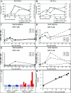
Viral replication during primary SIVsm infection of RMs and SMs. (A and B) SIV plasma viremia in the three SMs (A) and the three RMs (B). (C and D) SIV replication was detected by ISH in a lymph node biopsy from a SM (FGu) and a RM (RZw4) at day 14 postinfection.
FIG. 2.
FIG. 2.
T-cell activation and/or proliferation and viral replication during acute SIVsm infection of SM and RM. (A and B) Sequential activation of CD4 T cells, SIV replication, and CD8 T-cell activation in a representative RM (RHt4) and SM (FLn). For CD4+ HLA DR+ T cells and CD8+ CD69+ T cells, the numbers represent absolute cell counts per cubic millimeter. Plasma viremia is expressed as the log10 SIV RNA copies/milliliter of plasma. (C to F) Rates of T-cell proliferation in CD4+ (C and D) and CD8+ (E and F) T cells from three SMs (C and E) and three RMs (D and F) measured as percentage of Ki67 expression between day 0 and day 40 postinfection. (G) Average percentage Ki67+ CD4 and CD8 T cells on the day of infection and at the time of peak viremia. Peak viremia occurred at either day 10 (animals RHt4, FLn), 14 (animals RZw4, FCo, and FGu), or day 28 (animal RQl4). For RQl4, measures of Ki67+ T cells were not available at day 28; therefore, the next measurement at day 40 was taken. (H) Direct correlation between peak of HLA-DR expression on CD4+ T cells and peak plasma viremia in all six animals.
FIG. 3.
FIG. 3.
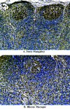
Divergent lymphocyte proliferation profiles in lymph nodes during SIVsm infection of SM and RM. (A) Immunohistochemical staining with Ki67 antibody of a lymph node biopsy obtained from a representative SM (FGu) at day 14 postinfection. The vast majority of the Ki67+ cells are localized in the B-cell areas, i.e., folliculi and germinal centers, whereas minimal Ki67 expression was observed in the T-cell area (i.e., paracortex). (B) Immunohistochemical staining with Ki67 antibody of a lymph node biopsy obtained from a representative RM (RZw4) at day 14 postinfection.
FIG. 4.
FIG. 4.
Acute CD8+-T-cell proliferation and establishment of postpeak levels of virus replication. (A and B) Postpeak decline of plasma viremia and establishment of levels of set-point viremia in SMs (A) and RMs (B) as measured between days 7 and 450 postinfection. (C) Correlation between rates of peak CD8+-T-cell proliferation, measured as Ki67 expression, and magnitude of postpeak decline in plasma viremia in three SIVsm-infected SMs and three RMs.
FIG. 5.
FIG. 5.
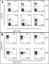
Persistence of chronic high levels of T-cell proliferation during chronic SIVsm infection of RMs. (A and B) Rates of CD4+ (A) and CD8+ (B) T-cell proliferation as measured by Ki67 expression in three SIVsm-infected SMs and three RMs at day 720 postinfection. The numbers in the dot plots represent the percentages of Ki67+ cells on total CD4+ or CD8+ T lymphocytes.
FIG. 6.
FIG. 6.
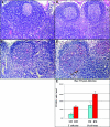
Elevated T-cell activation and apoptosis in lymph nodes from chronically SIVsm-infected RMs. (A and B) Hematoxylin-eosin staining of a lymph node biopsy obtained from a representative RM (RZw4) (A) and SM (FGu) (B) and at day 273 postinfection. (C and D) Levels of apoptosis, as measured by TUNEL staining, in a lymph node biopsy obtained from a representative RM (RZw4) (C) and SM (FGu) (D) and at day 273 postinfection. (E) Frequency of apoptotic cells in lymph nodes.
FIG. 7.
FIG. 7.
CD4+ T-cell depletion and AIDS during SIVsm infection of RMs. (A and B) Measurements over time of CD4+ T cells in three SIVsm-infected RMs (A) and three SMs (B) between days 0 and 720 postinfection. Each line represents an individual animal.

References

    1. Borrow, P., H. Lewicki, B. H. Hahn, G. M. Shaw, and M. B. Oldstone. 1994. Virus-specific CD8+ cytotoxic T-lymphocyte activity associated with control of viremia in primary human immunodeficiency virus type 1 infection. J. Virol. 68:6103-6110. - PMC - PubMed
    1. Broussard, S. R., S. I. Staprans, R. White, E. M. Whitehead, M. B. Feinberg, and J. S. Allan. 2001. Simian immunodeficiency virus replicates to high levels in naturally infected African green monkeys without inducing immunologic or neurologic disease. J. Virol. 75:2262-2275. - PMC - PubMed
    1. Callan, M. F., L. Tan, N. Annels, G. S. Ogg, J. D. Wilson, C. A. O'Callaghan, N. Steven, A. J. McMichael, and A. B. Rickinson. 1998. Direct visualization of antigen-specific CD8+ T cells during the primary immune response to Epstein-Barr virus in vivo. J. Exp. Med. 187:1395-1402. - PMC - PubMed
    1. Carlson, J. R., T. P. McGraw, E. Keddie, J. L. Yee, A. Rosenthal, A. J. Langlois, R. Dickover, R. Donovan, P. A. Luciw, and M. B. Jennings. 1990. Vaccine protection of rhesus macaques against simian immunodeficiency virus infection. AIDS Res. Hum. Retrovir. 6:1239-1246. - PubMed
    1. Chakrabarti, L. A., S. R. Lewin, L. Zhang, A. Gettie, A. Luckay, L. N. Martin, E. Skulsky, D. D. Ho, C. Cheng-Mayer, and P. A. Marx. 2000. Normal T-cell turnover in sooty mangabeys harboring active simian immunodeficiency virus infection. J. Virol. 74:1209-1223. - PMC - PubMed

Publication types

MeSH terms