. 2005 Apr;79(7):4238-45.
doi: 10.1128/JVI.79.7.4238-4245.2005.

Hepatitis B virus X protein stimulates viral genome replication via a DDB1-dependent pathway distinct from that leading to cell death

Affiliations

Hepatitis B virus X protein stimulates viral genome replication via a DDB1-dependent pathway distinct from that leading to cell death

Olivier Leupin et al. J Virol. 2005 Apr.

Abstract

The hepatitis B virus (HBV) X protein (HBx) is essential for virus infection and has been implicated in the development of liver cancer associated with chronic infection. HBx can interact with a number of cellular proteins, and in cell culture, it exhibits pleiotropic activities, among which is its ability to interfere with cell viability and stimulate HBV replication. Previous work has demonstrated that HBx affects cell viability by a mechanism that requires its binding to DDB1, a highly conserved protein implicated in DNA repair and cell cycle regulation. We now show that an interaction with DDB1 is also needed for HBx to stimulate HBV genome replication. Thus, HBx point mutants defective for DDB1 binding fail to complement the low level of replication of an HBx-deficient HBV genome when provided in trans, and one such mutant regains activity when directly fused to DDB1. Furthermore, DDB1 depletion by RNA interference specifically compromises replication of wild-type HBV, indicating that HBx produced from the viral genome also functions in a DDB1-dependent fashion. We also show that HBx in association with DDB1 acts in the nucleus and stimulates HBV replication mainly by enhancing viral mRNA levels, regardless of whether the protein is expressed from the HBV genome itself or supplied in trans. Interestingly, whereas HBx induces cell death in both HepG2 and Huh-7 hepatoma cell lines, it enhances HBV replication only in HepG2 cells, suggesting that the two activities involve distinct DDB1-dependent pathways.


Figures

FIG. 1.
FIG. 1.
HBx expressed in trans stimulates HBV genome replication through interaction with DDB1. HepG2 cells were transfected with a greater-than-unit-length HBV genomic construct (HBV) or with a derivative bearing a stop codon in the X gene to abolish expression of HBx [HBV(ΔX)], and equal amounts of the indicated HBx expression plasmids or the corresponding empty vector (vect). A GFP gene was cotransfected to assess comparable transfection efficiencies by FACS analysis (data not shown). Three days after transfection, cytoplasmic HBV core particles were isolated from equal numbers of cells, and the amount of associated HBV DNA replicative intermediates was assessed by Southern blot analysis. Hybridization was performed with 32P-labeled probes prepared from full-length HBV genomic DNA. (A) HBx and the DDB1-binding-defective HBx(R96E) point mutant were individually tested for their effects on replication of the wild-type (left) or the HBX-deficient (right) HBV genome when expressed in trans from a recombinant vector. The single-stranded (ssDNA) and double-stranded (dsDNA) HBV DNA replicative forms are indicated on the right. The results are representative of three independent transfection experiments. (B) HBx mutants with single or double amino acid substitutions were tested for complementation of the mutation in HBV(ΔX). The DDB1-binding abilities of the wild-type and mutant proteins as measured in a yeast two-hybrid assay were determined previously (21) and are summarized below the panel. All mutants were shown before to accumulate to protein levels similar to those of the wild type (21). Symbols: +++, wild-type interaction; +/−, very weak interaction; −, no detectable interaction. (C) The nonfunctional HBx(R96E) mutant, which cannot interact with the endogenous DDB1 protein, was tested for activity in the complementation assay when expressed as a fusion to wild-type DDB1 (R96E-DDB1) or to the DDB1(i947) insertion mutant that is selectively defective for HBx binding [R96E-DDB1(i947)] (20). Previous work has shown that a covalent link between HBx(R96E) and DDB1 restores cytotoxic activity to the HBx mutant by acting as a clamp forcing the two protein moieties into their natural interaction and that the i947 mutation in DDB1 prevents this from occurring (20). Results from one of three independent transfection experiments are shown. On the right is a Western blot analysis of the fusion proteins. Equal amounts of whole-cell extracts prepared from HepG2 cells transfected with the indicated plasmids were resolved by gel electrophoresis, and immunoblot analysis was performed with antibodies to DDB1. The signal corresponding to endogenous DDB1 serves as a control for loading. The relatively weak signals obtained for the fusion proteins compared to endogenous DDB1 are due to low (∼10%) transfection efficiencies of HepG2 cells.
FIG. 2.
FIG. 2.
HBx produced from the HBV genome shows decreased activity upon RNAi-mediated downregulation of endogenous DDB1 gene expression. (A) HepG2 cells were transduced with a control lentivirus vector expressing GFP alone (-) or with a derivative (+) that directs the synthesis of a DDB1-specific small interfering RNA (DDB1-siRNA). FACS analysis performed on day 3 (d.3) showed similar transduction efficiencies ranging from 95 to close to 100% (data not shown). On day 4, the cells were transfected with wild-type HBV or the HBV(ΔX) genomic construct. A red fluorescent protein (RFP) gene was cotransfected to assess comparable transfection efficiencies by FACS analysis 1 day later (data not shown). Viral DNA replication was assessed by Southern blot analysis 3 days posttransfection on day 7 as described in the legend to Fig. 1. Results from one of two independent experiments are shown. The single-stranded (ssDNA) and double-stranded (dsDNA) HBV DNA replicative forms are indicated on the right. (B) Western blot analysis demonstrating selective depletion of DDB1 by siRNA. HepG2 cells were transduced with a control lentivirus vector containing GFP alone (lane 1) or with a derivative expressing an siRNA against p53 (p53-siRNA) (lane 2) or DDB1 (DDB1-SiRNA) (lane 3). Cell lysates were prepared 6 days after transduction, and equal amounts of protein per sample were resolved by gel electrophoresis. Immunoblot analysis was performed with antibodies to DDB1 (top) or p53 (bottom). Downregulation of endogenous p53 expression had no effect on the replication of the wild-type or HBx mutant HBV genome (data not shown).
FIG. 3.
FIG. 3.
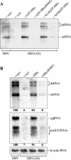
HBx functions in the nucleus and acts mostly on HBV RNA levels. (A) HepG2 cells were cotransfected with wild-type HBV or HBV(ΔX) genomic DNA and equal amounts of empty vector (vect) or the indicated GFP-HBx fusion proteins and variants bearing, respectively, a heterologous nuclear localization signal (NLSGFP-HBx) or nuclear export signal (NESGFP-HBx) at the amino terminus. Transfection efficiencies were comparable as assessed by cotransfection of a GFP gene and FACS analysis (data not shown). The amount of core particle-associated HBV DNA replicative intermediates was assessed 3 days after transfection as described in the legend to Fig. 1. The single-stranded (ssDNA) and double-stranded (dsDNA) HBV DNA replicative forms are indicated on the right. (B) Quantitative analysis of viral genomic DNA and RNA levels. HepG2 cells were cotransfected with the indicated HBV genomic constructs and HBx expression plasmids. Transfection efficiencies were similar based on cotransfection of a GFP gene (data not shown). Three days after transfection, equal numbers of cells were collected, and one-half of each culture was used to purify and analyze core particle-associated HBV DNA as before (upper blot). Total cellular RNA was extracted from the other half of the culture and used to determine HBV RNA levels by Northern blot analysis (lower blot). Hybridization was performed with a 32P-labeled full-length HBV probe. The positions of the HBV RNA species are indicated on the right (pgRNA, pregenomic RNA; preS/S RNAs, HBV surface antigen mRNAs). The small mRNA encoding HBx has migrated out of the gel. Quantitation was performed by phosphorimager analysis. The values indicated below each lane are relative to the levels of DNA and RNA seen with wild-type HBV, which were assigned a value of 100. Intensity values from Northern blotting of HBV RNA were normalized to β-actin. Representative results from one of three independent transfection experiments are shown.
FIG. 4.
FIG. 4.
HBx promotes HBV replication and induces cell death through distinct DDB1-dependent pathways. (A) Human hepatoma HepG2 and Huh-7 cells were transfected with wild-type HBV or HBV(ΔX) genomic DNA together with a GFP gene to assess transfection efficiencies by FACS analysis. The amount of core particle-associated HBV DNA replicative intermediates was assessed by Southern blot analysis 3 days after transfection as described in the legend to Fig. 1. Transfection efficiencies were similar for the two genomic constructs in each cell line, but important differences were noticed between the two cell lines (∼5% in HepG2 cells versus ∼20% in Huh-7 cells [data not shown]). Hence, the amounts of sample analyzed were corrected accordingly. One of two independent transfection experiments is shown. The single-stranded (ssDNA) and double-stranded (dsDNA) HBV DNA replicative forms are indicated on the right. (B) Western blot analysis. Whole-cell extracts prepared from HepG2 or Huh-7 cells transfected with GFP-HBx or empty vector (vect) (top) or from the indicated untransfected cell lines (bottom) were separated by gel electrophoresis. Immunoblot analyses were performed with antibodies to HBx (top), DDB1 (bottom), and, as a control for loading, α-tubulin. In the upper gel, fourfold-larger amounts of HepG2 protein extract were loaded on the gel to correct for transfection efficiencies. (C) Clonogenic cell survival assay. HeLa, Huh-7, and HepG2 cells were either mock transduced (Mock) or transduced with lentivirus vectors expressing the indicated GFP-HBx fusion proteins. Transduction efficiencies were comparable, as assessed by FACS analysis (data not shown). Cells were then seeded at appropriate dilutions in six-well culture dishes. After 16 days of undisturbed growth at 37°C, the surviving cells were fixed and stained with crystal violet. (D) Huh-7 and HepG2 cells were transfected with a GFP-expressing plasmid bearing a hygromycin resistance gene either alone (vect) or together with equal amounts of the indicated HBV genomic DNA. The transfected cells were seeded at appropriate dilutions in a six-well culture dish and cultured in medium containing hygromycin. Drug-resistant colonies were fixed and stained with crystal violet 20 (HepG2) or 15 (Huh-7) days after transfection. Note that the HBV replication assay presented in panel A was performed 3 days after transfection, at which time the HBx-expressing HepG2 and Huh-7 cells do not show any of the obvious changes in morphology that typically precede HBx-mediated cell death (data not shown).

References

    1. Arbuthnot, P., A. Capovilla, and M. Kew. 2000. Putative role of hepatitis B virus X protein in hepatocarcinogenesis: effects on apoptosis, DNA repair, mitogen-activated protein kinase and JAK/STAT pathways. J. Gastroenterol. Hepatol. 15:357-368. - PubMed
    1. Bondar, T., A. Ponomarev, and P. Raychaudhuri. 2004. Ddb1 is required for the proteolysis of the Schizosaccharomyces pombe replication inhibitor Spd1 during S phase and after DNA damage. J. Biol. Chem. 279:9937-9943. - PubMed
    1. Bontron, S., N. Lin-Marq, and M. Strubin. 2002. Hepatitis B virus X protein associated with UV-DDB1 induces cell death in the nucleus and is functionally antagonized by UV-DDB2. J. Biol. Chem. 277:38847-38854. - PubMed
    1. Bouchard, M. J., L. H. Wang, and R. J. Schneider. 2001. Calcium signaling by HBx protein in hepatitis B virus DNA replication. Science 294:2376-2378. - PubMed
    1. Bressac, B., K. M. Galvin, T. J. Liang, K. J. Isselbacher, J. R. Wands, and M. Ozturk. 1990. Abnormal structure and expression of p53 gene in human hepatocellular carcinoma. Proc. Natl. Acad. Sci. USA 87:1973-1977. - PMC - PubMed

Publication types

MeSH terms

LinkOut - more resources