. 2005 Apr;79(7):4246-56.
doi: 10.1128/JVI.79.7.4246-4256.2005.

Silencing of integrated human papillomavirus type 18 oncogene transcription in cells expressing SerpinB2

Affiliations

Silencing of integrated human papillomavirus type 18 oncogene transcription in cells expressing SerpinB2

Grant A Darnell et al. J Virol. 2005 Apr.

Abstract

The serine protease inhibitor SerpinB2 (PAI-2), a major product of differentiating squamous epithelial cells, has recently been shown to bind and protect the retinoblastoma protein (Rb) from degradation. In human papillomavirus type 18 (HPV-18)-transformed epithelial cells the expression of the E6 and E7 oncoproteins is controlled by the HPV-18 upstream regulatory region (URR). Here we illustrate that PAI-2 expression in the HPV-18-transformed cervical carcinoma line HeLa resulted in the restoration of Rb expression, which led to the functional silencing of transcription from the HPV-18 URR. This caused loss of E7 protein expression and restoration of multiple E6- and E7-targeted host proteins, including p53, c-Myc, and c-Jun. Rb expression emerged as sufficient for the transcriptional repression of the URR, with repression mediated via the C/EBPbeta-YY1 binding site (URR 7709 to 7719). In contrast to HeLa cells, where the C/EBPbeta-YY1 dimer binds this site, in PAI-2- and/or Rb-expressing cells the site was occupied by the dominant-negative C/EBPbeta isoform liver-enriched transcriptional inhibitory protein (LIP). PAI-2 expression thus has a potent suppressive effect on HPV-18 oncogene transcription mediated by Rb and LIP, a finding with potential implications for prognosis and treatment of HPV-transformed lesions.


Figures

FIG. 1.
FIG. 1.
PAI-2 expression in HeLa cells resulted in the recovery of multiple E6- and E7-targeted proteins and loss of E7 expression. (A) Western blot analysis of whole-cell lysates from the parental HeLa cell line (HeLa), HeLa lines stably expressing antisense PAI-2 (A2/7), and HeLa cell lines stably expressing PAI-2 (S1a and S1b). Resolved proteins were probed using antibodies specific for PAI-2, Rb, p53, Waf1/p21, c-Jun, c-Myc, and GAPDH. (B) The C-D interhelical region and RSL of PAI-2 are required for repression of E7 expression and restoration of Rb and p53. Shown are results of Western blot analysis of lysates from parental HeLa cells, S1a cells, HeLa cell lines stably expressing the PAI-2 C-D interhelical mutant (C-D PAI-2a and C-D PAI-2b), and HeLa cells stably expressing the RSL mutant of PAI-2 (PAI-2 Ala380a and PAI-2 Ala380b). Resolved proteins were probed using antibodies specific for PAI-2, the E7 protein from HPV-18 (E7), Rb, p53, and actin (as a protein loading control). (C) Quantitative real-time reverse transcription-PCR expression analysis of E7 mRNA levels. E7 cDNA from HeLa, A2/7, S1a, and S1b cells was quantified by PCR based on a standard curve established using dilutions of GAPDH cDNA prepared from the parental HeLa cell line. Data from three experiments are expressed as the mean fold change ± the standard deviation (SD) compared with parental HeLa cells. *, expression levels were undetectable.
FIG. 2.
FIG. 2.
PAI-2 expression repressed HPV-18 URR transcriptional activity via the C/EBPβ-YY1 binding site in HeLa cells. (A) The LUC HPV-18 URR reporter construct. The HPV-18 URR promoter region is fused to a LUC reporter gene. The arrangement of the transcription factor sites that were mutated is indicated. (B) The indicated cell lines (abbreviated as in Fig. 1) were transiently transfected with the LUC reporter gene containing the wild-type HPV-18 URR promoter (wild-type URR), the same promoter with a mutation within the URR 34 to 40 SP1 site (SP1 mutant), a mutation within the URR 7846-13 YY1 site (YY1 mutant), or a mutation in the URR 7709-7719 C/EBPβ-YY1 binding site (C/EBPβ-YY1 mutant). Transcriptional activity was measured as LUC activity, which was normalized to a constant β-galactosidase activity as a transfection control. For the wild-type URR experiments the β-galactosidase activity was as follows: HeLa, 3.6 ± 0.4 (standard deviation [SD]); A2/7, 3.2 ± 0.01; S1a, 2 ± 0.07; S1b, 2.2 ± 0.07; C-D PAI-2a, 3.05 ± 0.2; and PAI-2Ala380a, 2.9 ± 0.2 mU. For the experiments with the mutated URR plasmids the β-galactosidase activities were similar (data not shown). Data represent the means of at least three independent experiments performed in duplicate. Results of Student's t test analysis of normalized LUC activity comparing S1a and S1b cells with HeLa and A2/7 controls, which gave P values of <0.05, are indicated (*).
FIG. 3.
FIG. 3.
Binding of LIP to C/EBPβ-YY1 binding site in PAI-2-expressing HeLa cells. (A) Biotin-labeled C/EBPβ-YY1 binding site dsDNA probe (C/EBPβ-YY1), unlabeled C/EBPβ-YY1 binding site dsDNA probe (Unlabeled), or labeled mutant C/EBPβ-YY1 binding site dsDNA probe (C/EBPβ-YY1 mut) was incubated with nuclear lysates from HeLa cells (lanes 1 to 3), S1a cells (lanes 4 to 6), S1a cells treated with Rb siRNA (lanes 7 to 9), and S1a cells treated with control scrambled siRNA (lanes 10 to 12). Streptavidin-agarose beads were used to capture and pull down the probe and probe-bound transcription factors, which were then analyzed by SDS-PAGE and Western blotting with antibodies specific for YY1, C/EBPβ/LIP, Rb, and PAI-2. Molecular mass markers are indicated. (B) The nuclear lysates used in panel A were analyzed by Western blotting with the same antibodies with equal amounts of protein added to each lane. Molecular masses corresponding to panel A are indicated by small dashes.
FIG. 4.
FIG. 4.
EMSA of complexes binding to the C/EBPβ-YY1 binding site in HeLa and PAI-2-expressing HeLa cells. (A) Loss of C/EBPβ-YY1 binding to the C/EBPβ-YY1 binding site in PAI-2-expressing HeLa cells. A biotin-labeled C/EBPβ-YY1 binding site dsDNA probe (C/EBPβ-YY1, lanes 5 and 7) or a mutated C/EBPβ-YY1 binding site probe (C/EBPβ-YY1 mut, lanes 6 and 8) was used in EMSA experiments with nuclear lysates from either HeLa or S1a cells. Lanes 3 and 4 represent HeLa cell lysates incubated with labeled C/EBPβ-YY1 binding site probe and the mutated probe plus a 100-fold excess of the same unlabeled probe. Lanes 1 and 2 represent the same labeled probes in the absence of cell lysate. Complex I representing C/EBPβ-YY1 is indicated by an arrow. Free probe indicates unbound probe. (B) Formation of a novel complex bound to the C/EBPβ-YY1 binding site in HeLa cells expressing PAI-2. EMSA experiments were performed as described for panel A except that 10 times more biotin-labeled C/EBPβ-YY1 binding site probe (C/EBPβ-YY1) or mutant probe (C/EBPβ-YY1 mut) was used. Nuclear lysates from HeLa cells were incubated with C/EBPβ-YY1 probe (lane 3). S1a cell lysates (lanes 4 to 9) were incubated with either mutant C/EBPβ-YY1 binding site probe (C/EBPβ-YY1 mut, lane 4), C/EBPβ-YY1 binding site probe with no antibody (C/EBPβ-YY1, lane 5), or labeled C/EBPβ-YY1 binding site probe or mutant probe plus a 100-times excess of cold probe (lanes 10 and 11). Lanes 1 and 2 represent the same labeled probes in the absence of cell lysate. S1a lysates were also incubated with anti-C/EBPβ antibody (lane 6), anti-YY1 antibody (lane 7), anti-Rb antibody (lane 8), or anti-PAI-2 antibody (lane 9) prior to addition of labeled C/EBPβ-YY1 binding site probe. The new complex is indicated by an arrow and is present in lanes 5, 7, 8, and 9.
FIG. 5.
FIG. 5.
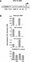
Rb expression in Weri and Y79 retinoblastoma lines repressed HPV-18 URR transcriptional activity via the C/EBPβ-YY1 binding site. (A) The LUC HPV-18 URR reporter construct, as described for Fig. 2A. (B) Parental Rb-null Weri cells (Weri), Weri cells stably expressing Rb (Weri Rb), parental Rb-null Y79 cells (Y79), and Y79 cells stably expressing Rb (Y79 Rb) were transiently transfected with the LUC reporter gene containing the wild-type HPV-18 URR promoter (wild-type URR), the same promoter with a mutation within the SP1 site (SP1 mutant), a mutation within the YY1 site (YY1 mutant), or a mutation in the C/EBPβ-YY1 binding site (C/EBPβ-YY1 mutant). Transcriptional activity was measured as described for Fig. 2. For the wild-type URR experiments the β-galactosidase activity was as follows: Weri, 1.4 ± 0.2 (standard deviation [SD]); Weri Rb, 1.3 ± 0.2; Y79, 0.8 ± 0.07; Y79 Rb, 0.9 ± 0.4 mU. For the experiments with the mutated URR plasmids the β-galactosidase activities were similar (data not shown). Data represent the means of at least three independent experiments performed in duplicate. Results of Student's t test analysis of LUC activity comparing Weri with Weri Rb cells giving P values of <0.05 are indicated (*). Differences in the LUC values for the different cell lines transfected with the C/EBPβ-YY1 mutant were not significant.
FIG. 6.
FIG. 6.
Binding of LIP to the C/EBPβ-YY1 binding site in Rb-expressing retinoblastoma cells. (A) Biotin-labeled C/EBPβ-YY1 binding site dsDNA probe (C/EBPβ-YY1), unlabeled C/EBPβ-YY1 binding site dsDNA probe (Unlabeled), or labeled mutant C/EBPβ-YY1 binding site probe (C/EBPβ-YY1 mut) was incubated with nuclear lysates from either Weri cells (lanes 1 to 3) or Weri Rb cells (lanes 4 to 6). Streptavidin-agarose beads were used to capture and pull down probe-bound transcription factors, which were then analyzed by SDS-PAGE and Western blotting with the antibodies to the indicated proteins. Molecular mass markers are indicated as for Fig. 3. (B) Nuclear lysates used in panel A were analyzed by Western blotting with the same antibodies, with equal protein loading for each lane. Molecular masses corresponding to panel A are indicated by small dashes.
FIG. 7.
FIG. 7.
EMSA of complexes binding to the C/EBPβ-YY1 binding site in retinoblastoma cells. (A) A biotin-labeled C/EBPβ-YY1 binding site dsDNA probe (C/EBPβ-YY1) or a mutated C/EBPβ-YY1 binding site probe (C/EBPβ-YY1 mut) was used in EMSA experiments with nuclear lysates from either Weri or Weri Rb cells. Lanes 1 and 2 show probes in the absence of cell lysates. Lanes 3 and 4 represent Weri cell lysates incubated with labeled probe and a 100-fold excess of unlabeled probe. The arrow indicates the position of complex I, which is a slow-migrating doublet in Fig. 4, lane 5, but is only a single band here (lane 5). Free probe indicates unbound probe. (B) Formation of a novel complex binding to the C/EBPβ-YY1 binding site in Rb-expressing retinoblastoma cell lines. EMSAs were performed as described for panel A with nuclear lysates from Weri cells (lane 3) or Rb-expressing Weri cells (Weri Rb) (lanes 4 to 8). Weri Rb nuclear lysates were also preincubated with an anti-C/EBPβ antibody (lane 6) prior to addition of labeled C/EBPβ-YY1 binding site probe. Weri Rb nuclear lysate incubated with a 100-fold excess of unlabeled probe is shown in lane 8. The novel complex is indicated by an arrow and is present only in lane 5.

Similar articles

References

    1. Antalis, T. M., M. La Linn, K. Donnan, L. Mateo, J. Gardner, J. L. Dickinson, K. Buttigieg, and A. Suhrbier. 1998. The serine proteinase inhibitor (serpin) plasminogen activation inhibitor type 2 protects against viral cytopathic effects by constitutive interferon alpha/beta priming. J. Exp. Med. 187:1799-1811. - PMC - PubMed
    1. Badal, S., V. Badal, I. E. Calleja-Macias, M. Kalantari, L. S. Chuang, B. F. Li, and H. U. Bernard. 2004. The human papillomavirus-18 genome is efficiently targeted by cellular DNA methylation. Virology 324:483-492. - PubMed
    1. Bauknecht, T., P. Angel, H. D. Royer, and H. zur Hausen. 1992. Identification of a negative regulatory domain in the human papillomavirus type 18 promoter: interaction with the transcriptional repressor YY1. EMBO J. 11:4607-4617. - PMC - PubMed
    1. Bauknecht, T., F. Jundt, I. Herr, T. Oehler, H. Delius, Y. Shi, P. Angel, and H. Zur Hausen. 1995. A switch region determines the cell type-specific positive or negative action of YY1 on the activity of the human papillomavirus type 18 promoter. J. Virol. 69:1-12. - PMC - PubMed
    1. Bauknecht, T., R. H. See, and Y. Shi. 1996. A novel C/EBP β-YY1 complex controls the cell-type-specific activity of the human papillomavirus type 18 upstream regulatory region. J. Virol. 70:7695-7705. - PMC - PubMed

Publication types

MeSH terms

Substances