. 2005 Apr;79(7):4347-56.
doi: 10.1128/JVI.79.7.4347-4356.2005.

Kinetic factors control efficiencies of cell entry, efficacies of entry inhibitors, and mechanisms of adaptation of human immunodeficiency virus

Affiliations

Kinetic factors control efficiencies of cell entry, efficacies of entry inhibitors, and mechanisms of adaptation of human immunodeficiency virus

Emily J Platt et al. J Virol. 2005 Apr.

Abstract

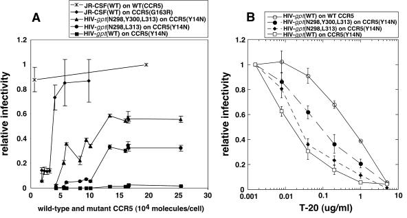
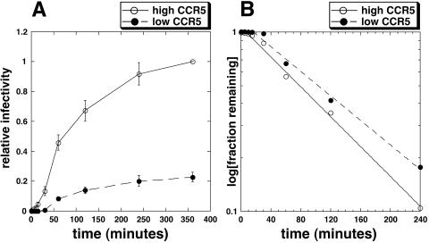
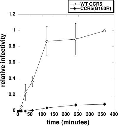
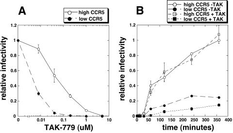
Replication of human immunodeficiency virus type 1 (HIV-1) in diverse conditions limiting for viral entry into cells frequently leads to adaptive mutations in the V3 loop of the gp120 envelope glycoprotein. This has suggested that the V3 loop limits the efficiencies of HIV-1 infections, possibly by directly affecting gp120-coreceptor affinities. In contrast, V3 loop mutations that enable HIV-1(JR-CSF) to use the low-affinity mutant coreceptor CCR5(Y14N) are shown here to have negligible effects on the virus-coreceptor affinity but to dramatically accelerate the irreversible conformational conversion of the envelope gp41 subunits from a three-stranded coil into a six-helix bundle. This slow step is blocked irreversibly by the inhibitor T-20. To further evaluate the role of entry rates in controlling infection efficiencies and viral adaptations, we developed methods to quantitatively measure viral entry kinetics. The virions were adsorbed by spinoculation at 4 degrees C onto HeLa-CD4/CCR5 cell clones that either had limiting or saturating concentrations of CCR5. After warming to 37 degrees C, the completion of entry was monitored over time by the resistance of infections to the competitive CCR5 inhibitor TAK-779. Our results suggest that the efficiency of entry of cell-attached infectious HIV-1 is principally controlled by three kinetic processes. The first is a lag phase that is caused in part by the concentration-dependent reversible association of virus with CD4 and CCR5 to form an equilibrium assemblage of complexes. Second, this assembly step lowers but does not eliminate a large activation energy barrier for a rate-limiting, CCR5-dependent conformational change in gp41 that is sensitive to blockage by T-20. The rate of infection therefore depends on the fraction of infectious virions that are sufficiently saturated with CCR5 to undergo this conformational change and on the magnitude of the activation energy barrier. Although only a small fraction of fully assembled viral complexes overcome this barrier per hour, the ensuing steps of entry are rapidly completed within 5 to 10 min. Thus, this barrier limits the overall flow rate at which the attached virions enter cells, but it has no effect on the lag time that precedes this entry flow. Third, a relatively rapid and kinetically dominant process of viral inactivation, which may partly involve endocytosis, competes with infectious viral entry. Our results suggest that the V3 loop of gp120 has a major effect on the rate-limiting coreceptor-dependent conformational change in gp41 and that adaptive viral mutations, including V3 loop mutations, function kinetically by accelerating this inherently slow step in the entry pathway.


Figures

FIG. 1.
FIG. 1.
Infectivities and T-20 sensitivities of wild-type and mutant CCR5(Y14N)-adapted HIV-1 in HeLa-CD4 cells expressing wild-type or mutant CCR5 coreceptors. (A) Infectivities of wild-type and CCR5(Y14N)-adapted HIV-1JR-CSF. The upper two curves show wild-type replication-competent HIV-1JR-CSF [JR-CSF(WT)] infectivity in HeLa-CD4 cells expressing wild-type CCR5 or in a HeLa-CD4 panel expressing varying discrete amounts of the mutant coreceptor CCR5(G163R) (28, 43). The lower three curves show infectivity data for HIV-gpt virions in a HeLa-CD4 panel expressing varying discrete amounts of the mutant coreceptor CCR5(Y14N) (28). The HIV-gpt virions were pseudotyped with either wild-type JR-CSF envelope [HIV-gpt(WT)] or JR-CSF envelopes that are moderately [HIV-gpt(N298, L313)] or highly [HIV-gpt(N298, N300, L313)] adapted to use CCR5(Y14N). Relative infectivity values for each virus were calculated by normalizing titers in a given cell clone to titers in HeLa-CD4 cells expressing a large amount of wild-type CCR5. Data are the averages from three to eight independent experiments, and error bars are standard errors of the means. (B) Efficiency of T-20 inhibition in HeLa-CD4/CCR5 and HeLa-CD4/CCR5(Y14N) cells. A HeLa-CD4/CCR5(Y14N) clone expressing large amounts of coreceptor was infected by HIV-gpt viruses pseudotyped with wild-type JR-CSF envelopes [HIV-gpt(WT)] or with envelopes adapted to use CCR5(Y14N) [(HIV-gpt(N298, N300, L313)] or [HIV-gpt(N298, L313)] in the presence of serial fivefold dilutions of T-20. The T-20 sensitivity of wild-type JR-CSF envelope-pseudotyped virions in HeLa-CD4 cells expressing a large amount of wild-type CCR5 [HIV-gpt(WT)] was also measured. Relative infectivity values were calculated for each virus by dividing titers generated in a given cell line at each T-20 concentration by titers obtained in the same cell line in the absence of inhibition. The averages of replicate experiments are displayed (n = 4); error bars are standard errors of the means.
FIG. 2.
FIG. 2.
Infection kinetics of HIV-1JR-CSF in HeLa-CD4/CCR5 cells. (A) Infection kinetic data in HeLa-CD4 cells expressing high or low concentrations of wild-type CCR5. Relative infectivity values were generated by dividing the titer from a given time point by the titer in HeLa-CD4 cells expressing a high concentration of CCR5 at the final time point (360 min). The averages of replicate experiments are displayed (n = 11), and error bars are standard errors of the means. (B) Mathematical analysis of the infection kinetic data. The data in panel A were analyzed according to equation 7 in Materials and Methods. Titers obtained at the final (360-min) time point were used as the ifinal value. The value for the final 360-min time point is not plotted because where ifinal = it, equation 7 is undefined. Therefore, the number of points is not the same as that in panel A, but all of the informative data available are represented. The intercept of the upper axis indicates the lag times for the two cell lines, with lags of ∼7 min in cells expressing high concentrations of CCR5 and ∼16 min in cells expressing low concentrations of CCR5. Measuring the slopes generated with HeLa-CD4 cells expressing high or low concentrations of CCR5 and then using equations 7 and 8 in Materials and Methods allows us to calculate the rate constant of viral inactivation, k2, as ∼0.47/h and the rate constant at which completely assembled virus-CD4-CCR5 complexes complete cellular entry, ki, as ∼0.09/h. The proportion of virions in competent assembly complexes, α, is ∼0.23 in cells expressing low concentrations of CCR5.
FIG. 3.
FIG. 3.
Infection kinetics of HIV-gpt virions in HeLa-CD4 cells expressing wild-type or mutant coreceptors. Infection kinetics of HIV-gpt virions pseudotyped with wild-type JR-CSF envelope were measured in HeLa-CD4 cells expressing CCR5(G163R) or in HeLa-CD4 cells expressing a similar amount of wild-type (WT) CCR5 (28, 43). Error bars are standard errors of the means for CCR5(G163R) (n = 4) or the range for wild-type CCR5 (n = 2). Relative infectivity values were obtained by dividing the titer from a given time point by the titer obtained in cells with wild-type CCR5 at the final time point (360 min). The data were analyzed according to equation 7, and lag times for the viruses were ∼5 min in cells with wild-type CCR5 and ∼48 min in cells with CCR5(G163R). It was not possible to calculate the other kinetic parameters since the ki values are different for the two coreceptors.
FIG. 4.
FIG. 4.
TAK-779 effects on infection efficiencies and infection kinetics. (A) Efficacy of TAK-779 inhibition. HeLa-CD4 cells expressing high or low concentrations of CCR5 were infected by wild-type HIV-1JR-CSF in the absence or presence of serial fivefold TAK-779 dilutions. Relative infectivities were calculated by dividing the titer in a given cell line at each inhibitor concentration by the titer in the same cell line in the absence of inhibitor. Error bars are standard errors of the means (n = 6). (B) Effect of TAK-779 on infection kinetics. A moderately inhibiting concentration (i.e., the 50% inhibitory concentration obtained in low-CCR5 cells in our inhibitor efficiency assays) of TAK-779 was included in medium added to HeLa-CD4 cells expressing low or high CCR5 concentrations after spinoculation at 4°C with wild-type HIV-1JR-CSF. Control cultures that lacked TAK-779 were analyzed in parallel. Relative infectivity values were generated by dividing the titer obtained at each time point by the titer obtained in the absence of inhibitor in HeLa-CD4 cells expressing a large amount of CCR5 at the final time point (360 min). Data points are the averages from a single representative experiment performed in duplicate, and error bars are the range.
FIG. 5.
FIG. 5.
T-20 effects on infectivity efficiencies and infection kinetics. (A) Efficacy of T-20 inhibition. Infections of HeLa-CD4 cells expressing high or low concentrations of CCR5 were performed in the absence or presence of serial fivefold dilutions of T-20. Relative infectivities were calculated by dividing the titer from a given cell line at each T-20 concentration by the titer in the same cell line in the absence of T-20. Results of a representative experiment performed in duplicate are shown; error bars are the range. (B) Effect of T-20 on infection kinetics. A moderately inhibiting concentration of T-20 was included in the medium of cells expressing low or high concentrations of CCR5 after virus spinoculation and washing. Control cultures that lacked T-20 were analyzed in parallel. Relative infectivity values were obtained by dividing the titer at a given time point by the titer obtained in the absence of T-20 at the final time point (360 min) in cells expressing large amounts of CCR5. Data points represent the averages from a single representative experiment performed in duplicate, and error bars are the range.

Similar articles

Cited by

References

    1. Baba, M., O. Nishimura, N. Kanzaki, M. Okamoto, H. Sawada, Y. Iizawa, M. Shiraishi, Y. Aramaki, K. Okonogi, Y. Ogawa, K. Meguro, and M. Fujino. 1999. A small-molecule, nonpeptide CCR5 antagonist with highly potent and selective anti-HIV-1 activity. Proc. Natl. Acad. Sci. USA 96:5698-5703. - PMC - PubMed
    1. Berger, E. A., P. M. Murphy, and J. M. Farber. 1999. Chemokine receptors as HIV-1 coreceptors: roles in viral entry, tropism, and disease. Annu. Rev. Immunol. 17:657-700. - PubMed
    1. Cavrois, M., C. De Noronha, and W. C. Greene. 2002. A sensitive and specific enzyme-based assay detecting HIV-1 virion fusion in primary T lymphocytes. Nat. Biotechnol. 20:1151-1154. - PubMed
    1. Cavrois, M., J. Neidleman, M. Bigos, and W. C. Greene. 2004. Fluorescence resonance energy transfer-based HIV-1 virion fusion assay. Methods Mol. Biol. 263:333-344. - PubMed
    1. Chan, D. C., and P. S. Kim. 1998. HIV entry and its inhibition. Cell 93:681-684. - PubMed

Publication types

MeSH terms