. 2005 Apr;79(7):4382-95.
doi: 10.1128/JVI.79.7.4382-4395.2005.

Effects of foot-and-mouth disease virus nonstructural proteins on the structure and function of the early secretory pathway: 2BC but not 3A blocks endoplasmic reticulum-to-Golgi transport

Affiliations

Effects of foot-and-mouth disease virus nonstructural proteins on the structure and function of the early secretory pathway: 2BC but not 3A blocks endoplasmic reticulum-to-Golgi transport

Katy Moffat et al. J Virol. 2005 Apr.

Abstract

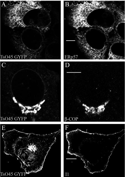
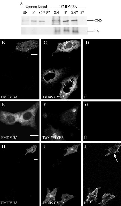
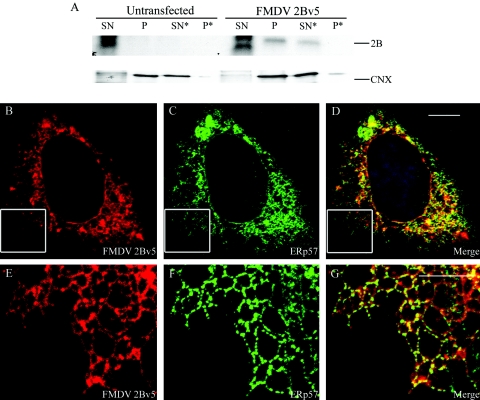
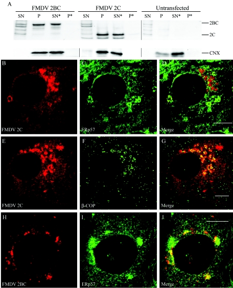
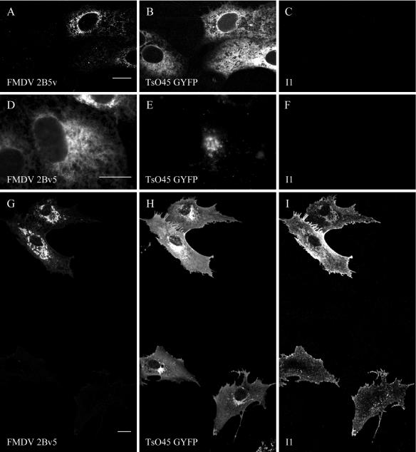
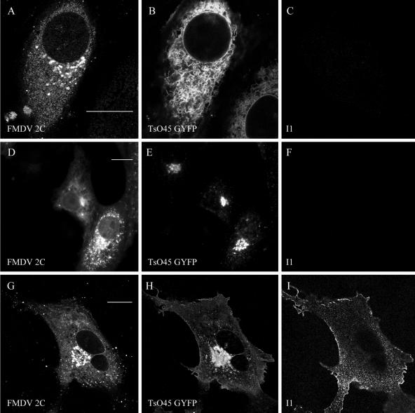
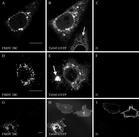
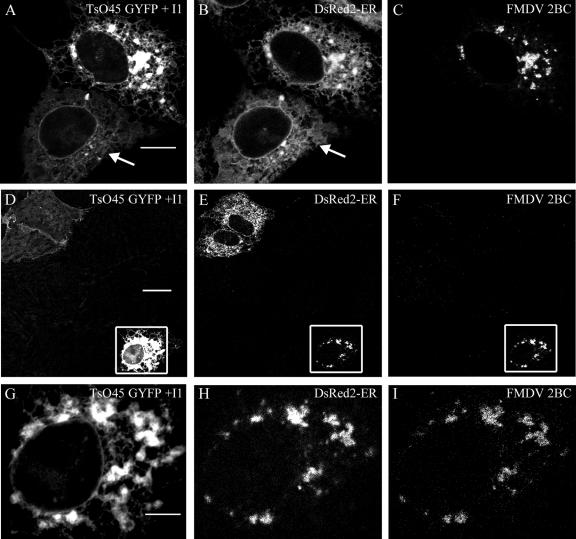
Infection of cells by picornaviruses leads to the generation of intracellular membrane vesicles. The expression of poliovirus (PV) 3A protein causes swelling of the endoplasmic reticulum (ER) and inhibition of protein trafficking between the ER and the Golgi apparatus. Here, we report that the nonstructural proteins of a second picornavirus, foot-and-mouth disease virus (FMDV), also perturb the secretory pathway. FMDV proteins 3A, 2B, 2C, and 2BC expressed alone in cells were recovered from crude membrane fractions, indicating membrane association. Immunofluorescence microscopy showed that 3A was located in a reticular structure and 2B was located in the ER, while 2C was located in both the ER and the bright punctate structures within the Golgi apparatus. 2BC gave punctate cytoplasmic staining and also caused accumulation of ER proteins in large vesicular structures located around the nuclei. The effect of the FMDV proteins on the trafficking of the vesicular stomatitis virus glycoprotein (G protein) from the ER to the cell surface was determined. Unlike its PV counterpart, the 3A protein of FMDV did not prevent trafficking of the G protein to the cell surface. Instead, surface expression of the G protein was blocked by 2BC, with retention of the G protein in a modified ER compartment staining for 2BC. The results suggest that the nonstructural proteins of different picornaviruses may vary in their ability to perturb the secretory pathway. Since FMDV 2BC can block the delivery of proteins to the cell surface, it may, as shown for PV 3A, play a role in immune evasion and contribute to the persistent infections observed in ruminants.


Figures

FIG. 1.
FIG. 1.
Distribution of VSV TsO45 GYFP protein at permissive and nonpermissive temperatures. Vero cells expressing TsO45 GYFP were incubated for 48 h at 40oC (A and B). Cells were also incubated for a further 30 min (C and D) or 3 h (E and F) at 32oC. For intracellular staining (panels B and D), cells were fixed, permeabilized, and counterstained using antibodies specific for ERp57 or using β-COP as indicated in the panel. For surface staining (panel F), cells were incubated with an antibody (I1) specific for the ectodomain of the G protein before permeabilization. Intracellular TsO45 GYFP (panels A, C, and E) was visualized through the natural fluorescence of YFP. Primary antibodies were detected using goat antibody conjugated to Alexa-568 (panels B, D, and F). Bars, 8 μm (panels B and D) or 16 μm (panel F).
FIG. 2.
FIG. 2.
Amino acid sequence comparison between the 2BC through 3A regions of picornaviruses. PV type 1, bovine enterovirus (BEV), human rhinovirus type 1 (HRV-1), FMDV, encephalomyocarditis virus (EMCV), Theiler's murine encephalitis virus (TMEV), and hepatitis A virus (HAV) 2B, 2C, and 3A regions were aligned using MACAW (41). Blocks of similarity were detected with a pairwise cutoff score of 25 (minimum of two sequences). Sequence similarities of 70 to 100% are shown by dark grey bars, while similarities of 30 to 70% are shown by light grey bars.
FIG. 3.
FIG. 3.
FMDV 3A is membrane associated but does not block transport of VSV TsO45 GYFP protein to the cell surface. (A) Membrane association of FMDV protein 3A. Mock-transfected Vero cells or Vero cells expressing FMDV 3A were metabolically labeled with [35S]methionine-cysteine and then homogenized. Postnuclear supernatants were separated by centrifugation into crude cytosol (SN) and membrane (P) fractions. Half the membrane fraction was solubilized in 2% Triton X-100 and recentrifuged to produce a solubilized membrane fraction (SN*) and a pellet of insoluble aggregated protein (P*). Equivalent fractions were immunoprecipitated using antibodies specific for 3A (2C2) or calnexin (CNX). Proteins were resolved by SDS-PAGE and detected by autoradiography. (B to J) Transport of VSV TsO45 GYFP protein to the surfaces of cells transfected with FMDV 3A. Vero cells were transiently cotransfected with plasmids which express TsO45 GYFP and FMDV 3A and incubated for 48 h at 40oC (panels B to D). A set of cells was also incubated for a further 30 min (panels E to G) or 3 h (panels H to J) at 32oC. All cells were fixed with 4% paraformaldehyde, and surface expression of the G protein was detected by adding an antibody (I1), recognizing the ectodomain of the G protein, prior to permeabilization (panels D, G, and J). Cells were then permeabilized, and intracellular staining of FMDV 3A was visualized using antibody 2C2 (panels B, E, and H). Intracellular TsO45 GYFP was visualized directly using the signal from YFP (panels C, F, and I). Primary antibodies were visualized with appropriate goat antibodies conjugated to Alexa-568 (panels B, E, and H) or Alexa-633 (panels D, G, and J). Bars, 10 μm. Arrow indicates surface staining of G protein in cells expressing FMDV 3A.
FIG. 4.
FIG. 4.
Subcellular location of FMDV 2B. (A) Membrane association of FMDV protein 2B. Vero cells were mock transfected or transfected with a vector encoding 2Bv5. After 48 h, cells were fractionated as described in the legend to Fig. 3A. The V5-epitope-tagged 2B (2Bv5) was immunoprecipitated using antibody specific for the V5 tag (V5). Calnexin was detected using an antipeptide antibody (CNX). Proteins were resolved by SDS-PAGE and detected by autoradiography. (B to G) Intracellular distribution of FMDV 2B. Vero cells expressing 2Bv5 were fixed in 4% paraformaldehyde and then permeabilized. 2Bv5 was detected using the epitope tag (panels B and E). Cells were counterstained using antibody specific for ERp57 (panels C and F). Primary antibodies were visualized with appropriate goat antibodies conjugated to Alexa-568 (panels A and E) or Alexa-488 (panels C and F). Panels D and G are digitally merged images. Panels E to G are higher magnifications of the insets defined in panels B to D, respectively. Bars, 8 μm (panel D) or 4 μm (panel G).
FIG. 5.
FIG. 5.
Subcellular location of FMDV 2BC and 2C. (A) Membrane association of FMDV proteins 2BC and 2C. Vero cells were mock transfected or transfected with a vector encoding 2BC or 2C. After 48 h, cells were fractionated as described in the legend to Fig. 3A. Both 2C and 2BC were immunoprecipitated using the DM12 antibody specific for 2C. Calnexin was detected using an antipeptide antibody (CNX). Proteins were resolved by SDS-PAGE and detected by autoradiography. (B to J) Intracellular distribution of FMDV 2BC and 2C. Vero cells expressing 2C (panels B to G) or 2BC (panels H to J) were fixed in 4% paraformaldehyde and then permeabilized. 2C and 2BC were both detected using antibody 3F7 (panels B, E, and H), and cells were counterstained using antibodies against ERp57 (panels C and I) or β-COP (panel F). Primary antibodies were visualized with appropriate goat antibodies conjugated to Alexa-568 (panels B, E, and H) or Alexa-488 (panels C, F, and I). Panels D, G, and J are digitally merged images. Bars, 8 μm.
FIG. 6.
FIG. 6.
FMDV 2B does not block transport of VSV TsO45 GYFP protein to the cell surface. Vero cells were transiently cotransfected with TsO45 GYFP and FMDV 2Bv5 and incubated for 48 h at 40oC (A to C). Some cells were also incubated for a further 30 min (D to F) or 3 h (G to I) at 32oC. All cells were fixed with 4% paraformaldehyde, and surface expression of the G protein was detected by adding an antibody (I1), recognizing the ectodomain of the G protein, prior to permeabilization (panels C, F, and I). Cells were then permeabilized, and 2Bv5 was detected using the epitope tag (panels A, D, and G). Intracellular TsO45 GYFP was visualized directly using the signal from YFP (panels B, E, and H). Primary antibodies were visualized with appropriate goat antibodies conjugated to Alexa-568 (panels A, D, and G) or Alexa-633 (panels C, F, and I). Bars, 10 μm.
FIG. 7.
FIG. 7.
FMDV 2C does not block transport of VSV TsO45 GYFP protein to the cell surface. Vero cells were transiently cotransfected with TsO45 GYFP and FMDV 2C and incubated for 48 h at 40oC (A to C). Some cells were also incubated for a further 30 min (D to F) or 3 h (G to I) at 32oC. All cells were fixed, and surface expression of the G protein was detected by adding an antibody (I1), recognizing the ectodomain of the G protein, prior to permeabilization (panels C, F and I). Cells were then permeabilized, and 2C was detected using antibody 3F7 (panels A, D, and G). Intracellular TsO45 GYFP was visualized directly using the signal from YFP (panels B, E, and H). Primary antibodies were visualized with appropriate goat antibodies conjugated to Alexa-568 (panels A, D, and G) or Alexa-633 (panels C, F, and I). Bars, 10 μm.
FIG. 8.
FIG. 8.
FMDV 2BC blocks transport of VSV TsO45 GYFP protein to the cell surface. Vero cells were transiently cotransfected with TsO45 GYFP and FMDV 2BC and incubated for 48 h at 40oC (A to C). Some cells were also incubated for a further 30 min (D to F) or 3 h (G to I) at 32oC. All cells were fixed with 4% paraformaldehyde, and surface expression of the G protein was detected by adding an antibody (I1), recognizing the ectodomain of the G protein, prior to permeabilization (panels C, F, and I). Cells were then permeabilized, and 2BC was detected using antibody 3F7 (panels A, D and G). Intracellular TsO45 GYFP was visualized directly using the signal from YFP (panels B, E, and H). Primary antibodies were visualized with appropriate goat antibodies conjugated to Alexa-568 (panels A, D, and G) or Alexa-633 (panels C, F, and I). Bars, 10 μm. Arrows indicate cells transfected with TsO45 GYFP only and show the reticular pattern of the G protein in cells at 40oC (panel B) and the Golgi appearance of the G protein after moving the cells to 32oC (panel E) for 30 min.
FIG. 9.
FIG. 9.
VSV TsO45 GYFP cellular location in FMDV 2BC-transfected cells. (A to C) Vero cells were transiently cotransfected with vectors expressing TsO45 GYFP, DsRed2-ER, and FMDV 2BC proteins and incubated for 48 h at 40oC. (D to I) Some cells were also incubated for a further 3 h at 32oC. Surface expression of TsO45 GYFP protein was determined by immunofluorescence analysis of fixed, but not permeabilized, cells by using the antibody I1, specific for the ectodomain of the G protein (panels A, D, and G). Panels A, D, and G also show the internal location of TsO45 GYFP detected from the fluorescence of the YFP in transfected cells. Panels B, E, and H show the location of DsRed2-ER, a marker for the ER, detected from the DsRed2 fluorescence. Following I1 staining, cells were permeabilized and 2BC was detected using antibody DM12 (C, F, and I). Primary antibodies were visualized with appropriate goat antibodies conjugated to Alexa-488 (panels A, D, and G) or Alexa-633 (panels C, F, and I). Bars, 8 μm (panel A), 20 μm (panel D), and 4 μm (panel G). Arrows indicate reticular patterns of the G protein and the ER marker.

Similar articles

Cited by

References

    1. Aldabe, R., A. Barco, and L. Carrasco. 1996. Membrane permeabilization by poliovirus proteins 2B and 2BC. J. Biol Chem. 271:23134-23137. - PubMed
    1. Alexandersen, S., Z. Zhang, and A. I. Donaldson. 2002. Aspects of the persistence of foot-and-mouth disease virus in animals—the carrier problem. Microbes Infect. 4:1099-1110. - PubMed
    1. Barco, A., and L. Carrasco. 1998. Identification of regions of poliovirus 2BC protein that are involved in cytotoxicity. J. Virol. 72:3560-3570. - PMC - PubMed
    1. Barlowe, C., L. Orci, T. Yeung, M. Hosobuchi, S. Hamamoto, N. Salama, M. F. Rexach, M. Ravazzola, M. Amherdt, and R. Schekman. 1994. COPII: a membrane coat formed by Sec proteins that drive vesicle budding from the endoplasmic reticulum. Cell 77:895-907. - PubMed
    1. Bergmann, J. E. 1989. Using temperature-sensitive mutants of VSV to study membrane protein biogenesis. Methods Cell Biol. 32:85-110. - PubMed

Publication types

MeSH terms

Substances

LinkOut - more resources