. 2005 Apr;79(7):4492-505.
doi: 10.1128/JVI.79.7.4492-4505.2005.

The Epstein-Barr virus EBNA-LP protein preferentially coactivates EBNA2-mediated stimulation of latent membrane proteins expressed from the viral divergent promoter

Affiliations

The Epstein-Barr virus EBNA-LP protein preferentially coactivates EBNA2-mediated stimulation of latent membrane proteins expressed from the viral divergent promoter

Rongsheng Peng et al. J Virol. 2005 Apr.

Abstract

The mechanistic contribution of the Epstein-Barr virus (EBV) EBNA-LP protein to B-cell immortalization remains an enigma. However, previous studies have indicated that EBNA-LP may contribute to immortalization by enhancing EBNA2-mediated transcriptional activation of the LMP-1 gene. To gain further insight into the potential role EBNA-LP has in EBV-mediated B-cell immortalization, we asked whether it is a global or gene-specific coactivator of EBNA2 and whether coactivation requires interaction between these proteins. In type I Burkitt's lymphoma cells, we found that EBNA-LP strongly coactivated EBNA2 stimulation of LMP-1 and LMP2B RNAs, which are expressed from the viral divergent promoter. Surprisingly, the viral LMP2A gene and cellular CD21 and Hes-1 genes were induced by EBNA2 but showed no further induction after EBNA-LP coexpression. We also found that EBNA-LP did not stably interact with EBNA2 in coimmunoprecipitation assays, even though the conditions were adequate to observe specific interactions between EBNA2 and its cellular cofactor, CBF1. Colocalization between EBNA2 and EBNA-LP was not detectable in EBV-transformed cell lines or transfected type I Burkitt's cells. Finally, no significant interactions between EBNA2 and EBNA-LP were found with mammalian two-hybrid assays. From this data, we conclude that EBNA-LP is not a global coactivator of EBNA2 targets, but it preferentially coactivates EBNA2 stimulation of the viral divergent promoter. While this may require specific transient interactions between these proteins that only occur in the context of the divergent promoter, our data strongly suggest that EBNA-LP also cooperates with EBNA2 through mechanisms that do not require direct or indirect complex formation between these proteins.


Figures
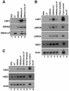
FIG. 1.
FIG. 1.
EBNA-LP preferentially coactivates EBNA2 stimulation of the viral divergent promoter. (A) Eli-BL cells were transfected with pSG5 vector (lane 1) or plasmids expressing LP (lane 2), EBNA2 (lane 3), EBNA2 plus EBNA-LP (lane 4), or EBNA2 plus a mutant version of EBNA-LP (S35/101A) (lane 5). All versions of EBNA-LP had 2 W repeats. Approximately 20% of the transfected cells were processed for Western blotting as described in the Materials and Methods. Replicate blots were probed with anti-LMP-1 (S12), anti-EBNA2 (R3), and anti-EBNA-LP (JF186) monoclonal antibodies as indicated. (B) RT-PCR and Southern blotting of RNA derived from transfected samples in panel A. The specific genes amplified and probed for each blot are indicated to the left of each panel. Primers and probes are indicated in Table 1. In addition, RNA from EBNA2- and EBNA-LP-cotransfected cells (lane 4) was subjected to 5 to 10 additional PCR cycles to make sure that the reactions in lanes 1 to 5 had not yet plateaued (lane 7). PCRs were also performed with water as a template or RNA that had not been reverse transcribed (lanes 6 and 8, respectively). (C) DG75 cells, an EBV-negative cell line, were transfected as described for Eli-BL cells in the legend to panel A. However, the EBNA2 effector plasmid pEΔA6 was used instead of an SG5-EBNA2 expression plasmid, since EBNA-LP induces protein levels of the SG5-driven EBNA2 in these cells and pEΔA6 is refractory to this induction. RT-PCR and Southern blotting was performed as described for panel B.
FIG. 2.
FIG. 2.
Association of EBNA2 and EBNA-LP in cells is minimal. (A) DG75 cells were cotransfected with the following pairs of expression plasmids. Lane 1, wild-type EBNA2 (pPDL151) and HA-CBF1; lane 2, mutant EBNA2 (pPDL152) and HA-CBF1; lane 3, wild-type EBNA2 (pPDL151) and SG5LP; lane 4, wild-type EBNA2-HA (pAG155) and SG5LP. The transfected cells were solubilized in either radioimmunoprecipitation assay or NP-40 lysis buffer (see Materials and Methods), resolved by SDS-PAGE, and blotted, and replicate blots were probed with anti-EBNA2, anti-HA (for CBF1), or anti-EBNA-LP monoclonal antibody as indicated for each blot. (B) Extracts described for panel A containing wild-type (wt) EBNA2 and CBF1 (lanes 1 to 2), mutant (mut) EBNA2 and CBF1 (lanes 3 to 4), and wild-type EBNA2 and Flag-EBNA-LP (lanes 5 to 6) were analyzed by immunoprecipitation (IP) followed by Western blotting (W). The extracts were precipitated with an anti-HA antibody (lanes 1 and 3) or anti-Flag (lane 5) followed by incubation with protein G-Sepharose beads or beads only (lanes 2, 4, and 6). The precipitates were divided into two parts, and replicate blots were probed with anti-EBNA2, anti-HA (for CBF1), or anti-EBNA-LP antibody as indicated below each panel. A longer exposure of the blot probed with anti-EBNA2 is shown in the panel on the right, and the asterisks indicate trace amounts of EBNA2 that were detected after maximum exposures to the X-ray film. Cross-reactivity with the immunoglobulin heavy chain (H) by the secondary antibody is indicated for some of the immunoblots. Fl, Flag. (C) Extracts described for panel A (lane 4) containing wild-type EBNA2-HA and Flag-EBNA-LP were analyzed by immunoprecipitation followed by Western blotting. The panel on the left shows immunoprecipitation with anti-HA followed by immunoblotting with anti-HA (lane1) or precipitation with protein G beads alone (lane 2). The other half of the immunoprecipitation was immunoblotted with an anti-EBNA-LP antibody (IP anti-HA in lane 3 or beads alone lane 4). Cross-reactivity with the immunoglobulin heavy chain (H) by the secondary antibody is indicated for some of the immunoblots. (D) DG75 cells were transfected with a wild-type EBNA-LP (2W repeats) and EBNA2-HA (lanes 1 and 2) or a mutant EBNA-LP (S35/101A, 2 W repeats) and EBNA2-HA (lanes 3 and 4). The extracts were prepared as described in Materials and Methods. The panels on top show the results of extracts precipitated with anti-HA or anti-Flag antibody followed by Western blotting with an anti-EBNA2 antibody. The panels below are identical but instead were probed with an anti-EBNA-LP antibody. Cross-reactivity with the immunoglobulin heavy chain (H) by the secondary antibody is indicated for some of the immunoblots.
FIG. 3.
FIG. 3.
CFP-EBNA-LP and EBNA2-YFP fusion proteins are transcriptionally competent but do not colocalize in cells. (A) Eli-BL cells were transfected with plasmids expressing EBNA-LP (lane 1), wild-type (wt) EBNA2 (lane 2), both effectors (lane 3), CFP-EBNA-LP (lane 4), EBNA2-YFP (lane 5), or both fusion proteins (lane 6). The cells were processed for Western blot analysis, and replicate blots were probed for LMP-1, EBNA2, and EBNA-LP with the appropriate antibodies. Bands corresponding to LMP-1 expression or each of the wild-type or fusion proteins are indicated on the right of each blot. The migration of molecular weight standards is shown on the left of each blot. α, anti. (B) Using high-resolution deconvolution microscopy, merged images of EBNA2 (green) and EBNA-LP (blue) cellular localization are shown. Images are single Z planes from four different cells.
FIG. 4.
FIG. 4.
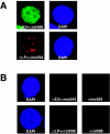
Specific detection of EBNA2 and EBNA-LP in EBV-immortalized B cells. (A) The anti-EBNA2 (αE2) monoclonal antibody R3 and the secondary anti-rat Alexa 488 (αrat488)-conjugated antibody (Molecular Probes) were used to detect EBNA2 in IB4 cells. Indirect immunofluorescence with an EBNA-LP-specific antibody JF186 (αLP) and an anti-mouse Alexa 594 (αmo594)-conjugated antibody (Molecular Probes) was used to detect EBNA-LP in IB4 cells. DAPI staining for each cell is shown in the panel on the right. (B) In the top row of panels, IB4 cells were stained with DAPI (first panel) or the rat anti-EBNA2 antibody followed by the anti-mouse Alexa 594-conjugated antibody or the anti-mouse Alexa 594-conjugated antibody alone (second and third panels, respectively). In the lower set of panels, a DAPI-stained cell is shown (first panel). The second and third panels show the same cell stained with the mouse anti-EBNA-LP monoclonal and a secondary anti-rat Alexa 488-conjugated antibody or the anti-rat Alexa 488-conjugated antibody alone.
FIG. 5.
FIG. 5.
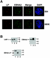
EBNA protein expression and subcellular localization in LCLs. (A) High-resolution images from deconvolution microscopy are shown for MHK or IB4 cells stained with anti-EBNA2 or anti-EBNA-LP antibody followed by anti-rat Alexa 488 (green) and anti-mouse Alexa 594 (red) secondary antibodies, respectively. Images are representative of the population and are single Z planes. (B) Western blot analysis of MHK (lane 1 in all panels), IB4 (lane 2 in all panels), and DG75 (lane 3 in all panels) cells. Cell extracts from each cell line were resolved by SDS-PAGE, and replicate blots were produced. The blots were probed for LMP-1, EBNA2, or EBNA-LP with S12, R3, and JF186 monoclonal antibodies, respectively. The specific proteins detected on each blot are indicated by the arrow(s) on the left. IB4 cells expressed at least 3 EBNA-LP isoforms.
FIG. 6.
FIG. 6.
EBNA2 does not functionally interact with a Gal4-EBNA-LP fusion protein in mammalian two-hybrid assays. Plasmids expressing Gal4, a Gal4-CBF1 fusion protein, or a Gal4-EBNA-LP fusion protein were cotransfected with pSG5 vector, wild-type (wt) EBNA2, or mutant (mut) EBNA2 and a Gal4-responsive luciferase reporter plasmid. An internal control plasmid driving the Renilla luciferase protein under the control of an SV40 promoter was also included. At 48 h posttransfection, luciferase activity was measured from transfected cell extracts and normalized to Renilla luciferase to control for transfection efficiencies. Results are reported as activation relative to the activity detected from the bait protein alone (e.g., cotransfected with pSG5 vector). Error bars indicate standard deviations from the means from at least three independent experiments. The levels of EBNA2 were detected by Western blot analysis from the transfected cells as shown in the panel below the graph with the R3 anti-EBNA2 monoclonal antibody. Cells were transfected with prey plasmids pSG5 (lanes 1, 4, 7), wild-type EBNA2 (lanes 2, 5, 8), and mutant EBNA2 (lanes 3, 5, 9) and bait plasmids Gal4 (lanes 1 to 3), Gal4-CBF1 (lanes 4 to 6), and Gal4-LP (lanes 7 to 9). Levels of the Gal4-LP fusion protein were also detected by immunoblotting with a monoclonal antibody to EBNA-LP and are shown in the panel above the bars corresponding to its activity in these assays.
FIG. 7.
FIG. 7.
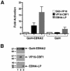
EBNA-LP and VP16-CBF1 enhance Gal4-EBNA2 stimulation of a Gal4-responsive reporter. (A) Gal4-EBNA2 or Gal4 was cotransfected with an SG5-VP16 vector, VP16-CBF1, or EBNA-LP in DG75 cells together with a Gal4-responsive reporter plasmid (5xGal4E1bLuc). Results are shown as activation relative to that of vector-transfected cells. Error bars indicate standard deviations from the means from at least three independent experiments. (B) A portion of the lysates from panel A were analyzed by Western blotting. Three identical blots were probed with anti-EBNA2, anti-HA, or anti-EBNA-LP antibody. Lane 1, Gal4-EBNA2 and VP16-SG5; lane 2, Gal4-EBNA2 and VP16-CBF1; lane 3 Gal4-EBNA2 and EBNA-LP.
FIG. 8.
FIG. 8.
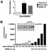
The bulk of EBNA-LP effects on a Gal4-EBNA2 bait protein are due to down-regulation of most internal control plasmids and induction of bait protein levels. (A) Gal4-EBNA2 was cotransfected with the indicated plasmids expressing various prey proteins. Cotransfections were performed as described for Fig. 7A, but only the RL-null internal control was used to control for transfection efficiency. (B) The Gal4-EBNA2 bait plasmid was transfected with the amounts indicated below the graph or cotransfected with 5 μg of EBNA-LP. Luminescence (in relative luciferase units [RLU]) levels are indicated for each sample on the left. The inset shows a Western blot from the transfected cell lysates probed with an EBNA2 antibody to detect the Gal4-EBNA2 bait protein. Samples transfected with 0.5 μg (lane 1 and 6), 1.0 μg (lanes 2 and 7), 2.0 μg (lanes 3 and 8), 4.0 μg (lanes 4 and 9), and 8.0 μg (lanes 5 and 10) are shown. Five micrograms of EBNA-LP was cotransfected in samples shown in lanes 6 to 10.

Similar articles

Cited by

References

    1. Allan, G. J., G. J. Inman, B. D. Parker, D. T. Rowe, and P. J. Farrell. 1992. Cell growth effects of Epstein-Barr virus leader protein. J. Gen. Virol. 73:1547-1551. - PubMed
    1. Allday, M. J., D. H. Crawford, and J. A. Thomas. 1993. Epstein-Barr virus (EBV) nuclear antigen 6 induces expression of the EBV latent membrane protein and an activated phenotype in Raji cells. J. Gen. Virol. 74:361-369. - PubMed
    1. Allday, M. J., and P. J. Farrell. 1994. Epstein-Barr virus nuclear antigen EBNA3C/6 expression maintains the level of latent membrane protein 1 in G1-arrested cells. J. Virol. 68:3491-3498. - PMC - PubMed
    1. Bornkamm, G. W., and W. Hammerschmidt. 2001. Molecular virology of Epstein-Barr virus. Philos. Trans. R. Soc. Lond. B 356:437-459. - PMC - PubMed
    1. Castillo, J. P., and T. F. Kowalik. 2002. Human cytomegalovirus immediate early proteins and cell growth control. Gene 290:19-34. - PubMed

Publication types

MeSH terms

Substances

LinkOut - more resources