Comparative Study
. 2005 Mar 14;168(6):855-62.
doi: 10.1083/jcb.200411026.

The AAA+ protein torsinA interacts with a conserved domain present in LAP1 and a novel ER protein

Affiliations
Comparative Study

The AAA+ protein torsinA interacts with a conserved domain present in LAP1 and a novel ER protein

Rose E Goodchild et al. J Cell Biol. .

Abstract

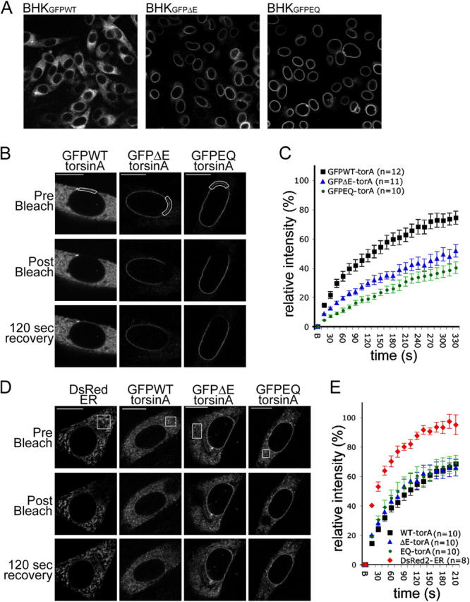
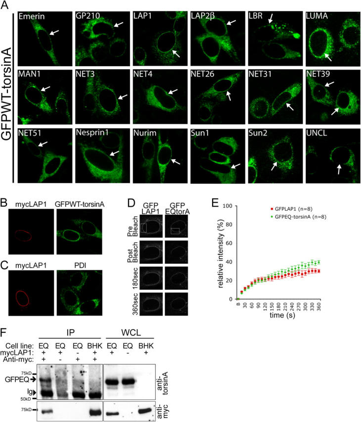
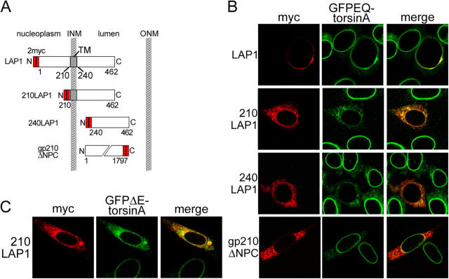
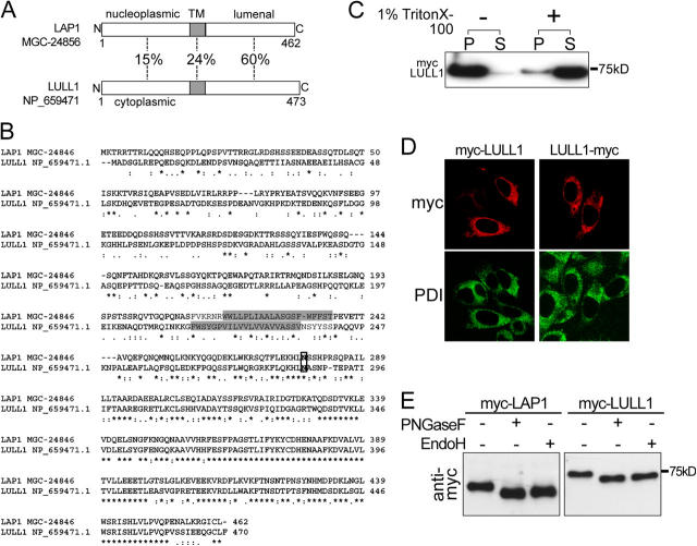
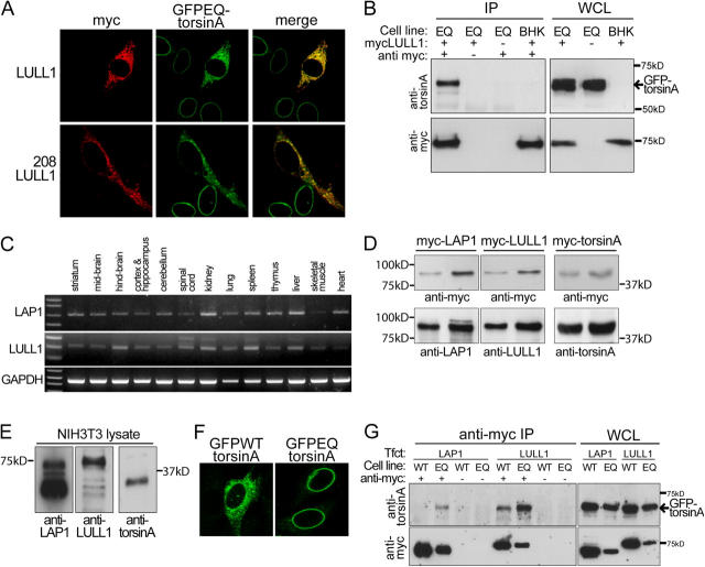
A glutamic acid deletion (DeltaE) in the AAA+ protein torsinA causes DYT1 dystonia. Although the majority of torsinA resides within the endoplasmic reticulum (ER), torsinA binds a substrate in the lumen of the nuclear envelope (NE), and the DeltaE mutation enhances this interaction. Using a novel cell-based screen, we identify lamina-associated polypeptide 1 (LAP1) as a torsinA-interacting protein. LAP1 may be a torsinA substrate, as expression of the isolated lumenal domain of LAP1 inhibits the NE localization of "substrate trap" EQ-torsinA and EQ-torsinA coimmunoprecipitates with LAP1 to a greater extent than wild-type torsinA. Furthermore, we identify a novel transmembrane protein, lumenal domain like LAP1 (LULL1), which also appears to interact with torsinA. Interestingly, LULL1 resides in the main ER. Consequently, torsinA interacts directly or indirectly with a novel class of transmembrane proteins that are localized in different subdomains of the ER system, either or both of which may play a role in the pathogenesis of DYT1 dystonia.


Figures

Figure 1.
Figure 1.
Pathogenic and substrate trap forms of torsinA display reduced mobility in the NE. (A) GFP immunolabeling of BHKGFPWT, BHKGFPΔE, and BHKGFPEQ stable cell lines. (B and D) GFP fluorescence of BHK21 cells transiently transfected with GFPWT-, GFPΔE-, or GFPEQ-torsinA and DsRed fluorescence of control cells transfected with DsRed2-ER (CLONTECH Laboratories, Inc.). Images show representative cells immediately before (top), immediately after (middle), and 120 s after (bottom) bleaching a ROI (boxed areas) in the NE (B) or ER (D). Bars, 10 μm. (C and E) Relative fluorescence intensity in the ROI as a function of time after photobleaching at time point “B” (B, bleach; see Materials and methods). Points show mean values and SEM.
Figure 2.
Figure 2.
LAP1 recruits torsinA to the NE. (A) GFPWT-torsinA in BHKGFPWT cells transfected with different NE proteins. Arrows show transfected GFPWT-torsinA cells expressing candidate NE proteins, which were identified by colabeling for myc or β-galactosidase reporters (see online supplemental material for more details). (B) Immunolabeling of myc-LAP1–transfected BHKGFPWT cells with anti-myc and anti-GFP. (C) Immunolabeling of myc-LAP1–transfected BHK21 cells with anti-myc and anti-PDI. (D) GFP fluorescence of HeLa cells transiently transfected with GFPEQ-torsinA and GFP-LAP1 immediately before (prebleach), immediately after (postbleach), and at 180 and 360 s after photobleaching in the ROI (boxed area). (E) Relative fluorescence intensity in the ROI as a function of time after photobleaching. FRAP analysis was performed as described in Materials and methods. Points represent mean and SEM. (F) Coimmunoprecipitation of GFPEQ-torsinA with myc-LAP1. Immunoprecipitations with anti-myc antibody were performed with whole cell lysates (WCL) of BHKGFPEQ cells (Cell line: EQ) or BHK21 cells (Cell line: BHK) transfected with myc-LAP1. Immunoblots of WCL and immunoprecipitated proteins were probed with anti-torsinA (top panel) and anti-myc (bottom panel). GFPEQ-torsinA coimmunoprecipitates with myc-LAP1 from transfected BHKGFPEQ cells (position of GFPEQ-torsinA indicated by arrow) but not in the absence of anti-myc antibody (second lane), with mock-transfected BHKGFPEQ cells (third lane), or with myc-LAP1–transfected BHK21 cells (fourth lane). The position of immunoglobulin heavy chains is indicated (Ig, arrowhead).
Figure 3.
Figure 3.
TorsinA interacts with the conserved lumenal domain of LAP1. (A) Schematic illustration of LAP1 protein structure and the deletion mutants used in this study. (B) Immunofluorescent labeling of transfected BHKGFPEQ cells with anti-GFP and anti-myc antibodies. GFPEQ-torsinA is displaced by either LAP1 lumenal fragment but not by the lumenal fragment of gp210. (C) Immunofluorescent labeling of BHKGFPΔE cells transfected with myc-tagged 210LAP1 with anti-GFP and anti-myc antibodies.
Figure 4.
Figure 4.
LULL1 is an ER resident protein with homology to LAP1. (A) The percentage of amino acid sequence identity of predicted nucleoplasmic (LAP1), cytoplasmic (LULL1), and transmembrane and lumenal portions of human LULL1 and LAP1. (B) CLUSTAL W alignment of human LAP1 and LULL1 amino acid sequences. Asterisk indicates position with identical amino acid residues, colon indicates conserved amino acid residues, and period indicates semi-conserved amino acid residues. Predicted membrane spanning domains (determined with TMPred) are shaded and a conserved potential N-linked glycosylation site is boxed. (C) BHK21 cells transfected with myc-LULL1 were lysed in buffer with or without 1% Triton X-100 and centrifuged to separate lysates into soluble (S) and insoluble (P) fractions. Immunoblots of equal amounts of soluble and insoluble fractions were probed with anti-myc antibodies. (D) BHK21 cells transfected with myc-LULL1 and LULL1-myc were labeled with anti-myc and anti-PDI. (E) Immunoblotting of lysates from BHK21 cells transfected with myc-LAP1 or myc-LULL1 digested with PNGase F or endoglycosidase H and probed with anti-myc antibody.
Figure 5.
Figure 5.
TorsinA interacts with LULL1. (A) TorsinA interacts with the conserved lumenal domain of LULL1. Immunofluorescent labeling of transfected BHKGFPEQ cells with anti-GFP and anti-myc antibodies. Full-length LULL1 (top) and the LULL1 lumenal domain (bottom) recruit GFPEQ-torsinA to the ER. (B) TorsinA coimmunoprecipitates with myc-LULL1. Immunoprecipitations and immunoblotting were performed as in Fig. 2 F except that transfections were performed with myc-LULL1. Immunoglobulin heavy chains were not visible with the exposure time needed to visualize GFPEQ-torsinA. (C) RT-PCR of mouse LAP1 and LULL1 from whole tissue RNA. (D) Rabbit polyclonal antibodies against LAP1, LULL1, and torsinA similarly detect their respective antigens. BHK21 cells were transfected with myc-tagged mouse forms of LAP1, LULL1, and torsinA; and WCL was probed with anti-myc to confirm that similar amounts of transfected protein were loaded (top panel). Immunoblots were subsequently probed (bottom panel) with anti-LAP1, anti-LULL1, and anti-torsinA at concentrations that generated similar levels of immunoreactivity. Comparative images are from a simultaneous exposure of a single immunoblot. (E) Immunoblots of NIH-3T3 WCL probed with rabbit polyclonal antibodies. 15 μg of 1% SDS NIH-3T3 WCL were probed with rabbit polyclonal antibodies at the concentrations used in D. Images are from a simultaneous 2-s exposure of a single immunoblot. (F) NIH-3T3 cells transfected with GFPWT-torsinA (left) or GFPEQ-torsinA (right) and labeled with anti-GFP. (G) LAP1 and LULL1 interact more strongly with substrate trap EQ-torsinA. WCL were prepared from BHKGFPWT cells (Cell line: WT) and BHKGFPEQ cells (Cell line: EQ) transfected with myc-LAP1 (Tfct: LAP1) or myc-LULL1 (Tfct: LULL1). Proteins were immunoprecipitated from WCL with anti-myc antibody, eluted from protein G agarose beads and immunoblotted. Parallel control precipitations were performed in the absence of anti-myc antibody. Immunoblots of immunoprecipitated proteins and 2% of WCL were probed with anti-torsinA and anti-myc. WCL from BHKGFPWT cells contained more GFP-torsinA, myc-LAP1, and myc-LULL1 than BHKGFPEQ cells because this was necessary to visualize coprecipitated GFPWT-torsinA. The position of the 65-kD GFP-torsinA is indicated by an arrow (top). Neither myc-LAP1, myc-LULL1, or GFP-torsinA proteins were immunoprecipitated in the absence of anti-myc antibody.

Similar articles

Cited by

References

    1. Berardelli, A., J.C. Rothwell, M. Hallett, P.D. Thompson, M. Manfredi, and C.D. Marsden. 1998. The pathophysiology of primary dystonia. Brain. 121:1195–1212. - PubMed
    1. Burke, B., and C.L. Stewart. 2002. Life at the edge: the nuclear envelope and human disease. Nat. Rev. Mol. Cell Biol. 3:575–585. - PubMed
    1. Daigle, N., J. Beaudouin, L. Hartnell, G. Imreh, E. Hallberg, J. Lippincott-Schwartz, and J. Ellenberg. 2001. Nuclear pore complexes form immobile networks and have a very low turnover in live mammalian cells. J. Cell Biol. 154:71–84. - PMC - PubMed
    1. De Sandre-Giovannoli, A., R. Bernard, P. Cau, C. Navarro, J. Amiel, I. Boccaccio, S. Lyonnet, C.L. Stewart, A. Munnich, M. Le Merrer, and N. Levy. 2003. Lamin a truncation in Hutchinson-Gilford progeria. Science. 300:2055. - PubMed
    1. Ellenberg, J., E.D. Siggia, J.E. Moreira, C.L. Smith, J.F. Presley, H.J. Worman, and J. Lippincott-Schwartz. 1997. Nuclear membrane dynamics and reassembly in living cells: targeting of an inner nuclear membrane protein in interphase and mitosis. J. Cell Biol. 138:1193–1206. - PMC - PubMed

Publication types

MeSH terms