Multiple mechanisms allow Mycobacterium tuberculosis to continuously inhibit MHC class II-mediated antigen presentation by macrophages
- PMID: 15767567
- PMCID: PMC555518
- DOI: 10.1073/pnas.0500362102
Multiple mechanisms allow Mycobacterium tuberculosis to continuously inhibit MHC class II-mediated antigen presentation by macrophages
Abstract
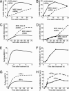
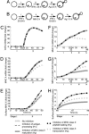
Previous experimental studies suggest that Mycobacterium tuberculosis inhibits a number of macrophage intracellular processes associated with antigen presentation, including antigen processing, MHC class II expression, trafficking of MHC class II molecules, and peptide-MHC class II binding. In this study, we investigate why multiple mechanisms have been observed. Specifically, we consider what purpose multiple mechanisms may serve, whether experimental protocols favor the detection of some mechanisms over others, and whether alternative mechanisms exist. By using a mathematical model of antigen presentation in macrophages that tracks levels of various molecules, including peptide-MHC class II complexes on the cell surface, we show that mechanisms targeting MHC class II expression are effective at inhibiting antigen presentation, but only after a delay of at least 10 h. By comparison, the effectiveness of mechanisms targeting other cellular processes is immediate, but may be attenuated under certain conditions. Therefore, targeting multiple cellular processes may represent an optimal strategy for M. tuberculosis (and other pathogens with relatively long doubling times) to maintain continuous inhibition of antigen presentation. In addition, based on a sensitivity analysis of the model, we identify other cellular processes that may be targeted by such pathogens to accomplish the same effect, representing potentially novel mechanisms.
Figures


Similar articles
-
Recombinant Lipoprotein Rv1016c Derived from Mycobacterium tuberculosis Is a TLR-2 Ligand that Induces Macrophages Apoptosis and Inhibits MHC II Antigen Processing.Front Cell Infect Microbiol. 2016 Nov 18;6:147. doi: 10.3389/fcimb.2016.00147. eCollection 2016. Front Cell Infect Microbiol. 2016. PMID: 27917375 Free PMC article.
-
Effect of multiple genetic polymorphisms on antigen presentation and susceptibility to Mycobacterium tuberculosis infection.Infect Immun. 2008 Jul;76(7):3221-32. doi: 10.1128/IAI.01677-07. Epub 2008 Apr 28. Infect Immun. 2008. PMID: 18443099 Free PMC article.
-
Prolonged toll-like receptor signaling by Mycobacterium tuberculosis and its 19-kilodalton lipoprotein inhibits gamma interferon-induced regulation of selected genes in macrophages.Infect Immun. 2004 Nov;72(11):6603-14. doi: 10.1128/IAI.72.11.6603-6614.2004. Infect Immun. 2004. PMID: 15501793 Free PMC article.
-
Immunoevasion and immunosuppression of the macrophage by Mycobacterium tuberculosis.Immunol Rev. 2015 Mar;264(1):220-32. doi: 10.1111/imr.12268. Immunol Rev. 2015. PMID: 25703562 Review.
-
Bacterial antigen delivery systems: phagocytic processing of bacterial antigens for MHC-I and MHC-II presentation to T cells.Behring Inst Mitt. 1997 Feb;(98):197-211. Behring Inst Mitt. 1997. PMID: 9382741 Review.
Cited by
-
Evaluation of fendiline treatment in VP40 system with nucleation-elongation process: a computational model of Ebola virus matrix protein assembly.Microbiol Spectr. 2024 Apr 2;12(4):e0309823. doi: 10.1128/spectrum.03098-23. Epub 2024 Feb 26. Microbiol Spectr. 2024. PMID: 38407984 Free PMC article.
-
Variability in tuberculosis granuloma T cell responses exists, but a balance of pro- and anti-inflammatory cytokines is associated with sterilization.PLoS Pathog. 2015 Jan 22;11(1):e1004603. doi: 10.1371/journal.ppat.1004603. eCollection 2015 Jan. PLoS Pathog. 2015. PMID: 25611466 Free PMC article.
-
A Validated Mathematical Model of the Cytokine Release Syndrome in Severe COVID-19.Front Mol Biosci. 2021 Jul 20;8:639423. doi: 10.3389/fmolb.2021.639423. eCollection 2021. Front Mol Biosci. 2021. PMID: 34355020 Free PMC article.
-
Host defense mechanisms against Mycobacterium tuberculosis.Cell Mol Life Sci. 2020 May;77(10):1859-1878. doi: 10.1007/s00018-019-03353-5. Epub 2019 Nov 13. Cell Mol Life Sci. 2020. PMID: 31720742 Free PMC article. Review.
-
Both ligand- and cell-specific parameters control ligand agonism in a kinetic model of g protein-coupled receptor signaling.PLoS Comput Biol. 2007 Jan 12;3(1):e6. doi: 10.1371/journal.pcbi.0030006. PLoS Comput Biol. 2007. PMID: 17222056 Free PMC article.
References
-
- Fenton, M. J. (1998) Curr. Opin. Hematol. 5, 72–78. - PubMed
-
- Chan, E. D., Chan, J. & Schluger, N. W. (2001) Am. J. Respir. Cell Mol. Biol. 25, 606–612. - PubMed
-
- Kaufmann, S. H. E. (1999) Curr. Biol. 9, R97–R99. - PubMed
-
- Pancholi, P., Mirza, A., Bhardwaj, N. & Steinman, R. M. (1993) Science 260, 984–986. - PubMed
Publication types
MeSH terms
Substances
Grants and funding
LinkOut - more resources
Full Text Sources
Other Literature Sources
Research Materials

