. 2005 Mar 22;102(12):4482-7.
doi: 10.1073/pnas.0500172102. Epub 2005 Mar 14.

Juxtaposition of C(2)M and the transverse filament protein C(3)G within the central region of Drosophila synaptonemal complex

Affiliations

Juxtaposition of C(2)M and the transverse filament protein C(3)G within the central region of Drosophila synaptonemal complex

Lorinda K Anderson et al. Proc Natl Acad Sci U S A. .

Abstract

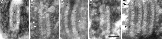
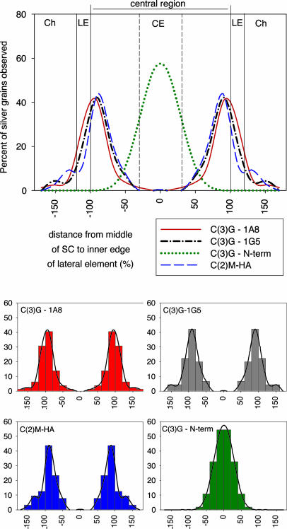
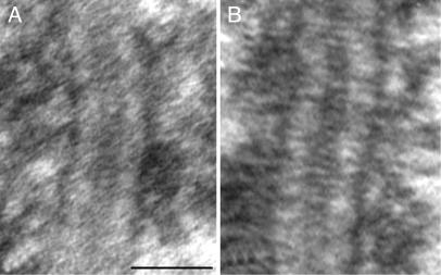
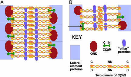
The synaptonemal complex (SC) is intimately involved in the process of meiotic recombination in most organisms, but its exact role remains enigmatic. One reason for this uncertainty is that the overall structure of the SC is evolutionarily conserved, but many SC proteins are not. Two putative SC proteins have been identified in Drosophila: C(3)G and C(2)M. Mutations in either gene cause defects in SC structure and meiotic recombination. Although neither gene is well conserved at the amino acid level, the predicted secondary structure of C(3)G is similar to that of transversefilament proteins, and C(2)M is a distantly related member of the alpha-kleisin family that includes Rec8, a meiosis-specific cohesin protein. Here, we use immunogold labeling of SCs in Drosophila ovaries to localize C(3)G and C(2)M at the EM level. We show that both C(3)G and C(2)M are components of the SC, that the orientation of C(3)G within the SC is similar to other transverse-filament proteins, and that the N terminus of C(2)M is located in the central region adjacent to the lateral elements (LEs). Based on our data and the known phenotypes of C(2)M and C(3)G mutants, we propose a model of SC structure in which C(2)M links C(3)G to the LEs.


Figures

Fig. 1.
Fig. 1.
EM micrographs of frontal sections of Drosophila SCs (AD) and a polycomplex (E) labeled with antibodies to the N-terminal domain of C(3)G (affinity-purified rabbit polyclonal) (A), the C-terminal end of the coiled-coil of C(3)G (mAb 1G5) (B), the C-terminal globular domain of C(3)G (mAb 1A8) (C), or the HA tag on the N terminus of C(2)M (mAb 3F10) (D and E). Some of the dark gold/silver particles are indicated by arrowheads. Although the light formaldehyde fixation necessary for immunolabeling is not as good for cellular preservation as are traditional EM fixation techniques, the basic structure of the SC is retained in these preparations (compare with Fig. 3A). (Scale bar, 100 nm.)
Fig. 2.
Fig. 2.
Localization of C(3)G and C(2)M protein domains within the SC. (Upper) Smoothed histograms of gold/silver particle distribution after labeling with antibodies to C(3)G and HA-tagged C(2)M. The distance from the middle of the CE to the near edge of the LE was defined as 100%, and the number of gold/silver particles was counted by using 20% intervals. The parts of a SC are indicated by vertical lines (Ch, chromatin). The same gold/silver particle distribution is mirrored from the middle of the CE to show the distribution over a complete SC. (Lower) Four graphs showing the underlying raw data used to generate the smoothed histograms shown above. The titles for the x and y axes on the small graphs are the same as shown for the large graph.
Fig. 3.
Fig. 3.
EM micrographs of SCs from conventionally prepared (glutaraldehyde fixation and osmium postfixation) ovaries from flies, wild type for SCs (A) and overexpressing HA-tagged C(2)M (B). The structure of the SC is similar in both types of flies. (Scale bar, 100 nm.)
Fig. 4.
Fig. 4.
Model of SC structure in Drosophila females based on immunolocalization of C(3)G and HA-tagged C(2)M in frontal (A) and transverse (B) sections. The LEs are probably composed of a number of different proteins (including SMC3 and ORD). The ORD protein is present on the inner side of the LEs and provides sister-chromatid cohesion. The C(2)M protein may be located intermittently along the SC. The N terminus of C(2)M is located near the inner side of the LE close to the C termini of C(3)G proteins. Here, we have shown the C terminus of C(2)M within the LEs, but it is also possible that both N and C termini are positioned along the inner side of the LEs (arrow in B). The N termini of the C(3)G dimers are located in the middle of the CE and are connected by interacting coiled-coil segments to the C termini that are located near or in the LEs. Our data are not sufficient to distinguish whether the N termini of two dimers are overlapping (as shown) or positioned end-to-end. One or more additional proteins may be involved in forming the longitudinal components of the CE.

Similar articles

Cited by

References

    1. Zickler, D. & Kleckner, N. (1999) Annu. Rev. Genet. 33, 603–754. - PubMed
    1. Schmekel, K., Skoglund, U. & Daneholt, B. (1993) Chromosoma 102, 682–692. - PubMed
    1. McKim, K. S., Green-Marroquin, B. L., Sekelsky, J. J., Chin, G., Steinberg, C., Khodosh, R. & Hawley, R. S. (1998) Science 279, 876–878. - PubMed
    1. Dernburg, A. F., McDonald, K., Moulder, G., Barstead, R., Dresser, M. & Villeneuve, A. M. (1998) Cell 94, 387–398. - PubMed
    1. Page, S. L. & Hawley, R. S. (2004) Annu. Rev. Cell Dev. Biol. 20, 525–558. - PubMed

Publication types

MeSH terms

LinkOut - more resources