. 2005 Mar 22;102(12):4330-5.
doi: 10.1073/pnas.0407252102. Epub 2005 Mar 14.

The polyglutamine neurodegenerative protein ataxin 3 regulates aggresome formation

Affiliations

The polyglutamine neurodegenerative protein ataxin 3 regulates aggresome formation

Barrington G Burnett et al. Proc Natl Acad Sci U S A. .

Abstract

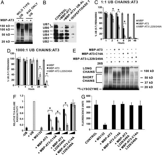
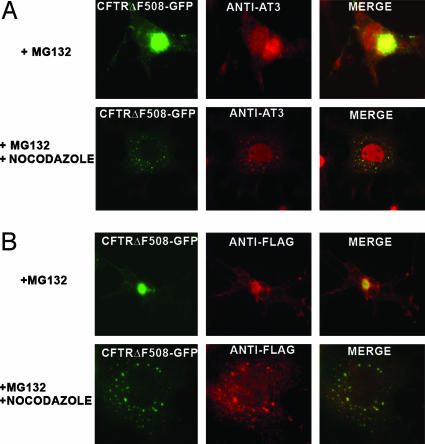
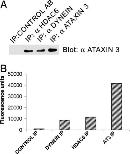
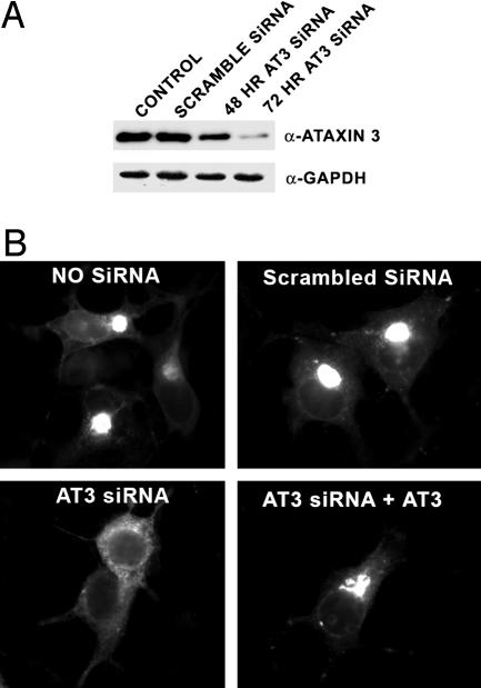
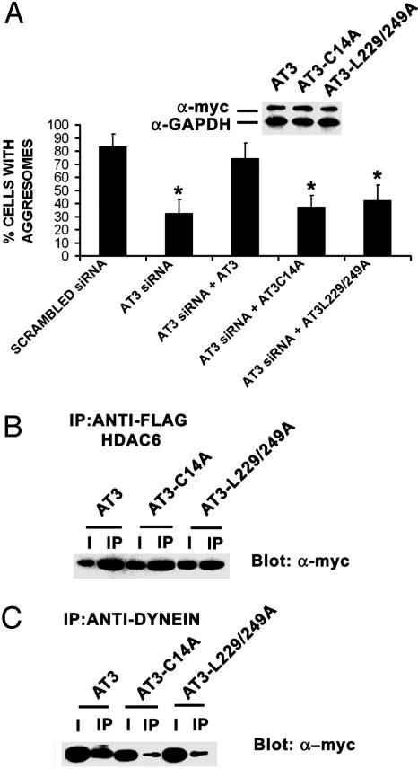
The polyglutamine-containing neurodegenerative protein ataxin 3 (AT3) has deubiquitylating activity and binds ubiquitin chains with a preference for chains of four or more ubiquitins. Here we characterize the deubiquitylating activity of AT3 in vitro and show it trims/edits K48-linked ubiquitin chains. AT3 also edits polyubiquitylated (125)I-lysozyme and decreases its degradation by proteasomes. Cellular studies show that endogenous AT3 colocalizes with aggresomes and preaggresome particles of the misfolded cystic fibrosis transmembrane regulator (CFTR) mutant CFTRDeltaF508 and associates with histone deacetylase 6 and dynein, proteins required for aggresome formation and transport of misfolded protein. Small interfering RNA knockdown of AT3 greatly reduces aggresomes formed by CFTRDeltaF508, demonstrating a critical role of AT3 in this process. Wild-type AT3 restores aggresome formation; however, AT3 with mutations in the active site or ubiquitin interacting motifs cannot restore aggresome formation in AT3 knockdown cells. These same mutations decrease the association of AT3 and dynein. These data indicate that the deubiquitylating activity of AT3 and its ubiquitin interacting motifs as well play essential roles in CFTRDeltaF508 aggresome formation.


Figures

Fig. 1.
Fig. 1.
AT3 deubiquitylating activity. (A) AT3 trims anchored K48-linked ubiquitin (UB) chains. Wild-type ubiquitin or mutated ubiquitin lacking all but one lysine (K48 ONLY UB) was used to ubiquitylate 125I-lysozyme, which was incubated with MBP-AT3. Similar to wild-type ubiquitin, K48 ubiquitin forms high-molecular-weight polyubiquitin chains that are cleaved to shorter chains by AT3. kD, kilodalton. (B) AT3 trims unanchored K48-linked ubiquitin chains. Polyubiquitin immunoblot (FK1 antibody) showing that MBP-AT3 preferentially cleaves free polyubiquitin chains containing five to seven ubiquitins (lane 2; 1:1 ratio AT3:chains) compared with isopeptidase T (lane 4), which completely disassembles chains. Ubiquitin aldehyde inhibits AT3 DUB activity (lane 3). (C and D) UIMs of AT3 modulate ubiquitin chain cleavage. Quantitation of cleavage of free polyubiquitin chains containing five to seven ubiquitins. At equimolar concentrations of polyubiquitin chains and AT3 (0.25 μM), MBP-AT3-L229-249A (the UIM mutant) significantly increases cleavage of chains of five to seven ubiquitins than is MBP-AT3. At a ratio of 1,000:1 (ubiquitin chains to 0.25 nM AT3), MBP-AT3 containing functional UIMs cleaves chains slightly better than does MBP-AT3-L229-249A. Data represent mean ± SEM of three experiments. *, P < 0.05. (E and F) AT3 inhibits degradation of ubiquitylated 125I-lysozyme by proteasomes. (E) Ubiquitylated 125I-lysozyme is degraded by 26S proteasomes, corresponding to a loss of high-molecular-weight polyubiquitylated 125I-lysozyme in the presence of 26S proteasome (compare lanes 1 and 2). MBP/AT3 trims long polyubiquitin chains and blocks degradation of polyubiquitylated 125I-lysozyme by 26S proteasomes (lanes 3 and 4), whereas MBP-AT3-L229-249A removes both short and long ubiquitin chains and inhibits degradation (lanes 7 and 8). MBP-AT3-C14A does not trim ubiquitin chains but blocks degradation of polyubiquitylated 125I-lysozyme (lanes 5 and 6). (F) Quantitation of 125I-lysozyme degradation. 26S proteasomes degrade (TCA-soluble radioactivity) polyubiquitylated 125I-lysozyme (+UB) but not nonubiquitylated 125I-lysozyme (–UB). The proteasome inhibitor MG132 (10 μM) inhibits degradation of polyubiquitylated 125I-lysozyme. Different concentrations (25, 150, or 250 nM, represented by filled, open, and stippled bars, respectively) of MBP-AT3, MBP-AT3-L229-249A, or MBP-AT3-C14A, but not AT3-C14A-L229/249A, decrease the degradation of polyubiquitylated 125I-lysozyme incubated with 26S proteasomes. Data represent mean ± SEM from four experiments. *, P < 0.05 compared with polyubiquitylated 125I-lysozyme (+UB). (G) AT3 does not affect proteolytic activity of the 26S proteasome. MBP-AT3 fusion proteins (250 nM) do not affect chymotryptic-like (or tryptic-like; data not shown) activity of 26S proteasomes against a fluorogenic peptide substrate.
Fig. 2.
Fig. 2.
Endogenous AT3 is present in aggresomes formed by CFTRΔF508. (A) Endogenous AT3 colocalizes with aggresomes formed by CFTRΔF508-GFP in COS 7 cells after proteasome inhibition with MG132 as well as preaggresome particles after nocodazole disruption of microtubules. (B) FLAG-tagged HDAC6 colocalizes with aggresomes and preaggresome particles of CFTRΔF508-GFP.
Fig. 3.
Fig. 3.
Endogenous AT3 associates with dynein and HDAC6. (A) Endogenous AT3 coimmunoprecipitates with endogenous dynein and transfected FLAG-tagged HDAC6. (B) Immunoprecipitated (IP) dynein, HDAC6, and AT3 all contain ubiquitin protease activity against the fluorogenic substrate ubiquitin-AMC. Control antibody does not immunoprecipitate AT3 or ubiquitin protease activity.
Fig. 4.
Fig. 4.
siRNA knockdown of AT3 decreases aggresomes formed by CFTRΔF508. (A) An siRNA vector targeting AT3 was transfected into COS 7 cells and cells either collected after 48 h or transfected again and collected 24 h later and blotted for AT3 or GAPDH as a control. AT3 siRNA greatly reduces AT3 after 72 h and has no apparent effect on cell morphology or viability. (B) CFTRΔF508-GFP aggresomes are dramatically decreased in AT3 knockdown cells compared with untransfected cells or cells transfected with scrambled AT3 siRNA. Cotransfecting CFTRΔF508-GFP with an siRNA-resistant AT3 restores aggresome formation in AT3 knockdown cells.
Fig. 5.
Fig. 5.
The deubiquitylating activity and UIMs of AT3 regulate formation of aggresomes and association with dynein. (A) AT3 knockdown cells were cotransfected with CFTRΔF508-GFP and one of the following: pcDNA3 vector, myc-AT3, myc-AT3-C14A, or myc-AT3-L229/249A. myc-AT3 restores CFTRΔF508 aggresome formation, whereas neither myc-AT3-C14A nor myc-AT3-L229/249A restores formation of aggresomes. (Inset) Transfected myc-AT3 constructs are not targets of siRNA and show similar expression. Data represent mean ± SEM of three independent experiments with at least 600 cells counted for each condition. *, P < 0.05 compared with scrambled siRNA or AT3 siRNA plus AT3. (B) FLAG-tagged HDAC6 coimmunoprecipitates myc-AT3, myc-AT3-C14A, and myc-AT3-L229/249A from COS 7 cells. (C) Dynein intermediate chain coimmunoprecipitates myc-AT3 but considerably less myc-AT3-C14A or myc-AT3-L229/249A. I, 10% input; IP, immunoprecipitate.

Similar articles

Cited by

References

    1. Takiyama, Y., Nishizawa, M., Tanaka, H., Kawashima, S., Sakamoto, H., Karube, Y., Shimazaki, H., Soutome, M., Endo, K., Ohta, S., et al. (1993) Nat. Genet. 4, 300–304. - PubMed
    1. Paulson, H. L., Perez, M. K., Trottier, Y., Trojanowski, J. Q., Subramony, S. H., Das, S. S., Vig, P., Mandel, J. L., Fischbeck, K. H. & Pittman, R. N. (1997) Neuron 19, 333–344. - PubMed
    1. DiFiglia, M., Sapp, E., Chase, K. O., Davies, S. W., Bates, G. P., Vonsattel, J. P. & Aronin, N. (1997) Science 277, 1990–1993. - PubMed
    1. Skinner, P. J., Koshy, B. T., Cummings, C. J., Klement, I. A., Helin, K., Servadio, A., Zoghbi, H. Y. & Orr, H. T. (1997) Nature 389, 971–974. - PubMed
    1. Li, M., Miwa, S., Kobayashi, Y., Merry, D. E., Yamamoto, M., Tanaka, F., Doyu, M., Hashizume, Y., Fischbeck, K. H., et al. (1998) Ann. Neurol. 44, 249–254. - PubMed

Publication types

MeSH terms

LinkOut - more resources