. 2005 Apr;25(7):2593-606.
doi: 10.1128/MCB.25.7.2593-2606.2005.

Role of phosphoinositide 3-kinase regulatory isoforms in development and actin rearrangement

Affiliations

Role of phosphoinositide 3-kinase regulatory isoforms in development and actin rearrangement

Saskia M Brachmann et al. Mol Cell Biol. 2005 Apr.

Abstract

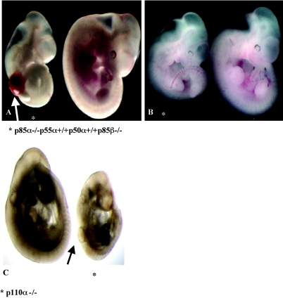
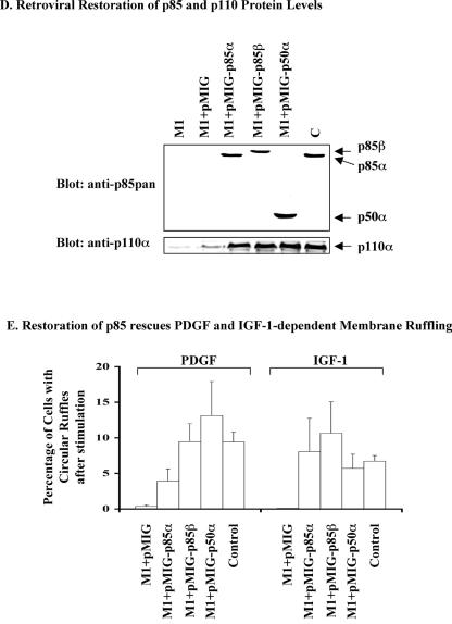
Class Ia phosphoinositide 3-kinases (PI3Ks) are heterodimers of p110 catalytic and p85 regulatory subunits that mediate a variety of cellular responses to growth and differentiation factors. Although embryonic development is not impaired in mice lacking all isoforms of the p85alpha gene (p85alpha-/- p55alpha-/- p50alpha-/-) or in mice lacking the p85beta gene (p85beta-/-) (D. A. Fruman, F. Mauvais-Jarvis, D. A. Pollard, C. M. Yballe, D. Brazil, R. T. Bronson, C. R. Kahn, and L. C. Cantley, Nat Genet. 26:379-382, 2000; K. Ueki, C. M. Yballe, S. M. Brachmann, D. Vicent, J. M. Watt, C. R. Kahn, and L. C. Cantley, Proc. Natl. Acad. Sci. USA 99:419-424, 2002), we show here that loss of both genes results in lethality at embryonic day 12.5 (E12.5). The phenotypes of these embryos, including subepidermal blebs flanking the neural tube at E8 and bleeding into the blebs during the turning process, are similar to defects observed in platelet-derived growth factor receptor alpha null (PDGFRalpha-/-) mice (P. Soriano, Development 124:2691-2700, 1997), suggesting that PI3K is an essential mediator of PDGFRalpha signaling at this developmental stage. p85alpha-/- p55alpha+/+ p50alpha+/+ p85beta-/- mice had similar but less severe defects, indicating that p85alpha and p85beta have a critical and redundant function in development. Mouse embryo fibroblasts deficient in all p85alpha and p85beta gene products (p85alpha-/- p55alpha-/- p50alpha-/- p85beta-/-) are defective in PDGF-induced membrane ruffling. Overexpression of the Rac-specific GDP-GTP exchange factor Vav2 or reintroduction of p85alpha or p85beta rescues the membrane ruffling defect. Surprisingly, reintroduction of p50alpha also restored PDGF-dependent membrane ruffling. These results indicate that class Ia PI3K is critical for PDGF-dependent actin rearrangement but that the SH3 domain and the Rho/Rac/Cdc42-interacting domain of p85, which lacks p50alpha, are not required for this response.


Figures

FIG. 1.
FIG. 1.
Deficiency in class Ia PI3K causes subepidermal blebbing. (A) Subepidermal blebbing in unturned E8 p85α−/− p55α−/− p50α−/− p85β−/− embryo. Shown are one unturned control littermate embryo (left) and one unturned p85α−/− p55α−/− p50α−/− p85β−/− embryo (right) with a subepidermal bleb on the trunk. (B and C) Blood-filled bleb in turned E9.5 p85α−/− p55α−/− p50α−/− p85β−/− embryos. Shown are one control littermate embryo (left) and two p85α−/− p85β−/− embryos (middle and right). Both embryos lacking p85α gene products and p85β display blood-filled blebs flanking their neural tube. (D) Facial abnormalities in E11.5 p85α−/− p55α−/− p50α−/− p85β−/− embryo. Shown are one control littermate embryo (left) and a p85α−/− p55α−/− p50α−/− p85β−/− embryo (right) which has a non-blood-filled subepidermal structure on the head. (E) Blood-filled, subepidermal bleb in turned E9 p85α−/− p55α−/− p50α−/− p85β−/− embryo. Shown are one control littermate embryo (left) and one p85α−/− p55α−/− p50α−/− p85β−/− embryo (right). Embryos were dissected at E9 and embedded in paraffin, and sagittal sections were stained with H&E to study their cell sructures.
FIG. 2.
FIG. 2.
Partial rescue in p85α−/− p55α+/+ p50α+/+ p85β−/− embryos. (A) Blood-filled bleb in turned E10.5 p85α−/− p55α+/+ p50α+/+ p85β−/− embryo. Shown are one control littermate embryo (right) and one p85α−/− p55α+/+ p50α+/+ p85β−/− embryo (left). Embryos lacking both 85-kDa isoforms (p85α and p85β) display a blood-filled bleb flanking their neural tube. The mutant embryo is smaller than the control littermate. (B) Minor defects in turned E10.5 p85α−/− p55α+/+ p50α+/+ p85β−/− embryo. Shown are one control littermate embryo (right) and one p85α−/− p55α+/+ p50α+/+ p85β−/− embryo (left). The embryo lacking both 85-kDa isoforms (p85α and p85β) is smaller than the control littermate but exhibits no blebs. (C) Subepidermal bleb in turned E10.5 p110α−/− embryo. Shown are one control littermate embryo (left) and one p110α−/− embryo (right). The embryo lacking p110α displays a subepidermal bleb flanking the neural tube. The mutant embryo is smaller than the control littermate.
FIG. 3.
FIG. 3.
Loss of all p85α and p85β gene products is associated with increased expression of p55γ but reduced overall PI3K activity. (A) p85 protein levels. HA-p55γ was transfected into a mutant cell pool (M1). The next day, growing MEFs of the indicated genotypes were lysed and equal amounts of protein were subjected to SDS-PAGE and immunoblotted with anti-p85pan (upper panel) or anti-p55γ antisera (lower panel). Panel A shows one out of three independent experiments with similar results. (B) p85-associated PI3K activity. Exponentially growing MEFs of the indicated genotypes were lysed, and equal amounts of protein were immunoprecipitated with anti-p85pan antibody. The immunoprecipitates were subjected to in vitro PI 3 -kinase assay with phosphoinositol (PI) as a substrate. The left panel in panel B shows a representative radiograph with one mutant cell pool (M2) and one control cell pool (C). A duplicate set was pretreated with 100 nM wortmannin (WM) for 20 min to inhibit the reaction. The right panel in panel B shows a quantification of three independent experiments described in the left panel, using three different mutant cell pools (M) and one control cell pool (C). Results are means ± standard error of the relative PI3K activity. (C) p110α protein levels. Exponentially growing MEFs of the indicated genotypes were lysed, and equal amounts of protein were immunoprecipitated with an antibody specific for p110α. The immunoprecipitates were subjected to SDS-PAGE and probed with anti-p110α antibody. Equal loading was verified by detecting similar amounts of Erk on the same membrane(data not shown). Panel 3C shows a representative immunoblot with one control pool (C) and two mutant pools (M1 and M3). (D) p110α-associated PI3K activity. Exponentially growing MEFs of the indicated genotypes were lysed, and equal amounts of protein were immunoprecipitated with anti-p110α antibody. The immunoprecipitates were subjected to in vitro PI3K assay with PI as a substrate. The left panel in panel D shows a representative radiograph on one control pool (C) and one mutant pool (M2). The right panel in panel D shows a quantification of three independent experiments described in the left panel, using three mutant pools (M) and one control pool (C). Results are means ± standard error of the relative PI3K activity.
FIG. 4.
FIG. 4.
Loss of all p85α and p85β gene products results in greatly reduced PDGF-stimulated PI3K activity in vitro and in vivo. (A) In vitro generation of PI3K products. Growing MEFs of the indicated genotypes were starved and then stimulated for 5 min with 10 ng of PDGF per ml (with or without pretreatment with 100 nM WM for 20 min). Equal amounts of protein were immunoprecipitated with antiphosphotyrosine (anti-PY) (4G10), and then an in vitro PI3K assay was performed. The left panel shows a representative experiment with one control clone (C) and one mutant clone (M1). The right panel shows the fold increase in PDGF-stimulated PI3K activity of three mutant pools in comparison to one control pool (result of three independent experiments). Results are means ± standard error of the relative PI3K activity. (B) In vivo generation of PI3K products. Growing MEFs of the indicated genotypes were starved, metabolically labeled with 32PO4, and then stimulated for 5 min with 10 ng of PDGF per ml (with or without pretreatment with 100 nM wortmannin for 20 min). Lipids were extracted and analyzed by HPLC. The panel shows one representative experiment out of two independent experiments with similar results.
FIG. 5.
FIG. 5.
Defective signaling in p85α−/− p55α−/− p50α−/− p85β−/− MEFs. (A and B) p85 is required for activation of Akt. MEFs with the indicated genotypes were starved overnight and then stimulated with 1 to 10 ng of PDGF per ml for 7 to 60 min (A) or saturating amounts of PDGF (10 ng/ml), IGF-1 (20 nM), or EGF (100 ng/ml) for 5 min (B). Lysates were resolved by SDS-PAGE and immunoblotted with anti-Akt and anti-phospho-Akt antibodies. Blots are representative of at least three independent experiments for each condition. (C) p85 is required for PDGF-induced activation of Erk. MEFs with indicated genotypes were starved overnight and then stimulated with 1, 3, or 10 ng of PDGF per ml for 5 min. Lysates were resolved by SDS-PAGE and immunoblotted with anti-Erk and anti-phospho-Erk antibodies. Note that the antibody that recognizes total Erk preferentially blots the nonphosphorylated Erk, so increased phosphorylation correlates with decreased blotting of total Erk. A second loading control for this experiment was added, a blot for total Akt. Blots are representative of at least three independent experiments. (D) Normal PDGF-dependent tyrosine phosphorylation of the PDGF receptor in mutant cell pools. MEFs with indicated genotypes were starved overnight and then stimulated with 10 or 20 ng of PDGF per ml for the indicated times. Lysates were resolved by SDS-PAGE and immunoblotted with anti-PDGF receptor and anti-Akt antibodies. The activation of these proteins wase assessed by antiphosphotyrosine (anti-PY) or phospho-specific antibodies. The blots are representative of three independent experiments.
FIG. 6.
FIG. 6.
P85 is required for PDGF-induced actin rearrangements. (A) P85 is required for PDGF-induced lamellipodium formation. MEFs with indicated genotypes were starved and then stimulated with 50 ng of PDGF per ml for 10 min (with or without pretreatment with 100 nM wortmannin [WM] for 20 min). The cells were fixed and actin structures were stained with rhodamine-phalloidin. The panels are representative of at least three independent experiments. (B) p85 is required for PDGF-induced activation of Rac. MEFs with the indicated genotypes were transfected with HA-Rac. The cells were starved overnight and then stimulated for 3 min with 30 ng of PDGF per ml. Equal amounts of lysates were subjected to in vitro binding assays with the immobilized CRIB domain of PAK65. Bound proteins were resolved by SDS-PAGE and immunoblotted with anti-Rac antibodies (Rac GTP). The levels of total Rac (Rac) and p85 are shown below. Equal stimulation was assessed by Erk activation. Blots are representative of at least three independent experiments. While 65% of the control MEFs exhibited PDGF-induced ruffling, 0% of the mutant MEFs ruffled upon PDGF treatment. (C) Wild-type Vav2 overexpression leads to membrane ruffling in the absensce of PI3K signaling. Control and mutant MEFs were transfected with T7-tagged wild-type Vav2. The transfected cells were grown for 24 h. Then the cells were fixed (with or without pretreatment with 100 nM wortmannin for 30 min), and the actin structures were stained with rhodamine-phalloidin. Vav2-expressing cells were identified with anti-T7 antibody staining. About 90% of either control or mutant MEFs (with or without wortmannin) that were overexpressing Vav2 exhibited lamellipodium formation. (D) Retroviral restoration of p85 isoforms. Mutant (M1) cell pools were infected with retroviral pMIG constructs to restore expression of p85α, p85β, or p50α similarly to the control (C) cell pool. The infected cells were lysed, and the protein levels were examined by Western blot analysis of the lysates, or, alternatively, p85/p110 complexes were immunoprecipitated with the anti-p85pan antibody and then examined by Western blot analysis. Blots are representative of at least three independent experiments. (E) The Rho-GAP domain of p85 is not necessary for PDGF-induced membrane ruffling. Various p85 isoforms mediate PDGF- or IGF-1-induced membrane ruffling. Mutant (p85α−/− p55α−/− p50α−/− p85β−/−) cell pools were infected with retroviral pMIG constructs to restore expression of p85α, p85β, or p50α similarly to the control (p85α+/− p55α+/− p50α+/− p85β−/−) cell pool. Mutant and control cells were plated on coverslips, starved overnight, and then stimulated with either 50 ng of PDGF-BB per ml or 20 nM IGF-1. Pictures were taken every 15 s for 2 h. The number of cells exhibiting circular ruffles were counted and expressed as a percentage of total cells. The data represent the means of at least three independent experiments ± standard error.
FIG. 6.
FIG. 6.
P85 is required for PDGF-induced actin rearrangements. (A) P85 is required for PDGF-induced lamellipodium formation. MEFs with indicated genotypes were starved and then stimulated with 50 ng of PDGF per ml for 10 min (with or without pretreatment with 100 nM wortmannin [WM] for 20 min). The cells were fixed and actin structures were stained with rhodamine-phalloidin. The panels are representative of at least three independent experiments. (B) p85 is required for PDGF-induced activation of Rac. MEFs with the indicated genotypes were transfected with HA-Rac. The cells were starved overnight and then stimulated for 3 min with 30 ng of PDGF per ml. Equal amounts of lysates were subjected to in vitro binding assays with the immobilized CRIB domain of PAK65. Bound proteins were resolved by SDS-PAGE and immunoblotted with anti-Rac antibodies (Rac GTP). The levels of total Rac (Rac) and p85 are shown below. Equal stimulation was assessed by Erk activation. Blots are representative of at least three independent experiments. While 65% of the control MEFs exhibited PDGF-induced ruffling, 0% of the mutant MEFs ruffled upon PDGF treatment. (C) Wild-type Vav2 overexpression leads to membrane ruffling in the absensce of PI3K signaling. Control and mutant MEFs were transfected with T7-tagged wild-type Vav2. The transfected cells were grown for 24 h. Then the cells were fixed (with or without pretreatment with 100 nM wortmannin for 30 min), and the actin structures were stained with rhodamine-phalloidin. Vav2-expressing cells were identified with anti-T7 antibody staining. About 90% of either control or mutant MEFs (with or without wortmannin) that were overexpressing Vav2 exhibited lamellipodium formation. (D) Retroviral restoration of p85 isoforms. Mutant (M1) cell pools were infected with retroviral pMIG constructs to restore expression of p85α, p85β, or p50α similarly to the control (C) cell pool. The infected cells were lysed, and the protein levels were examined by Western blot analysis of the lysates, or, alternatively, p85/p110 complexes were immunoprecipitated with the anti-p85pan antibody and then examined by Western blot analysis. Blots are representative of at least three independent experiments. (E) The Rho-GAP domain of p85 is not necessary for PDGF-induced membrane ruffling. Various p85 isoforms mediate PDGF- or IGF-1-induced membrane ruffling. Mutant (p85α−/− p55α−/− p50α−/− p85β−/−) cell pools were infected with retroviral pMIG constructs to restore expression of p85α, p85β, or p50α similarly to the control (p85α+/− p55α+/− p50α+/− p85β−/−) cell pool. Mutant and control cells were plated on coverslips, starved overnight, and then stimulated with either 50 ng of PDGF-BB per ml or 20 nM IGF-1. Pictures were taken every 15 s for 2 h. The number of cells exhibiting circular ruffles were counted and expressed as a percentage of total cells. The data represent the means of at least three independent experiments ± standard error.

References

    1. Arcaro, A., S. Volinia, M. J. Zvelebil, R. Stein, S. J. Watton, M. J. Layton, I. Gout, K. Ahmadi, J. Downward, and M. D. Waterfield. 1998. Human phosphoinositide 3-kinase C2beta, the role of calcium and the C2 domain in enzyme activity. J. Biol. Chem. 273:33082-33090. - PubMed
    1. Bazenet, C. E., and A. Kazlauskas. 1994. The PDGF receptor alpha subunit activates p21ras and triggers DNA synthesis without interacting with rasGAP. Oncogene 9:517-525. - PubMed
    1. Bi, L., I. Okabe, D. J. Bernard, and R. L. Nussbaum. 2002. Early embryonic lethality in mice deficient in the p110beta catalytic subunit of PI 3-kinase. Mamm. Genome 13:169-172. - PubMed
    1. Bi, L., I. Okabe, D. J. Bernard, A. Wynshaw-Boris, and R. L. Nussbaum. 1999. Proliferative defect and embryonic lethality in mice homozygous for a deletion in the p110alpha subunit of phosphoinositide 3-kinase. J. Biol. Chem. 274:10963-10968. - PubMed
    1. Bokoch, G. M., C. J. Vlahos, Y. Wang, U. G. Knaus, and A. E. Traynor-Kaplan. 1996. Rac GTPase interacts specifically with phosphatidylinositol 3-kinase. Biochem. J. 315:775-779. - PMC - PubMed

Publication types

MeSH terms

Substances

LinkOut - more resources