. 2005 Apr;25(7):2607-21.
doi: 10.1128/MCB.25.7.2607-2621.2005.

Kinase activation through dimerization by human SH2-B

Affiliations

Kinase activation through dimerization by human SH2-B

Masahiro Nishi et al. Mol Cell Biol. 2005 Apr.

Abstract

The isoforms of SH2-B, APS, and Lnk form a family of signaling proteins that have been described as activators, mediators, or inhibitors of cytokine and growth factor signaling. We now show that the three alternatively spliced isoforms of human SH2-B readily homodimerize in yeast two-hybrid and cellular transfections assays, and this is mediated specifically by a unique domain in its amino terminus. Consistent with previous reports, we further show that the SH2 domains of SH2-B and APS bind JAK2 at Tyr813. These findings suggested a model in which two molecules of SH2-B or APS homodimerize with their SH2 domains bound to two JAK2 molecules, creating heterotetrameric JAK2-(SH2-B)2-JAK2 or JAK2-(APS)2-JAK2 complexes. We further show that APS and SH2-B isoforms heterodimerize. At lower levels of SH2-B or APS expression, dimerization approximates two JAK2 molecules to induce transactivation. At higher relative concentrations of SH2-B or APS, kinase activation is blocked. SH2-B or APS homodimerization and SH2-B/APS heterodimerization thus provide direct mechanisms for activating and inhibiting JAK2 and other kinases from the inside of the cell and for potentiating or attenuating cytokine and growth factor receptor signaling when ligands are present.


Figures

FIG. 1.
FIG. 1.
Human SH2-B isoforms. (A) Sequences of human SH2-B isoforms and APS were aligned by using the program CLUSTAL W. The three SH2-B isoforms have identical sequences through residue 632; the C-terminal tails differ by sequence and length. Dimerization, PH, and SH2 domains are shaded gray. Residues buried at the interface between two molecules in the DDs are denoted with triangles. (B) Northern analyses of SH2-B show a 3.3-kb transcript. The probe hybridizes to a common region so all three isoforms are recognized. Tissue codes: HE, heart; BR, brain; PL, placenta; LU, lung; LI, liver; MU, skeletal muscle; KI, kidney; PA, pancreas; SP, spleen; TH, thymus; PR, prostate; TE, testis; OV, ovary; IN, small intestine; CO, colon; PB, peripheral blood leukocyte.
FIG. 2.
FIG. 2.
Tyrosine phosphorylation of SH2-Bβ requires a functional SH2 domain. HA-tagged SH2-Bβ (wt) and SH2-Bβ R555A (RA) were stably expressed in 3T3-L1 fibroblasts by retrovirus infection. Cultures were stimulated with GH, EGF, PDGF, serum, or pervanadate (VO4). SH2-Bβ was immunoprecipitated and analyzed by Western blotting with antiphosphotyrosine and anti-HA antibodies. M, transfected with empty vector.
FIG. 3.
FIG. 3.
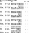
Yeast two-hybrid studies show that JAK2/SH2-B binding requires tyrosine phosphorylation of JAK2 and an intact SH2 domain in the SH2-B isoforms. Full-length wt JAK2 or kinase-deficient (KD; K882E) or Y813F variants were used as bait, and the indicated wt, mutated, or truncated versions of human SH2-Bα, SH2-Bβ, SH2-Bγ, or APS were used as prey. Y2H interactions between JAK2 and SH2-B were determined by growth of transformants in medium lacking leucine (for the reporter Leu2) and by filter lift color assays (for the reporter LacZ).
FIG. 4.
FIG. 4.
SH2-B dimerization. Y2H studies were further used to define a mechanism for SH2-B dimerization. Human SH2-Bγ was used as bait, and wt, mutated, or truncated human SH2-Bα, SH2-Bβ, or SH2-Bγ were used as prey. Homodimeric interactions were determined by growth of transformants in medium lacking leucine (Leu2) and by filter lift color assays (LacZ).
FIG. 5.
FIG. 5.
Dimerization of SH2-B and APS in cultured HEK293 cells. (A) HA- and Flag-tagged forms of each full-length protein were coexpressed by transient transfection in HEK293 cells. SH2-Bα-HA and SH2-Bα-Flag (lane 1), SH2-Bβ-HA and SH2-Bβ-Flag (lane 2), SH2-Bγ-HA and SH2-Bγ-Flag (lane 3), or APS-HA and APS-Flag (lane 4) proteins were coexpressed. Proteins were immunoprecipitated with anti-HA antibodies and immunoblotted with anti-Flag antibodies. (B) Flag-tagged, full-length proteins were coexpressed in HEK293 cells, along with an HA-tagged DD. Proteins were immunoprecipitated with anti-HA antibodies and immunoblotted with anti-Flag antibodies. (C) HA- and Flag-tagged APS DDs were coexpressed in 293 cells. Proteins were immunoprecipitated with anti-Flag antibodies and immunoblotted with anti-HA antibodies.
FIG. 6.
FIG. 6.
SH2-B and APS heterodimerization by yeast two-hybrid analysis. APS or SH2-Bγ were used as bait, and wt, mutated, and truncated APS, SH2-Bα, SH2-Bβ, and SH2-Bγ were used as prey. Interactions were determined independently using Leu2 and LacZ reporters. (B) HA- and Flag-tagged forms of SH2-B and APS were coexpressed in HEK293 cells, as described above, although in this case different proteins were coexpressed. For example, for the first lane SH2-Bβ-HA and SH2-Bα-Flag proteins were coexpressed, for the second lane SH2-Bγ-HA and SH2-Bα-Flag proteins were coexpressed, etc. Proteins were immunoprecipitated with anti-HA antibodies and immunoblotted with anti-Flag antibodies.
FIG. 6.
FIG. 6.
SH2-B and APS heterodimerization by yeast two-hybrid analysis. APS or SH2-Bγ were used as bait, and wt, mutated, and truncated APS, SH2-Bα, SH2-Bβ, and SH2-Bγ were used as prey. Interactions were determined independently using Leu2 and LacZ reporters. (B) HA- and Flag-tagged forms of SH2-B and APS were coexpressed in HEK293 cells, as described above, although in this case different proteins were coexpressed. For example, for the first lane SH2-Bβ-HA and SH2-Bα-Flag proteins were coexpressed, for the second lane SH2-Bγ-HA and SH2-Bα-Flag proteins were coexpressed, etc. Proteins were immunoprecipitated with anti-HA antibodies and immunoblotted with anti-Flag antibodies.
FIG. 7.
FIG. 7.
Molecular models of DDs. (A) The X-ray crystal structure of the APS DD (9) is detailed on the left. (B) A molecular model of the SH2-B domain was generated by using the program MODELLER and the coordinates from the APS domain structure. For both models, one molecule in the dimer is colored green and the other is red. Amino acids are numbered and colored accordingly.
FIG. 8.
FIG. 8.
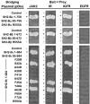
Bridging Y3H assays show that heterotetramers form between SH2-B isoforms and either JAK2 or insulin or IGF-1 receptors. The bridging Y3H system was developed as a modification of the pLexA yeast two-hybrid method. Either full-length JAK2, the IRK, or the IGF1RK was expressed as both bait (pLexA) and prey (pB42AD). wt and variants of SH2-Bα, SH2-Bβ, and SH2-Bγ were expressed from a third (pDis) plasmid. Growth on synthetic dextrose plates indicates an interaction (+) between bait and prey.
FIG. 9.
FIG. 9.
In vitro reconstitution showing JAK2 activation and inhibition by SH2-B. Recombinant JAK2 and recombinant SH2-B (indicated concentrations) were incubated with ATP (1.2 mM). Experiments were conducted with JAK2 at (A and B) 14 pM or (C and D) 118 pM concentrations. In panels A and C, JAK2 was visualized immunoblots with anti-pY and anti-JAK2 antibodies; in panels B and D, the results from three separate experiments were quantified by scanning densitometry and combined (mean ± the standard error of the mean; ✽, P < 0.05).
FIG. 10.
FIG. 10.
SH2-B and APS activate JAK2 in cultured cells. HA-tagged SH2-Bβ (0.0 or 1.0 μg of pCMV-Tag2-SH2-Bβ DNA) and Flag-tagged JAK2 (0.0 to 1.0 μg of pCMV-Tag2-JAK2 DNA) were coexpressed in HEK293 cells. Proteins were immunoprecipitated with anti-HA (A) or anti-Flag (B) antibodies and immunoblotted with anti-pY antibodies. (C) Experiments similar to those described in panel B were conducted with APS and the three isoforms of SH2-B. HA-tagged SH2-Bα, SH2-Bβ, SH2-Bγ, or APS (0.0 or 1.0 μg of pCMV-Tag2 DNA) and JAK2 (1.0 μg of pCMV-Tag2-JAK2 DNA) were coexpressed in 293 cells. Proteins were immunoprecipitated with antibodies to JAK2 and immunoblotted with pY antibodies.
FIG. 11.
FIG. 11.
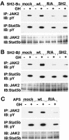
(A) Activation or inhibition of endogenous JAK2 and Stat5b by expression of wt or dominant inhibitory forms of SH2-B or APS, respectively. 3T3-L1 fibroblasts were infected with retrovirus vectors expressing wt SH2-B or APS proteins, SH2 domain-incompetent, full-length proteins (R/A; SH2-Bα R555A, SH2-Bβ R555A, and APS R455A), or the carboxyl-terminal segments of SH2-Bα(497-756) or SH2-Bβ(497-671) containing the SH2 domains and unique tails (SH2). Cells were stimulated (+) or not (−) with GH (200 ng/ml for 10 min) and lysed. Proteins were immunoprecipitated with the indicated antibodies, separated by SDS-PAGE, and identified by immunoblotting with antibodies to pTyr, JAK2, or STATb.
FIG. 12.
FIG. 12.
(A) The DD dominantly inhibits JAK2 phosphorylation. Proteins were coexpressed in 293 cells: JAK2 was expressed in variable amounts (0.0, 0.1, 0.3, and 0.5 μg of pCMV-Tag2-JAK2 DNA), HA-tagged SH2-Bβ was expressed in each experiment (0.7 μg of pCMV-Tag2-SH2-Bβ DNA), and the DDs from SH2-B (residues 24 to 85; 3 μg of pCMV-Tag2-SH2-B/DD DNA) or APS (residues 21 to 85; 3 μg of pCMV-Tag2-APS/DD DNA) were expressed as indicated (S or A, respectively). (B) Full-length SH2-Bβ (0.0 or 0.7 μg of pCMV-Tag2-SH2-Bβ DNA), JAK2 (1.0 μg of pCMV-Tag2-JAK2 DNA), and the APS DD (residues 21 to 85; 0.0 or 3.0 μg of pCMV-Tag2-APS/DD DNA) were coexpressed in 293 cells. Proteins were immunoprecipitated with anti-JAK2 antibodies and immunoblotted with antibodies to pY or JAK2. Cell lysates were blotted with antibodies to HA to detect SH2-Bβ or the APS DD.

Similar articles

Cited by

References

    1. Ahmed, Z., B. J. Smith, K. Kotani, P. Wilden, and T. S. Pillay. 1999. APS, an adapter protein with a PH and SH2 domain, is a substrate for the insulin receptor kinase. Biochem. J. 341:665-668. - PMC - PubMed
    1. Backer, J. M., S. E. Shoelson, M. A. Weiss, Q. X. Hua, B. Cheatham, E. Haring, D. C. Cahill, and M. F. White. 1992. The insulin receptor juxtamembrane region contains two independent tyrosine/β-turn internalization signals. J. Cell Biol. 118:831-839. - PMC - PubMed
    1. Bansal, A., and L. M. Gierasch. 1991. The NPXY internalization signal of the LDL receptor adopts a reverse-turn conformation. Cell 67:1195-1201. - PubMed
    1. Briddon, S. J., S. K. Melford, M. Turner, V. Tybulewicz, and S. P. Watson. 1999. Collagen mediates changes in intracellular calcium in primary mouse megakaryocytes through Syk-dependent and -independent pathways. Blood 93:3847-3855. - PubMed
    1. Carroll, D. J., D. T. Albay, M. Terasaki, L. A. Jaffe, and K. R. Foltz. 1999. Identification of PLCγ-dependent and -independent events during fertilization of sea urchin eggs. Dev. Biol. 206:232-247. - PubMed

Publication types

MeSH terms