. 2005 Apr;25(7):2722-32.
doi: 10.1128/MCB.25.7.2722-2732.2005.

Deficits in motor coordination with aberrant cerebellar development in mice lacking testicular orphan nuclear receptor 4

Affiliations

Deficits in motor coordination with aberrant cerebellar development in mice lacking testicular orphan nuclear receptor 4

Yei-Tsung Chen et al. Mol Cell Biol. 2005 Apr.

Abstract

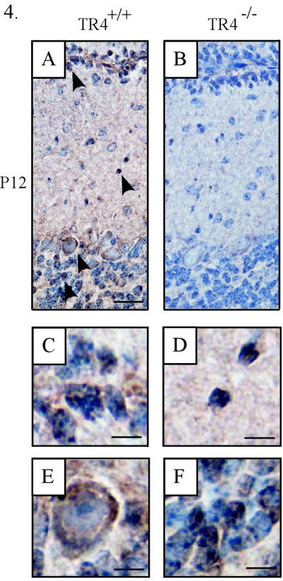
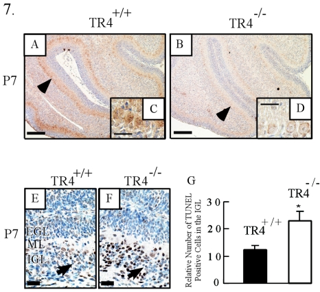
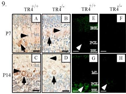
Since testicular orphan nuclear receptor 4 (TR4) was cloned, its physiological function has remained largely unknown. Throughout postnatal development, TR4-knockout (TR4-/-) mice exhibited behavioral deficits in motor coordination, suggesting impaired cerebellar function. Histological examination of the postnatal TR4-/- cerebellum revealed gross abnormalities in foliation; specifically, lobule VII in the anterior vermis was missing. Further analyses demonstrated that the laminations of the TR4-/- cerebellar cortex were changed, including reductions in the thickness of the molecular layer and the internal granule layer, as well as delayed disappearance of the external granule cell layer (EGL). These lamination irregularities may result from interference with granule cell proliferation within the EGL, delayed inward migration of postmitotic granule cells, and a higher incidence of apoptotis. In addition, abnormal development of Purkinje cells was observed in the postnatal TR4-/- cerebellum, as evidenced by aberrant dendritic arborization and reduced calbindin staining intensity. Expression of Pax-6, Sonic Hedgehog (Shh), astrotactin (Astn), reelin, and Cdk-5, genes correlated with the morphological development of the cerebellum, is reduced in the developing TR4-/- cerebellum. Together, our findings suggest that TR4 is required for normal cerebellar development.


Figures

FIG.1.
FIG.1.
Motor dysfunction and abnormal behavior in 21-day-old and adult TR4−/− mice. (A) Photographs of TR4+/+ and TR4−/− mice at P21, as they were suspended by the tail. TR4+/+ mice usually extended their hind and front limbs when in this position, whereas the limbs of TR4−/− mice were often crossed or clasped. (B) Results of the ledge (left) and wire hanging (right) tests. The latency to fall in both tests was significantly shorter for TR4−/− mice than for TR4+/+ controls. Solid bars, TR4+/+ mice (n = 7); open bars, TR4−/− mice (n = 7). Asterisks indicate significant differences between TR4+/+ and TR4−/− mice. Data shown are means ± standard errors of the means. *, P < 0.005 by Student's t test. (C) Images of inked footprints of TR4+/+ and TR4−/− mice. Normal adults place equal weight on all four limbs when walking, while adult TR4−/− mice appeared to drag their hind limbs. Arrow indicates the direction of movement. (D) Photographs of gross brain samples from P7 and P14 TR4+/+ and TR4−/− mice showing reduced cerebellar size in the TR4−/− mice. (E) The adult TR4−/− cerebellum in the midsagittal section is obviously smaller than that of the TR4+/+ control. Asterisk indicates correlated folium, which shows abnormal folium development in TR4−/− mice.
FIG. 2.
FIG. 2.
Cerebellar morphology in developing TR4+/+ and TR4−/− mice. Paraffin-embedded, Nissel-stained midsagittal sections reveal abnormal cerebellar structure at various postnatal stages in TR4−/− mice. Aberrant folium development in TR4−/− cerebellar sections is indicated by arrowheads. Insets in panels E and F show magnified views of the EGL, which is abnormally thick in the TR4−/− cerebellum. Mouse ages and genotypes are as indicated. Cerebellar lobules are indicated by Roman numerals. PCL, Purkinje cell layer. Bars in panels A and B, 250 μm; bars in panels C to H, 500 μm.
FIG. 3.
FIG. 3.
Differences in cortical layer thickness in TR4+/+ and TR4−/− cerebella. Nissel-stained sections from comparable lobules of TR4+/+ littermates and TR4−/− mice at P7, P14, and P21 reveal that both the lamination and the dispersion of the EGL are delayed in TR4−/− mice. Arrows designate migrating granule cells; arrowheads point to apoptotic bodies. PCL, Purkinje cell layer. Bars, 50 μm.
FIG. 4.
FIG. 4.
Expression of TR4 in the postnatal cerebellar cortex. Midsagittal cerebellar sections were immunostained with the anti-TR4 antibody at P12. Arrowheads point to the cells that show positive signals. (A and B) Cerebellar sections from TR4+/+ and TR4−/− mice, respectively. (C, D, E, and F) Higher-magnification views of the cells indicated by the arrowheads in panel A. Bars in panels A and B, 100 μm; bars in panels C to F, 10 μm.
FIG. 5.
FIG. 5.
Disrupted migration of granule cells in the TR4−/− cerebellum. Midsagittal sections of mouse cerebella after BrdU incorporation for different lengths of time revealed an abnormal granule cell migration pattern in the postnatal TR4−/− cerebellum. Arrowheads point to granule cells in the inner EGL (A and B) or to migrating granule cells in the ML (C and D). Arrows indicate granule cells in the IGL. BrdU incorporation time and genotypes are as indicated. PCL, Purkinje cell layer. Bars, 100 μm.
FIG. 6.
FIG. 6.
Short-term BrdU incorporation in the TR4+/+ and TR4−/− cerebella at different developmental stages. (A to H) Arrows point to proliferating granule cell precursors. Mouse ages and genotypes are as indicated. PCL, Purkinje cell layer. Bars, 50 μm. (I to L) The TR4−/− cerebellar primordium at a late embryonic stage exhibits germinal cell deficiency. (I and J) Nissel-stained parasagittal sections of E16.5 TR4+/+ (I) and TR4−/− (J) cerebella. The neuroepithelium (NE) and rhombic lip (RL) are designated in each micrograph. CP, choroid plexus. (K and L) BrdU staining in correlated germinal zones. Arrowheads point to proliferating granule cell precursors. Bars, 250 μm.
FIG. 7.
FIG. 7.
(A to D) Decreased Sonic Hedgehog RNA level in Purkinje cells in the TR4−/− cerebellum. (A and B) RT in situ PCR of Shh was performed on P7 midsagittal cerebellar sections from TR4+/+ and TR4−/− mice. Arrowheads indicate Purkinje cells with positive signals. Bars, 200 μm. (C and D) High-magnification views from panels A and B, respectively. Bars, 50 μm. (E to G) Increased apoptosis of granule cells in the IGL. (E and F) TUNEL staining of P7 cerebellar sections from TR4+/+ and TR4−/− mice. Arrows indicate cells with positive signals. Bars, 50 μm. (G) Relative numbers of TUNEL-positive cells in correlated areas of the IGL of TR4+/+ and TR4−/− cerebella at P7. Asterisks indicate significant differences between TR4+/+ and TR4−/− mice. Values are means ± standard errors of the means. *, P < 0.05.
FIG. 8.
FIG. 8.
Abnormal neonatal development of Purkinje cells in the TR4−/− cerebellum. Midsagittal cerebellar sections at different developmental stages were immunostained with an anticalbindin antibody. Mouse ages and genotypes are as indicated. Arrows point to Purkinje cells. Arrowheads indicate primary bundles of Purkinje cell dendritic trees. Bars, 200 μm (A and B), 50 μm (C, E, G, and I), and 30 μm (D, F, H, J, K, and L).
FIG. 9.
FIG. 9.
Decreased expression of Cdk-5 and reelin in the TR4−/− cerebellum. Midsagittal cerebellar sections at different developmental stages were immunostained with anti-Cdk-5 (A to D) or anti-Reelin (E to H) antibodies. Mouse ages and genotypes are as indicated. (A to D) Arrows indicate Purkinje cells; arrowheads point to migrating granule cells. (E, G, and H) Arrowheads indicate Purkinje cells. Bars, 50 μm.

References

    1. Adams, N. C., T. Tomoda, M. Cooper, G. Dietz, and M. E. Hatten. 2002. Mice that lack astrotactin have slowed neuronal migration. Development 129:965-972. - PubMed
    1. Aruga, J., O. Minowa, H. Yaginuma, J. Kuno, T. Nagai, T. Noda, and K. Mikoshiba. 1998. Mouse Zic1 is involved in cerebellar development. J. Neurosci. 18:284-293. - PMC - PubMed
    1. Beffert, U., E. J. Weeber, G. Morfini, J. Ko, S. T. Brady, L.-H. Tsai, J. D. Sweatt, and J. Herz. 2004. Reelin and cyclin-dependent kinase 5-dependent signals cooperate in regulating neuronal migration and synaptic transmission. J. Neurosci. 24:1897-1906. - PMC - PubMed
    1. Bock, H. H., Y. Jossin, P. May, O. Bergner, and J. Herz. 2004. Apolipoprotein E receptors are required for reelin-induced proteasomal degradation of the neuronal adaptor protein Disabled-1. J. Biol. Chem. 279:33471-33479. - PubMed
    1. Chang, C., S. L. Da Silva, R. Ideta, Y. Lee, S. Yeh, and J. P. Burbach. 1994. Human and rat TR4 orphan receptors specify a subclass of the steroid receptor superfamily. Proc. Natl. Acad. Sci. USA 91:6040-6044. - PMC - PubMed

Publication types

MeSH terms