. 2005 Apr;25(7):2744-56.
doi: 10.1128/MCB.25.7.2744-2756.2005.

ZPR1 is essential for survival and is required for localization of the survival motor neurons (SMN) protein to Cajal bodies

Affiliations

ZPR1 is essential for survival and is required for localization of the survival motor neurons (SMN) protein to Cajal bodies

Laxman Gangwani et al. Mol Cell Biol. 2005 Apr.

Abstract

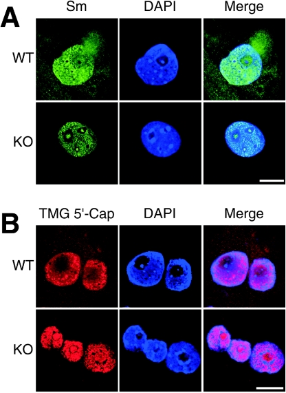
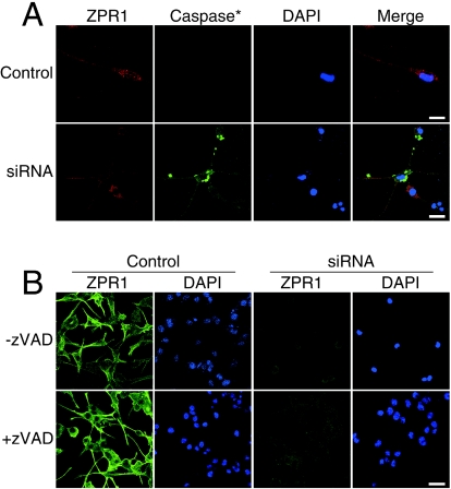
Mutation of the survival motor neurons 1 (SMN1) gene causes motor neuron apoptosis and represents the major cause of spinal muscular atrophy in humans. Biochemical studies have established that the SMN protein plays an important role in spliceosomal small nuclear ribonucleoprotein (snRNP) biogenesis and that the SMN complex can interact with the zinc finger protein ZPR1. Here we report that targeted ablation of the Zpr1 gene in mice disrupts the subcellular localization of both SMN and spliceosomal snRNPs. Specifically, SMN localization to Cajal bodies and gems was not observed in cells derived from Zpr1-/- embryos and the amount of cytoplasmic snRNP detected in Zpr1-/- embryos was reduced compared with that in wild-type embryos. We found that Zpr1-/- mice die during early embryonic development, with reduced proliferation and increased apoptosis. These effects of Zpr1 gene disruption were confirmed and extended in studies of cultured motor neuron-like cells using small interfering RNA-mediated Zpr1 gene suppression; ZPR1 deficiency caused growth cone retraction, axonal defects, and apoptosis. Together, these data indicate that ZPR1 contributes to the regulation of SMN complexes and that it is essential for cell survival.


Figures

FIG. 1.
FIG. 1.
Targeted disruption of mouse Zpr1. (A) Exon-intron structure (coding and noncoding exons are numbered and represented by solid and open boxes, respectively) and restriction map of the mouse Zpr1 gene, structure of the targeting vector, and structure of the mutated allele after homologous recombination. (B) The mutated Zpr1 allele was detected by Southern blot analysis and PCR. Genomic DNA from targeted embryonic stem cells was digested with EcoRI and detected with cDNA probe (exons 10 to 14; bp 954 to 1379). The 17.5-kb fragment corresponding to the wild-type (WT) allele and the 9.0-kb fragment corresponding to the mutated (knockout [KO]) allele are indicated. (C) Genomic DNA samples were also examined by PCR; the wild-type (WT) allele (250 bp) and mutated (KO) allele (444 bp) are shown. (D) Genomic DNA prepared from E3.5 embryos cultured in vitro for 96 h was genotyped by PCR.
FIG. 2.
FIG. 2.
Mutation in Zpr1 results in cell death and early embryonic death in mice. (A) Differential interference contrast images of wild-type (WT) and mutant (KO) blastocysts (E3.5) at 0 h and after 96 h of culture in vitro. The inner cell mass (ICM) is surrounded by trophoblast giant (TG) cells. Scale bar, 100 μm. (B) Scanning electron micrographs of WT and mutant embryos (KO) cultured in vitro for 96 h. The ICM is growing on the trophectoderm (TE) formed by trophoblast giant cells in WT embryos. The ICM and TE of mutant embryo (KO) display defective growth (top; magnification, ×200; scale bar, 100 μm). Microvilli (MV), projections at the apical surface of cells from wild-type embryos, are absent in mutant (KO) embryos (bottom; magnification, ×1,000; scale bar, 10 μm). (C) In situ detection of caspase activation, using FITC-VAD-fmk and confocal laser-scanning microscopy. Activated caspase (green) in WT and mutant (KO) embryos was observed. Nuclei were stained with DAPI (blue). The mutant embryo exhibited a large amount of caspase activation. Scale bar, 40 μm. (D) Transmission electron micrographs of trophoblast giant cells (top) and cells from the ICM (bottom). The mutant embryo (KO) shows apoptotic bodies (AB) phagocytosed by the trophoblast giant cells, as well as aggregation of heterochromatin and accumulation at nuclear membrane (bottom). Arrowheads show nucleolar accessory bodies (Cajal bodies). Scale bar, 1.0 μm.
FIG. 3.
FIG. 3.
ZPR1 deficiency results in defective embryo growth and causes mislocalization of SMN and coilin. (A) Blastocysts (E3.5) cultured in vitro were labeled with 50 μM BrdU for 1 h at 37°C. Wild-type (WT) and mutant (KO) embryos were stained with mouse monoclonal antibodies to ZPR1 (red), SMN (purple), and BrdU (green) and examined by indirect-immunofluorescence microscopy. Nuclei were stained with DAPI (blue). Scale bar, 100 μm. (B) Colocalization of ZPR1 and coilin in mouse embryos. Blastocysts were stained with antibodies to coilin (red) and ZPR1 (green). Colocalization of coilin (red) and ZPR1 (green) is represented by yellow. Nuclei were stained with DAPI (blue). Scale bar, 8.0 μm. (C) Colocalization of SMN and ZPR1 in mouse embryos. Blastocysts (E3.5) cultured for 96 h were stained with antibodies to SMN (red) and ZPR1 (green) and examined by confocal laser-scanning immunofluorescence microscopy. Colocalization of SMN (red) and ZPR1 (green) is represented by yellow. Scale bar, 8.0 μm.
FIG. 4.
FIG. 4.
ZPR1 deficiency causes defects in snRNP localization. (A) Loss of ZPR1 results in nuclear and nucleolar accumulation of Sm proteins. Wild-type (WT) and mutant (KO) blastocysts (E3.5) were cultured in vitro for 96 h and stained with antibodies to Sm proteins (green). All stained samples were examined by confocal laser-scanning immunofluorescence microscopy. Nuclei were stained with DAPI (blue). Scale bar, 20 μm. (B) Deficiency of ZPR1 causes nuclear and nucleolar accumulation of snRNPs. Embryos were stained with antibodies to TMG 5′-cap of U snRNA for detection of snRNPs (red) in wild-type and mutant embryos. Scale bar, 20 μm.
FIG. 5.
FIG. 5.
Loss of ZPR1 causes mislocalization of SMN and snRNPs in differentiated NSC-34 cells. (A) Colocalization of SMN with ZPR1 in differentiated NSC-34 cells that resemble motor neurons. The cells were stained with antibodies to SMN and ZPR1 and examined by confocal laser-scanning immunofluorescence microscopy. Colocalization of SMN (red) with ZPR1 (green) is represented by yellow in nuclear bodies (top, see inset; scale bar, 40 μm) and in the neuronal growth cone (bottom, see inset; scale bar, 8.0 μm). (B) A low level of ZPR1 results in the loss of nuclear bodies and mislocalization of nuclear SMN in motor neurons. Motor neurons were transfected with scrambled siRNA (Control) and ZPR1-specific siRNA (siRNA) and cultured for 48 h. Cellular distribution of SMN (red) and ZPR1 (green) were examined using antibodies to SMN and ZPR1. Scale bar, 16 μm. (C) Loss of ZPR1 causes nuclear accumulation of snRNPs. Cells were transfected with scrambled siRNA (Control) and ZPR1-specific siRNA (siRNA), were cultured for 72 h, and stained with antibodies to ZPR1 (green) and TMG 5′-cap (red). Scale bar, 8.0 μm. Nuclei were stained with DAPI (blue).
FIG. 6.
FIG. 6.
Deficiency of ZPR1 causes defects in axons of differentiated NSC-34 cells. (A) Loss of ZPR1 causes axonal defects in differentiated NSC-34 cells that resemble motor neurons. Cells transfected with scrambled siRNA (Control) and ZPR1-specific siRNA (siRNA) were cultured for 72 h and stained with antibodies to tubulin (red) and ZPR1 (green). The arrowhead indicates the kinks in the axon of motor neuron after knockdown of ZPR1 (siRNA). Scale bar, 20 μm. (B) ZPR1 deficiency results in the loss of growth cones of differentiated NSC-34 cells. ZPR1 was stained with antibodies to ZPR1 (green), and actin was stained with phalloidin conjugated with Alexa 546 (red). Nuclei were stained with DAPI (blue). All samples were examined by confocal laser-scanning fluorescence microscopy. Scale bar, 20 μm.
FIG. 7.
FIG. 7.
Loss of ZPR1 results in death of differentiated NSC-34 cells. (A) Low levels of ZPR1 causes caspase activation and death of differentiated NSC-34 cells that resemble motor neurons. Cells transfected with scrambled siRNA (Control) and ZPR1-specific siRNA (siRNA) were cultured for 72 h and labeled with FITC-VAD-fmk. Motor neurons stained with antibodies to ZPR1 (red) and activated caspase (green) were examined by confocal laser-scanning microscopy. Scale bar, 20 μm. (B) Inhibition of death of motor neurons that lack ZPR1 by zVAD. Cells were left untreated (−zVAD) or treated (+zVAD) with zVAD (10 μM) during transfection with scrambled siRNA (Control) and ZPR1-specific siRNA (siRNA). Samples were stained with antibodies to ZPR1 and examined by confocal laser-scanning microscopy. Nuclei were stained with DAPI (blue). Scale bar, 40 μm.

Similar articles

Cited by

References

    1. Andrade, L. E., E. M. Tan, and E. K. Chan. 1993. Immunocytochemical analysis of the coiled body in the cell cycle and during cell proliferation. Proc. Natl. Acad. Sci. USA 90:1947-1951. - PMC - PubMed
    1. Ansari, H., G. Greco, and J. Luban. 2002. Cyclophilin a peptidyl-prolyl isomerase activity promotes ZPR1 nuclear export. Mol. Cell. Biol. 22:6993-7003. - PMC - PubMed
    1. Boisvert, F. M., J. Cote, M. C. Boulanger, P. Cleroux, F. Bachand, C. Autexier, and S. Richard. 2002. Symmetrical dimethylarginine methylation is required for the localization of SMN in Cajal bodies and pre-mRNA splicing. J. Cell Biol. 159:957-969. - PMC - PubMed
    1. Brahms, H., L. Meheus, V. de Brabandere, U. Fischer, and R. Luhrmann. 2001. Symmetrical dimethylation of arginine residues in spliceosomal Sm protein B/B' and the Sm-like protein LSm4, and their interaction with the SMN protein. RNA 7:1531-1542. - PMC - PubMed
    1. Carmo-Fonseca, M., R. Pepperkok, M. T. Carvalho, and A. I. Lamond. 1992. Transcription-dependent colocalization of the U1, U2, U4/U6, and U5 snRNPs in coiled bodies. J. Cell Biol. 117:1-14. - PMC - PubMed

Publication types

MeSH terms

Substances