. 2005 Apr;25(7):2819-31.
doi: 10.1128/MCB.25.7.2819-2831.2005.

Characterization of E3Histone, a novel testis ubiquitin protein ligase which ubiquitinates histones

Affiliations

Characterization of E3Histone, a novel testis ubiquitin protein ligase which ubiquitinates histones

Zhiqian Liu et al. Mol Cell Biol. 2005 Apr.

Abstract

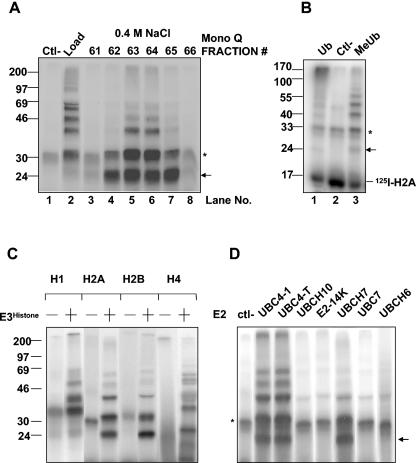
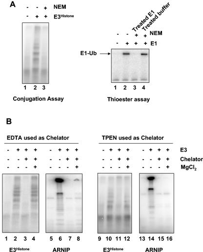
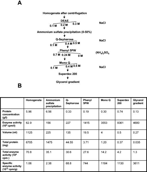
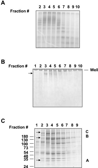
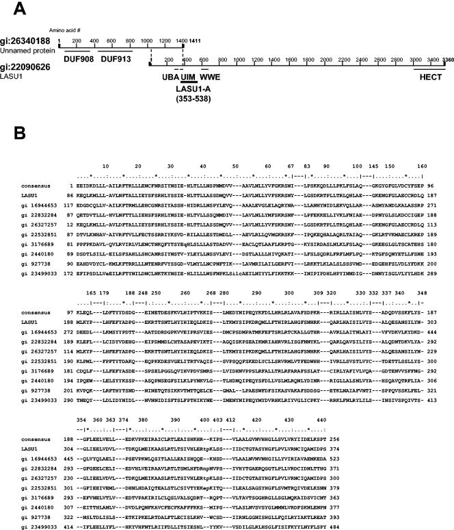
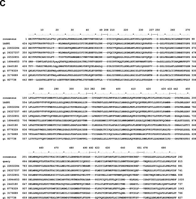
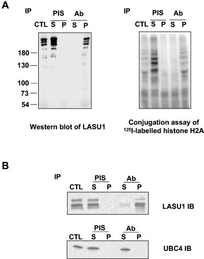
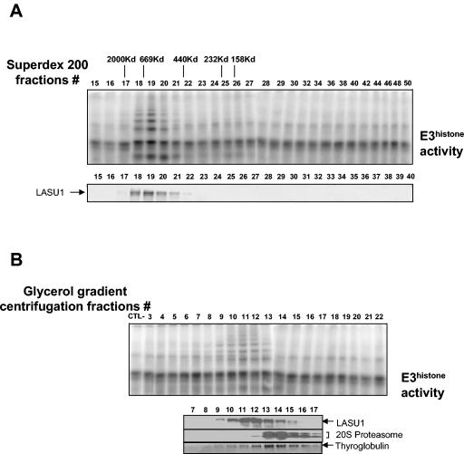
During spermatogenesis, a large fraction of cellular proteins is degraded as the spermatids evolve to their elongated mature forms. In particular, histones must be degraded in early elongating spermatids to permit chromatin condensation. Our laboratory previously demonstrated the activation of ubiquitin conjugation during spermatogenesis. This activation is dependent on the ubiquitin-conjugating enzyme (E2) UBC4, and a testis-particular isoform, UBC4-testis, is induced when histones are degraded. Therefore, we tested whether there are UBC4-dependent ubiquitin protein ligases (E3s) that can ubiquitinate histones. Indeed, a novel enzyme, E3Histone, which could conjugate ubiquitin to histones H1, H2A, H2B, H3, and H4 in vitro, was found. Only the UBC4/UBC5 family of E2s supported E3Histone-dependent ubiquitination of histone H2A, and of this family, UBC4-1 and UBC4-testis are the preferred E2s. We purified this ligase activity 3,600-fold to near homogeneity. Mass spectrometry of the final material revealed the presence of a 482-kDa HECT domain-containing protein, which was previously named LASU1. Anti-LASU1 antibodies immunodepleted E3Histone activity. Mass spectrometry and size analysis by gel filtration and glycerol gradient centrifugation suggested that E3Histone is a monomer of LASU1. Our assays also show that this enzyme is the major UBC4-1-dependent histone-ubiquitinating E3. E3Histone is therefore a HECT domain E3 that likely plays an important role in the chromatin condensation that occurs during spermatid maturation.


Figures

FIG. 1.
FIG. 1.
Identification and initial characterization of E3Histone. (A) A soluble testis extract was chromatographed on a Mono Q anion exchange column. The supernatant of the rat testis extract that was centrifuged at 100,000 × g (Load), water as a negative control (Ctl-), and eluted fractions were tested for the ability to support the conjugation of ubiquitin to 125I-labeled histone H2A in the presence of E1 and UBC4. Lanes 3 to 8 are the fractions eluting at approximately 0.4 M NaCl that contained the activity. (B) E3Histone (lanes 1 and 3) or water (lane 2) was used in a ubiquitin conjugation assay of 125I-labeled histone H2A with either wild-type Ub or MeUb. (C) Different 125I-labeled histones were tested in the conjugation assay in the presence (+) or absence (−) of E3Histone. (D) E3Histone-mediated ubiquitination was tested in the presence or absence (ctl-) of the indicated E2s. (A to D) Reaction products were resolved by SDS-PAGE and detected by autoradiography. *, background bands. Arrows show the single monoubiquitinated form of histone H2A. Note that in panels A, C, and D, the free histone substrates were run out of the gels because they created large shadows when the gels were exposed to film with the use of intensifying screens. In panel B, the gel was exposed to a phosphorimager screen, which does not result in the 125I-labeled histone substrate generating a large shadow. (B to D) The E3Histone used was partially purified as described in Materials and Methods.
FIG. 2.
FIG. 2.
E3Histone is probably a HECT domain-containing E3. (A) E3Histone requires a free thiol group for its E3 activity. (Left panel) In the conjugation assay, E3Histone was first treated with NEM or not treated. Excess NEM was then quenched by DTT, and the other components of the conjugation assay were added. Products of the assay were resolved by SDS-PAGE and detected by autoradiography. (Right panel) To confirm that both the NEM treatment and quenching by DTT were adequate, E1 was treated sequentially with NEM and DTT (lane 3) or added subsequently to buffer that had been similarly treated with NEM and DTT (lane 4) and then assayed for the ability to form a thioester bond with 125I-labeled ubiquitin. Reaction products were resolved by SDS-PAGE at 4°C under nonreducing conditions and detected by autoradiography. (B) E3Histone does not require divalent cations for its function. E3Histone was treated with EDTA (lanes 3 and 4) or TPEN (lanes 11 and 12). Remaining chelators were then quenched with excess Mg2+ (lanes 4 and 12) or not quenched (lanes 3 and 11) prior to assays for ubiquitination of histone H2A. To confirm the efficacy of the treatment, the RING finger E3 ARNIP was similarly treated and then assayed for its autoubiquitination ability by incubation with E1, UBC4-1, and 125I-labeled ubiquitin (lanes 5 to 8 and 13 to 16). Products were resolved by SDS-PAGE and detected by autoradiography.
FIG. 3.
FIG. 3.
Purification of E3Histone from bovine testis extract. (A) E3Histone purification scheme. The salts used to elute proteins in each step are shown on the right of the purification tree. Arrows indicate the salt concentration at which the enzyme eluted. The buffers used are described in the text. (B) Protein purification table. Aliquots of the pooled fractions from each protein purification step were assayed under quantification conditions (see the text) to measure E3Histone activity. The results were detected by phosphorimager and quantified as described in Materials and Methods.
FIG. 4.
FIG. 4.
Analysis of the fractions from the last purification step. (A) Fractions from the glycerol gradient centrifugation were assayed for E3Histone activity. As described in Materials and Methods, MeUb was used to start the reaction. The results were detected by autoradiography. (B) Protein fractions were resolved on a 5% native acrylamide gel and detected by colloidal blue staining. (C) Protein fractions were resolved by 7 to 15% gradient SDS-PAGE and detected by colloidal blue staining. Arrows in panels B and C show the bands cut for tandem mass spectrometric analysis. The presence of proteins in the A-, B-, and C-labeled bands in panel C that were also found in the band from the native gel in panel B is noted in Table 1.
FIG. 5.
FIG. 5.
Structure of LASU1. (A) Domain analysis of LASU1 and the unnamed protein. The region (amino acids 353 to 538) used to generate an anti-LASU1 antibody is shown. (B) Alignment of DUF908. (C) Alignment of DUF913. In panels B and C, the conserved residues in the domain are shown. gi 16944653, related to TOM1 protein; gi 22832284, CG8184-PB; gi 26327257, unnamed protein product; gi 22532851, hypothetical protein Y67D8C.5; gi 3176689, contains similarity to ubiquitin carboxyl-terminal hydrolase 14; gi 2440180, SPAC19D5.04; gi 927738, Tom1p; gi 23499033, ubiquitin-protein ligase 1; and gi 8778329, F14J16.10.
FIG. 5.
FIG. 5.
Structure of LASU1. (A) Domain analysis of LASU1 and the unnamed protein. The region (amino acids 353 to 538) used to generate an anti-LASU1 antibody is shown. (B) Alignment of DUF908. (C) Alignment of DUF913. In panels B and C, the conserved residues in the domain are shown. gi 16944653, related to TOM1 protein; gi 22832284, CG8184-PB; gi 26327257, unnamed protein product; gi 22532851, hypothetical protein Y67D8C.5; gi 3176689, contains similarity to ubiquitin carboxyl-terminal hydrolase 14; gi 2440180, SPAC19D5.04; gi 927738, Tom1p; gi 23499033, ubiquitin-protein ligase 1; and gi 8778329, F14J16.10.
FIG. 6.
FIG. 6.
E3Histone contains HECT domain protein LASU1. (A) E3Histone was immunoprecipitated (IP) with preimmune serum (PIS) or anti-LASU1 antiserum (Ab). (Left panel) Western blot of the indicated sample with anti-LASU1 antibody. (Right panel) Samples were assayed for E3Histone activity. (B) UBC4 was not coimmunoprecipitated with E3Histone. Crude testis extract was immunoprecipitated with the LASU1 antibody, and then the supernatants and pellets were subjected to immunoblotting with anti-LASU1 antibody or anti-UBC4 antibody. Multiple bands in the LASU1 blot likely represent degradation products of LASU1 during the procedure. CTL, the same amount of sample before IP; S, supernatant; P, pellet.
FIG. 7.
FIG. 7.
E3Histone appears to be a monomeric protein. (A) Separation of a crude testis extract using a Superdex 200 gel filtration column. (Top panel) Fractions were assayed for E3Histone activity as described in the legend to Fig. 1. Elution positions and molecular weights of the protein standards are shown. (Bottom panel) Western blot of the fractions with the anti-LASU1 antibody. The arrow shows the LASU1 band. (B) Separation of a crude testis extract by glycerol gradient centrifugation. The sample, purified 20S proteasome, or thyroglobulin was applied to a 4 to 40% glycerol gradient and subjected to the centrifugation at 30,000 rpm in an SW 40Ti rotor for 20 h at 4°C. (Top panel) Fractions from 4 to 40% glycerol gradient centrifugation were assayed for E3Histone activity. (Bottom panel) Detection of protein standards and LASU1. 20S proteasome and LASU1 were detected by Western blotting. Thyroglobulin was detected by Coomassie blue staining. (In both panels A and B, the unmodified 125I-labeled E3Histone substrate was run off the bottom of the gels.)

References

    1. Agell, N., M. Chiva, and C. Mezquita. 1983. Changes in nuclear content of protein conjugate histone H2A-ubiquitin during rooster spermatogenesis. FEBS Lett. 155:209-212. - PubMed
    1. Aravind, L. 2001. The WWE domain: a common interaction module in protein ubiquitination and ADP ribosylation. Trends Biochem. Sci. 26:273-275. - PubMed
    1. Baarends, W. M., J. W. Hoogerbrugge, H. P. Roest, M. Ooms, J. Vreeburg, J. H. Hoeijmakers, and J. A. Grootegoed. 1999. Histone ubiquitination and chromatin remodeling in mouse spermatogenesis. Dev. Biol. 207:322-333. - PubMed
    1. Bastians, H., L. M. Topper, G. L. Gorbsky, and J. V. Ruderman. 1999. Cell cycle-regulated proteolysis of mitotic target proteins. Mol. Biol. Cell 10:3927-3941. - PMC - PubMed
    1. Beitel, L. K., Y. A. Elhaji, R. Lumbroso, S. S. Wing, V. Panet-Raymond, B. Gottlieb, L. Pinsky, and M. A. Trifiro. 2002. Cloning and characterization of an androgen receptor N-terminal-interacting protein with ubiquitin-protein ligase activity. J. Mol. Endocrinol. 29:41-60. - PubMed

Publication types