. 2005 Apr;25(7):2846-52.
doi: 10.1128/MCB.25.7.2846-2852.2005.

Normal thyroid structure and function in rhophilin 2-deficient mice

Affiliations

Normal thyroid structure and function in rhophilin 2-deficient mice

Jens Behrends et al. Mol Cell Biol. 2005 Apr.

Abstract

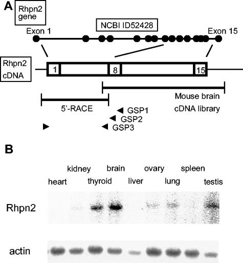
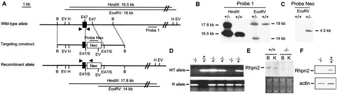
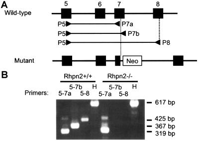
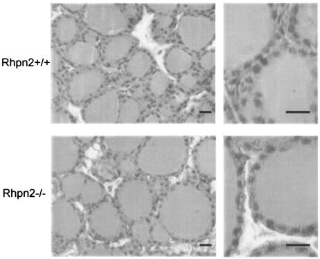
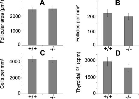
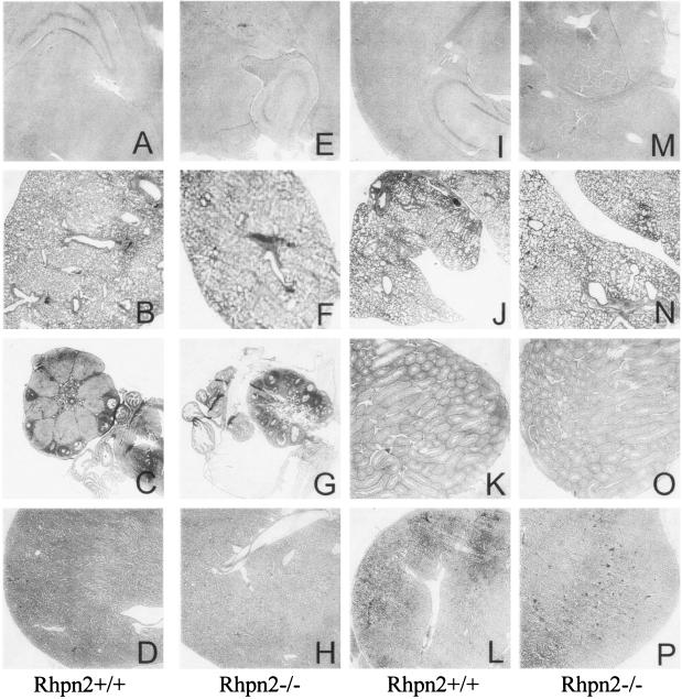
Rhophilin 2 is a Rho GTPase binding protein initially isolated by differential screening of a chronically thyrotropin (TSH)-stimulated dog thyroid cDNA library. In thyroid cell culture, expression of rhophilin 2 mRNA and protein is enhanced following TSH stimulation of the cyclic AMP (cAMP) transduction cascade. Yeast two-hybrid screening and coimmunoprecipitation have revealed that the GTP-bound form of RhoB and components of the cytoskeleton are protein partners of rhophilin 2. These results led us to suggest that rhophilin 2 could play an important role downstream of RhoB in the control of endocytosis during the thyroid secretory process which follows stimulation of the TSH/cAMP pathway. To validate this hypothesis, we generated rhophilin 2-deficient mice and analyzed their thyroid structure and function. Mice lacking rhophilin 2 develop normally, have normal life spans, and are fertile. They have no visible goiter and no obvious clinical signs of hyper- or hypothyroidism. The morphology of thyroid cells and follicles in these mice were normal, as were the different biological tests performed to investigate thyroid function. Our results indicate that rhophilin 2 does not play an essential role in thyroid physiology.


Figures

FIG. 1.
FIG. 1.
Mouse rhophilin 2 gene structure, cDNA, and expression in tissues. (A) Schematic representation of the exon-intron structure of the mouse rhophilin 2 gene on chromosome 7, as reported in NCBI databases (see above). The two DNA fragments isolated by screening of a mouse brain cDNA library and by 5′ RACE are depicted as well as the three specific primers (GSP1, GSP2, and GSP3, all located in exon 8 [arrowheads]) used in 5′ RACE (see below). (B) Hybridization analysis of RNA isolated from mouse tissues with a rhophilin 2 probe or an actin probe.
FIG. 2.
FIG. 2.
Targeted disruption of mouse Rhpn2. (A) Wild-type allele, targeting vector, predicted structure of the mutant allele, probes used in Southern blot analysis, DNA fragments generated after digestion with HindIII or EcoRV, and primers used for mouse genotyping by PCR (filled arrowheads). Filled boxes: exon 7 of the Rhpn2 gene. B, BamHI; EV, EcoRV; E47, Eco47III; E47/S, E47 DNA fragment ligated into SmaI-digested vector; H, HindIII. (B and C) Southern blots of the recombinant embryonic stem cell clone with probe 1 and the probe corresponding to the Neo cassette. Left and/or right margins, length of genomic DNA fragments generated after digestion with restriction enzymes (above blot). (D) Mouse genotyping by PCR amplification from tail DNA using primers specific for either the WT or the recombinant (R) allele. (E) RNA hybridization analysis of total brain (B) and kidney (K) RNA isolated from Rhpn2+/+ and Rhpn2−/− mice. The probe was a 2.2-kb dog rhophilin 2 cDNA fragment containing the complete coding region. (F) Western blot of brain protein extract from Rhpn2+/+ and Rhpn2−/− mice using anti-Rhpn2 or anti-actin antibody.
FIG. 3.
FIG. 3.
Absence of normal Rhpn2 mRNA in Rhpn2−/− thyroid. (A) Exons 5 to 8 (filled boxes) of the mouse Rhpn2 gene in wild-type and mutant alleles and localization of primers P5, P7a, P7b, and P8 (arrowheads). (B) Electrophoresis of PCR products generated with the indicated primers on cDNA isolated from Rhpn2+/+ and Rhpn2−/− thyroids. H, hypoxanthine phosphoribosyltransferase primers. Right margin, lengths of DNA fragments generated by PCR.
FIG. 4.
FIG. 4.
Normal histological structure of Rhpn2−/− thyroid. Thyroid lobes and follicles from Rhpn2+/+ and Rhpn2−/− mice are shown (scale bar, 15 μm).
FIG. 5.
FIG. 5.
Morphological parameters measured in thyroid glands of Rhpn2+/+ and Rhpn2−/− mice. (A) Average follicular area. (B) Follicular density. (C) Cellular density. (D) Thyroidal 131I uptake 2 days after injection of the isotope. Values are means ± standard errors of the means (five mice per group).
FIG. 6.
FIG. 6.
Normal histological structure of brain, lung, ovary, testis, and kidney in Rhpn2−/− mice: hematoxylin and eosin-stained sections from brain (A, E, I, and M), lung (B, F, J, and N), ovary (C and G), testis (K and O), and kidney (D, H, L, and P) of 12-week-old Rhpn2+/+ (first and third columns) and Rhpn2/ (second and fourth columns) mice. Low-magnification pictures are shown.

References

    1. Clement, S., S. Refetoff, B. Robaye, J. E. Dumont, and S. Schurmans. 2001. Low TSH requirement and goiter in transgenic mice overexpressing IGF-I and IGF-Ir receptor in the thyroid gland. Endocrinology 142:5131-5139. - PubMed
    1. Di Cunto, F., S. Imarisio, E. Hirsch, V. Broccoli, A. Bulfone, A. Migheli, C. Atzori, E. Turco, R. Triolo, G. P. Dotto, L. Silengo, and F. Altruda. 2000. Defective neurogenesis in citron kinase knockout mice by altered cytokinesis and massive apoptosis. Neuron 28:115-127. - PubMed
    1. Dremier, S., K. Coulonval, S. Perpete, F. Vandeput, N. Fortemaison, A. Van Keymeulen, S. Deleu, C. Ledent, S. Clement, S. Schurmans, J. E. Dumont, F. Lamy, P. P. Roger, and C. Maenhaut. 2002. The role of cyclic AMP and its effect on protein kinase A in the mitogenic action of thyrotropin on the thyroid cell. Ann. N. Y. Acad. Sci. 968:106-121. - PubMed
    1. Dumont, J. E., F. Lamy, P. Roger, and C. Maenhaut. 1992. Physiological and pathological regulation of thyroid cell proliferation and differentiation by thyrotropin and other factors. Physiol. Rev. 72:667-697. - PubMed
    1. Dumont, J. E., C. Maenhaut, F. Lamy, I. Pirson, S. Clement, and P. P. Roger. 2003. Growth and proliferation of the thyroid cell in normal physiology and in disease. Ann. Endocrinol. 64:10-11. - PubMed

Publication types

MeSH terms