. 2005 Apr;25(7):2853-60.
doi: 10.1128/MCB.25.7.2853-2860.2005.

Normal cell cycle and checkpoint responses in mice and cells lacking Cdc25B and Cdc25C protein phosphatases

Affiliations

Normal cell cycle and checkpoint responses in mice and cells lacking Cdc25B and Cdc25C protein phosphatases

Angela M Ferguson et al. Mol Cell Biol. 2005 Apr.

Abstract

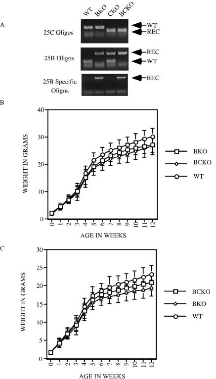
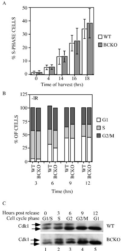
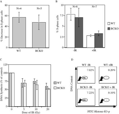
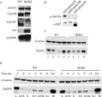
The Cdc25 family of protein phosphatases positively regulates cell division by activating cyclin-dependent protein kinases (CDKs). In humans and rodents, there are three Cdc25 family members--denoted Cdc25A, Cdc25B, and Cdc25C--that can be distinguished based on their subcellular compartmentalizations, their abundances and/or activities throughout the cell cycle, the CDKs that they target for activation, and whether they are overexpressed in human cancers. In addition, murine forms of Cdc25 exhibit distinct patterns of expression throughout development and in adult tissues. These properties suggest that individual Cdc25 family members contribute distinct biological functions in embryonic and adult cell cycles of mammals. Interestingly, mice with Cdc25C disrupted are healthy, and cells derived from these mice exhibit normal cell cycles and checkpoint responses. Cdc25B-/- mice are also generally normal (although females are sterile), and cells derived from Cdc25B-/- mice have normal cell cycles. Here we report that mice lacking both Cdc25B and Cdc25C are obtained at the expected Mendelian ratios, indicating that Cdc25B and Cdc25C are not required for mouse development or mitotic entry. Furthermore, cell cycles, DNA damage responses, and Cdc25A regulation are normal in cells lacking Cdc25B and Cdc25C. These findings indicate that Cdc25A, or possibly other phosphatases, is able to functionally compensate for the loss of Cdc25B and Cdc25C in mice.


Figures

FIG. 1.
FIG. 1.
Genotyping and growth rates of mice with Cdc25B and Cdc25C disrupted. (A) PCR analysis of mouse tail DNA from WT, Cdc25B/ (BKO), Cdc25C/ (CKO), and Cdc25B/C/ (BCKO) mice, using the indicated oligonucleotides. REC, recombinant. (B and C) WT, Cdc25B/ (BKO), and Cdc25B/C/ (BCKO) male (B) and female (C) mice were weighed beginning at birth and at weekly intervals up to 12 weeks. Weights (in grams) represent averages from at least 30 mice per genotype per time point. Standard deviations are shown as error bars along the y axis.
FIG. 2.
FIG. 2.
Cell cycle analysis of cells lacking Cdc25B and Cdc25C. (A) WT and BCKO MEFs (passages 4 to 6) were serum starved for 96 h to synchronize cells in G0. After the addition of serum, cells were incubated with BrdU for 1 h prior to harvest. Cells were stained with PI and for BrdU and were analyzed by flow cytometry. Each experiment was performed six times in duplicate with independent MEF strains, and standard deviations are shown as error bars along the y axis. Differences between WT and BCKO MEFs were not statistically different at any of the time points (0 h [P = 0.936], 4 h [P = 0.7374], 14 h [P = 0.9441], 16 h [P = 0.6278], and 18 h [P = 0.2667]). (B) MEFs at passages 4 to 6 were pulse labeled with BrdU for 1 h. Cells were harvested at the indicated times (hours) and stained with PI and for BrdU. The cellular DNA content of BrdU-positive cells was analyzed by flow cytometry. Graphs show the averages from six independent experiments. (C) Early-passage MEFs prepared from wild-type and BCKO mice were synchronized in early S phase. Cells were harvested prior to release (time zero) or at various times (hours) after release. Cdk1 precipitates were resolved on a 12% SDS gel, and Cdk1 was detected by Western blotting. The arrows indicate two electrophoretic forms of Cdk1.
FIG. 3.
FIG. 3.
The IR-induced DNA damage checkpoint is intact in BCKO cells. (A) Early-passage MEFs were serum starved for 96 h. Arrested cells were mock irradiated or gamma irradiated and then incubated in BrdU-containing complete medium. Cells were stained with PI and for BrdU and analyzed by flow cytometry 24 h later. The percent decreases in S-phase cells following irradiation are indicated. Differences between WT and BCKO cells were not significant (P = 0.2909) (B) WT and BCKO mice were mock irradiated (−IR) or gamma irradiated (+IR) with 10 Gy of IR and injected intraperitoneally with BrdU 2 h later. One hour after the BrdU injection, thymocytes were prepared and analyzed by flow cytometry. The percentages of S-phase cells are indicated. Standard deviations are shown as error bars along the y axis. (C) Radioresistant DNA synthesis was assessed 1 h after the exposure of early-passage MEFs to various doses of IR. Experiments were repeated four times. Differences between WT and BCKO MEFs were not statistically different at any dose of IR (5 Gy [P = 0.9035], 10 Gy [P = 0.2095], and 20 Gy [P = 0.1333]). Standard deviations are shown as error bars along the y axis. (D) Early-passage MEFs were mock irradiated or gamma irradiated and incubated in nocodazole 40 min later. Cells were costained for DNA content and histone H3 phosphorylation (p) and were analyzed by flow cytometry 2 h after IR. The percentages of mitotic cells are indicated. The panels are representative results obtained using WT (n = 2) and BCKO (n = 3) MEFs.
FIG. 4.
FIG. 4.
Regulation of Cdc25A is normal in cells lacking Cdc25B and Cdc25C. (A) RNA isolated from WT and BCKO MEFs was processed for Northern blot analysis using probes specific for mCdc25A, mCdc25B, mCdc25C, neomycin phosphotransferase (Neo), and GAPDH. (B) GST-tagged mouse Cdc25 proteins purified from overproducing insect cells (7) were analyzed for reactivity with the mouse Cdc25A (a-Cdc25A) monoclonal antibody and GST (a-GST) antibody. (C) Lysates derived from early-passage WT and BCKO MEFs were analyzed for Cdc25A and β-catenin levels by Western blotting. (D) Asynchronous early-passage MEFs (A) were synchronized at the G1/S border. G1/S-arrested cells (0) were released from the block and processed for Western blotting and flow cytometry at the indicated time points. +, samples that were incubated with nocodazole at the time of release.

References

    1. Abrieu, A., T. Brassac, S. Galas, D. Fisher, J. C. Labbe, and M. Doree. 1998. The Polo-like kinase Plx1 is a component of the MPF amplification loop at the G2/M-phase transition of the cell cycle in Xenopus eggs. J. Cell Sci. 111:1751-1757. - PubMed
    1. Atherton-Fessler, S., F. Liu, B. Gabrielli, M. S. Lee, C.-Y. Peng, and H. Piwnica-Worms. 1994. Cell cycle regulation of the p34cdc2 inhibitory kinases. Mol. Biol. Cell 5:989-1001. - PMC - PubMed
    1. Barlow, C., K. D. Brown, C. X. Deng, D. A. Tagle, and A. Wynshaw-Boris. 1997. Atm selectively regulates distinct p53-dependent cell-cycle checkpoint and apoptotic pathways. Nat. Genet. 17:453-456. - PubMed
    1. Bernardi, R., D. A. Lieberman, and B. Hoffman. 2000. Cdc25A stability is controlled by the ubiquitin-proteasome pathway during cell cycle progression and terminal differentiation. Oncogene 19:2447-2454. - PubMed
    1. Blomberg, I., and I. Hoffman. 1999. Ectopic expression of Cdc25A accelerates the G1/S transition and leads to premature activation of cyclin E- and cyclin A-dependent kinases. Mol. Cell. Biol. 19:6183-6194. - PMC - PubMed

Publication types

LinkOut - more resources