Comparative Study
. 2005 Apr 1;118(Pt 7):1437-47.
doi: 10.1242/jcs.01737. Epub 2005 Mar 15.

Potentiation of neutrophil cyclooxygenase-2 by adenosine: an early anti-inflammatory signal

Affiliations
Comparative Study

Potentiation of neutrophil cyclooxygenase-2 by adenosine: an early anti-inflammatory signal

Jean-Sébastien Cadieux et al. J Cell Sci. .

Abstract

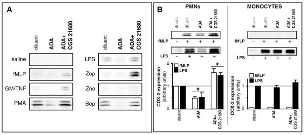
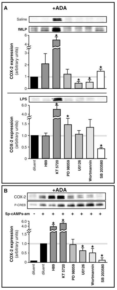
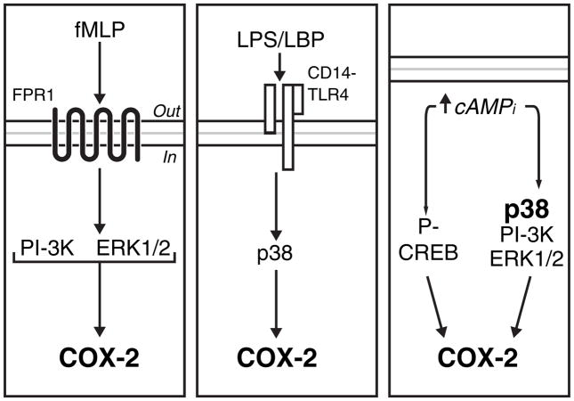
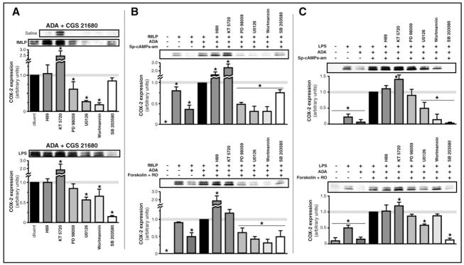
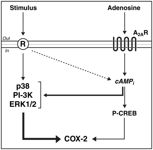
Neutrophils, which are often the first to migrate at inflamed sites, can generate leukotriene B(4) from the 5-lipoxygenase pathway and prostaglandin E(2) through the inducible cyclooxygenase-2 pathway. Adenosine, an endogenous autacoid with several anti-inflammatory properties, blocks the synthesis of leukotriene B(4) while it potentiates the cyclooxygenase-2 pathway in fMLP-treated neutrophils, following activation of the A(2A) receptor. Using the murine air pouch model of inflammation, we observed that inflammatory leukocytes from mice lacking the A(2A) receptor have less cyclooxygenase-2 induction than wild-type animals. In human leukocytes, A(2A) receptor activation specifically elicited potentiation of cyclooxygenase-2 in neutrophils, but not in monocytes. Signal transduction studies indicated that the cAMP, ERK1/2, PI-3K and p38K intracellular pathways are implicated both in the direct upregulation of cyclooxygenase-2 and in its potentiation. Together, these results indicate that neutrophils are particularly important mediators of adenosine's effects. Given the uncontrolled inflammatory phenotype observed in knockout mice and in view of the potent inhibitory actions of prostaglandin E(2) on inflammatory cells, an increased cyclooxygenase-2 expression resulting from A(2A) receptor activation, observed particularly in neutrophils, may take part in an early modulatory mechanism promoting anti-inflammatory activities of adenosine.


Figures

Fig. 1
Fig. 1
Leukocytes from wild-type (A2AR+/+) mice express more COX-2 in response to LPS than do knockout (A2AR−/−) mice. (A) Dorsal air pouches from wild-type (A2AR+/+) CD1 mice were injected with PBS alone or containing 500 ng LPS for 4 hours; migrated leukocytes were collected and processed for the determination of indicated mRNA expression by real-time PCR, as described in Materials and Methods. Typical real-time PCR results from one mouse are shown. (Inset) Enumeration of leukocytes which migrated into the air pouch of wild-type (+/+) or homozygote knockout mice (−/−). Results are expressed as the mean±s.e.m., with values between brackets being the number (n) of animals in each condition. *Statistically different from PBS-injected mice. (B) Comparative GAPDH, COX-1 and COX-2 mRNA expression in migrated leukocytes between LPS-injected A2AR+/+ and A2AR−/− mice. Results show the mean±s.e.m. from at least four animals. *Statistically different from GAPDH and COX-1. **Statistically different from A2AR+/+. (Inset) Comparative mRNA expression in resident cells (mean±s.e.m., n=4). (C) Air pouches were injected with LPS for the indicated times. Migrated leukocytes were collected, enumerated, and samples, equalized by cell numbers, were processed for the determination of COX-2 by western immunoblotting, as described in Materials and Methods. (D) Air pouches were injected with LPS for 60 minutes. Migrated leukocytes were collected, enumerated, and samples were processed for the determination of COX-2 by western immunoblotting. *Statistically different from A2AR+/+. In each panel, immunoblots from one experiment typical of at least three separate experiments are shown. Bar graph depicts the expression of COX-2 protein levels in leukocytes from LPS-injected mice, relative to that observed in A2AR+/+ mice, as determined by densitometric analysis of the bands in three independent experiments (mean±s.e.m.; n=3).
Fig. 2
Fig. 2
Activation of the A2A receptor specifically potentiates the expression of COX-2 from human neutrophils. (A) Human PMNs were stimulated for 60 minutes with indicated agonists, alone or in the presence of adenosine deaminase (ADA; 0.1 U/ml), or with ADA and the A2AR agonist CGS 21680 (1 μM). Reactions were stopped and samples were processed for the determination of COX-2 by western immunoblotting. Shown are immunoblots from one experiment, typical of at least n=3 experiments performed in identical conditions with separate donors. fMLP, formyl-methionyl-leucyl-phenylalanine (100 nM); GM/TNF, mixture of granulocyte-macrophage colony-stimulating factor and tumor necrosis factor-α (1 nM and 100 nM, respectively); PMA (10 nM), phorbol 12-myristate 13-acetate; LPS, lipopolysaccharide (100 ng/ml); Zop, opsonized zymosan (300 μg/ml); Zno, nonopsonized zymosan (300 μg/ml); Bop, opsonized bacteria (108 bacteria/ml). (B) Human leukocytes were stimulated with fMLP or LPS, for 1 hour (PMNs) or 4 hours (Monocytes) alone or in the presence of ADA or with ADA and CGS 21680; samples were processed for the determination of COX-2 by western immunoblotting. In each condition, one typical immunoblot is shown and is representative of at least n=3 experiments performed in identical conditions with different donors. Bar graphs depict the expression of COX-2 protein levels, relative to those observed in leukocytes stimulated in the absence of ADA and CGS 21680, as determined by densitometric analysis of the bands in three independent experiments (mean±s.e.m.; n=3). *Statistically different from diluent-treated cells.
Fig. 3
Fig. 3
The anti-inflammatory drug methotrexate does not influence the release of adenosine from fMLP-stimulated neutrophils. Human PMNs were stimulated with fMLP, alone or in the presence of CGS 21680, or with the anti-inflammatory compound methotrexate (MTX; 1 μM), or with adenosine deaminase (ADA), as described in Materials and Methods. At indicated time points, samples were processed for the measurement of extracellular adenosine by mass-spectrometry.
Fig. 4
Fig. 4
Distinct metabolic pathways can lead to the upregulation of neutrophil COX-2: implication of PI-3K, ERK1/2, p38 MAPK and cAMP pathways. (A) PMNs were stimulated in adenosine-free conditions (i.e. in the presence of ADA) with fMLP (upper) or LPS (lower), alone or in combination with the indicated metabolic inhibitors. Samples were processed for the determination of COX-2 by western immunoblotting. (B) PMNs were incubated in adenosine-free conditions (i.e. in the presence of ADA) with a cell-permeable stable analog of cAMP, Sp-cAMPs-am (50 μM), alone or in combination with indicated metabolic inhibitors. Samples were processed for the determination of COX-2 and phosphorylated (P-)CREB by western immunoblotting. In each panel, one typical immunoblot is shown and is representative of at least n=3 independent experiments performed in identical conditions with different donors. Bar graphs depict the expression of COX-2 protein levels, relative to those observed in A: PMNs stimulated with fMLP or LPS only (solid black bars) and in B: PMNs incubated with Sp-cAMPs-am only (solid black bar) as determined by densitometric analysis of the bands in three independent experiments (mean±s.e.m.; n=3). *Statistically different from diluent-treated cells. H89 and KT 5720 (two structurally distinct inhibitors of PKA; 10μM and 3 μM, respectively), PD 98059 (MEK-ERK1/2 inhibitor; 10 μM), U0126 (ERK1/2 inhibitor; 10 μM), wortmannin (PI-3K inhibitor; 200 nM), SB 203580 (p38 inhibitor; 10 μM).
Fig. 5
Fig. 5
Cellular pathways leading to upregulation of COX-2 in neutrophils. Left: In adenosine-free conditions, the PI-3K and ERK pathways are primarily involved in the induction of COX-2, following engagement of formylated peptide receptor-1 (FPR1). Center: In response to LPS, the p38 pathway is almost exclusively solicited. Right: An elevation of intracellular cAMP concentrations can cause COX-2 induction, through a classic PKA-dependent activation of CREB, or through activation of the p38, PI-3K and ERK1/2 pathways. LBP, LPS-binding protein; TLR4, Toll-like receptor-4.
Fig. 6
Fig. 6
Metabolic pathways implicated in potentiating COX-2 expression in neutrophils following A2A receptor activation. (A) PMNs were stimulated with fMLP (upper) or LPS (lower) in the presence of ADA and of CGS 21680 (A2AR agonist), alone or in combination with indicated metabolic inhibitors. Samples were processed for the determination of COX-2 by western immunoblotting. Bar graphs depict the expression of COX-2 protein levels, relative to those observed in PMNs stimulated in absence of metabolic inhibitors (solid black bars), as determined by densitometric analysis of the bands in three independent experiments (mean±s.e.m.; n=3). (B, C) PMNs were stimulated with fMLP (B) of LPS (C), in combination either with a membrane-permeable cAMP analog, Sp-cAMPs-am (upper), or with a mixture of an activator of adenylyl cyclase and an inhibitor of phosphodiesterase IV, RO-20-1724 and forskolin; 50 μM and 10 μM, respectively (lower), alone or in combination with the indicated metabolic inhibitors. Bar graphs depict the expression of COX-2 protein levels, relative to that observed in PMNs stimulated in absence of metabolic inhibitors, as determined by densitometric analysis of the bands in three independent experiments (mean±s.e.m.; n=3). In each panel, one typical immunoblot is shown and is representative of at least n=3 independent experiments performed in identical conditions with different donors. *Statistically different from cells treated with stimulus+ADA+cAMP-elevating agents.
Fig. 7
Fig. 7
The cellular pathways implicated in potentiating the expression of COX-2 in neutrophils, following activation of the A2A receptor. Different agonists cause the induction of COX-2 in human neutrophils by activating distinct classes of receptors, which in turn differentially activate the p38, PI-3K and ERK1/2 pathways. Some of these agonists can also transiently elevate cAMPi. Activation of A2AR elevates cAMPi, which has a positive impact on COX-2 by further activating the p38, PI-3K and ERK1/2 pathways and also through the classic PKA/CREB pathway.

Similar articles

Cited by

References

    1. Bertagnolli MM. Cyclooxygenase-2 as a target for prevention of colorectal cancer. Curr Oncol Rep. 1999;1:173–178. - PubMed
    1. Beullens M, Van Eynde A, Bollen M, Stalmans W. Inactivation of nuclear inhibitory polypeptides of protein phosphatase-1 (NIPP-1) by protein kinase A. J Biol Chem. 1993;268:13172–13177. - PubMed
    1. Bornfeldt KE, Campbell JS, Koyama H, Argast GM, Leslie CC, Raines EW, Krebs EG, Ross R. The mitogen-activated protein kinase pathway can mediate growth inhibition and proliferation in smooth muscle cells. Dependence on the availability of downstream targets. J Clin Invest. 1997;100:875–885. - PMC - PubMed
    1. Böyum A. Isolation of mononuclear cells and granulocytes from human blood: isolation of mononuclear cells by one centrifugation, and of granulocytes by combining centrifugation and sedimentation at 1 g. Scand J Clin Invest (Suppl) 1968;97:77–89. - PubMed
    1. Busse WW. Leukotrienes and inflammation. Am J Respir Crit Care Med. 1998;157:S210–S213. discussion S247–S248. - PubMed

Publication types

MeSH terms