. 2005 Apr;11(4):517-31.
doi: 10.1261/rna.7291205.

Yeast poly(A)-binding protein Pab1 shuttles between the nucleus and the cytoplasm and functions in mRNA export

Affiliations

Yeast poly(A)-binding protein Pab1 shuttles between the nucleus and the cytoplasm and functions in mRNA export

Christiane Brune et al. RNA. 2005 Apr.

Abstract

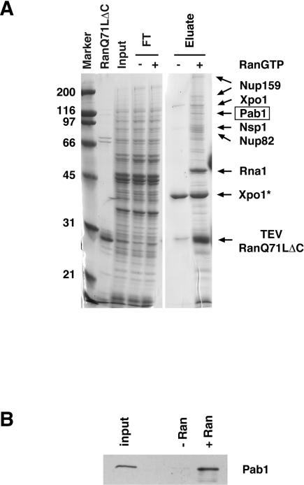
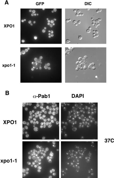
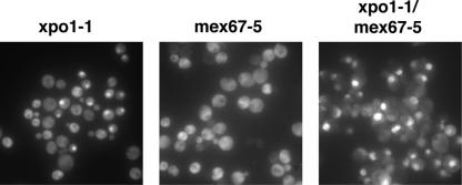
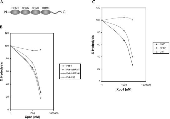
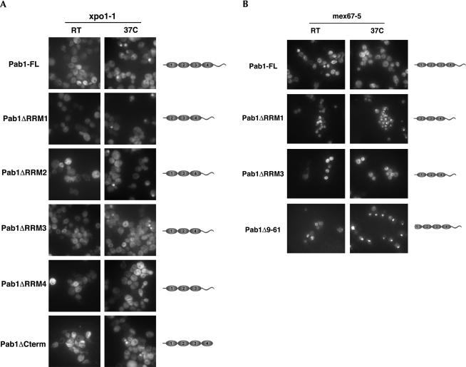
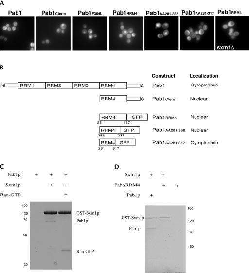
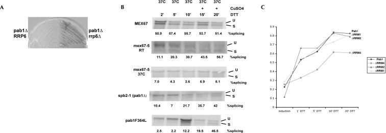
Pab1 is the major poly(A)-binding protein in yeast. It is a multifunctional protein that mediates many cellular functions associated with the 3'-poly(A)-tail of messenger RNAs. Here, we characterize Pab1 as an export cargo of the protein export factor Xpo1/Crm1. Pab1 is a major Xpo1/Crm1-interacting protein in yeast extracts and binds directly to Xpo1/Crm1 in a RanGTP-dependent manner. Pab1 shuttles rapidly between the nucleus and the cytoplasm and partially accumulates in the nucleus when the function of Xpo1/Crm1 is inhibited. However, Pab1 can also be exported by an alternative pathway, which is dependent on the MEX67-mRNA export pathway. Import of Pab1 is mediated by the import receptor Kap108/Sxm1 through a nuclear localization signal in its fourth RNA-binding domain. Interestingly, inhibition of Pab1's nuclear import causes a kinetic delay in the export of mRNA. Furthermore, the inviability of a pab1 deletion strain is suppressed by a mutation in the 5'-3' exoribonuclease RRP6, a component of the nuclear exosome. Therefore, nuclear Pab1 may be required for efficient mRNA export and may function in the quality control of mRNA in the nucleus.


Figures

FIGURE 1.
FIGURE 1.
(A) Purification of Xpo1/Crm1-interacting proteins. S-TEV-ZZ-tagged Xpo1/Crm1 was tandem affinity-purified from yeast extracts (as described in Materials and Methods) either in the absence or presence of 10 μM Gsp1Q71LΔC preloaded with GTP. Proteins that interacted with immobilized Xpo1/Crm1 were eluted with 1 M MgCl2, and the input, flowthrough (FT), and eluate were analyzed by SDS-PAGE and Coomassie blue staining. Proteins were identified by MALDI mass spectrometry. Xpo1* corresponds to a degradation product of Xpo1/Crm1. Gsp1Q71LΔC and TEV comigrated and could not be separated on this gel. Molecular mass is given in kilodaltons (kDa). (B) Binding of Pab1 to Xpo1/Crm1 is dependent on the presence of RanGTP. Eluates from protein S beads were analyzed by Western blot using the anti-Pab1 monoclonal antibody 1G1 (Anderson et al. 1993). Based on serial dilutions using recombinant Pab1, we estimate that Pab1 is ~200-fold enriched in purifications performed in the presence of Gsp1Q71LΔC (+Ran) compared to purifications without the addition of Gsp1Q71LΔC (−Ran).
FIGURE 2.
FIGURE 2.
Pab1 mislocalizes to the nucleus in xpo1-1 cells. (A) Wild-type and xpo1-1 cells expressing functional Pab1-GFP were grown at room temperature and then shifted to 37°C for 40 min. The localization of Pab1-GFP was analyzed by epifluorescence (GFP), and yeast cells were visualized by Nomarski microscopy (DIC). (B) Wild-type and xpo1-1 cells were shifted to 37°C for 40 min, fixed, prepared for immunofluorescence, and analyzed with the anti-Pab1 monoclonal antibody 1G1 (α-Pab1). Nuclear DNA was stained with DAPI, and the signal was visualized by epifluorescence.
FIGURE 3.
FIGURE 3.
Pab1 can be exported by two distinct pathways. xpo1-1-expressing Pab1-GFP, mex67-5, and xpo1-1 mex67-5 cells were grown in full medium at room temperature and then shifted to 37°C for 1 h. The localization of Pab in xpo1-1 cells was analyzed by fluorescence microscopy. The localization of Pab1 in mex67-5 and in xpo1-1 mex67-5 mutant cells was examined by immunofluorescence.
FIGURE 4.
FIGURE 4.
(A) Schematic representation of the domain organization of Pab1. (B) Pab1 binds directly to Xpo1/Crm1. Gsp1 [γ-32P]GTP (80 nM) was incubated with the indicated concentrations of recombinant Xpo1/Crm1 and 15 μM recombinant Pab1 wild-type (Pab1), Pab1 lacking RRM1 (Pab1ΔRRM1), Pab1 lacking RRM4 (Pab1ΔRRM4), or Pab1 lacking the carboxyl terminus (Pab1ΔC) for 15 min on ice. Gsp1 GTPase reactions were started by the addition of 40 nM Rna1, and the amount of hydrolysis was determined by measuring the release of [32P]phosphate using the charcoal method after 2 min. (C) GTPase hydrolysis assays were performed as described in B using 80 nM Gsp1 [γ-32P]GTP and increasing concentrations of Xpo1/Crm1 in the presence of 15 μM either recombinant wild-type Pab1 (Pab1), the RRM1 domain of Pab1 comprising amino acids 15–129 (RRM1), or BSA (Ctrl).
FIGURE 5.
FIGURE 5.
(A) xpo1-1 cells expressing full-length Pab1 (Pab1-FL), Pab1ΔRRM1, Pab1ΔRRM2, Pab1ΔRRM3, Pab1ΔRRM4, or Pab1ΔCterm were grown at room temperature (RT) or shifted to 37°C for 1 h, and the localization of the various Pab1 variants was analyzed by immunofluorescence using an anti-Pab1 polyclonal serum. (B) Wild-type and mex67-5 cells expressing either Pab1 wild-type, Pab1ΔRRM1, Pab1ΔRRM3, or a Pab1 mutant lacking amino acids 9–61 (Pab1Δ9–61) were grown at room temperature or shifted to 37°C for 1 h. The localization of Pab1 was visualized by immunofluorescence using a monospecific anti-Pab1 polyclonal serum.
FIGURE 6.
FIGURE 6.
(A) The localization of Pab1-GFP, a Pab1 truncation expressing RRM4 and the carboxyl terminus fused to GFP (Pab1Cterm), the Pab1 truncation mutant Pab1F364L (Sachs et al. 1987) fused to GFP, RRM4Pab1-GFP, amino acids 281–338 of Pab1 fused to GFP, and amino acids 281–317 of Pab1 fused to GFP were analyzed by fluorescence in wild-type cells. RRM4Pab1-GFP is no longer localized to the nucleus when expressed in kap108/sxm1 deletion cells. (B) Schematic diagram of the analyzed Pab1 truncation mutants and their subcellular localization. (C) Pab1 binds to Kap108/Sxm1 in vitro. Recombinant Sxm1-GST was incubated with Pab1 either in the absence or presence of RanGTP, and complexes were purified on glutathione beads and analyzed by SDS gel electrophoresis (lanes 2 and 3). Pab1 alone does not bind to glutathione beads (lane 1). (D) The interaction between Pab1 and Kap108/Sxm1 requires RRM4. Recombinant Sxm1-GST was incubated with full-length Pab1 and/or Pab1ΔRRM4. Complexes were purified on glutathione beads and analyzed by SDS gel electrophoresis.
FIGURE 7.
FIGURE 7.
(A) Deletion of pab1 is rescued by a deletion of rrp6. RRP6 was deleted in pab1Δ cells covered by a URA3-marked plasmid expressing Pab1. After growth on media containing 5-fluoro-orotic acid (5-FOA), viable colonies are obtained in the rrp6-deletion background but not in cells that are wild type for RRP6. (B,C) Pab1 mutants are deficient for Hac1 mRNA export. Hac1 expression was induced by the addition of copper in the presence of DTT in MEX67, mex67-5, pab1ΔRRM1, spb2-1 (pab1Δ rpl39-1), or in pab1F364L cells (B) and in PAB1, pab1ΔRRM1, pab1ΔRRM2, and in pab1ΔRRM4 cells (C). Samples were collected at the indicated time points, and the kinetics of Hac1 mRNA splicing was monitored by Northern blot analysis. Percent splicing indicates the ratio between the unspliced (U) and the spliced (S) isoform of Hac1 as quantified by PhosphorImager analysis.

Similar articles

Cited by

References

    1. Adam, S.A., Nakagawa, T., Swanson, M.S., Woodruff, T.K., and Drey-fuss, G. 1986. mRNA polyadenylate-binding protein: Gene isolation and sequencing and identification of a ribonucleoprotein consensus sequence. Mol. Cell. Biol. 6: 2932–2943. - PMC - PubMed
    1. Afonina, E., Stauber, R., and Pavlakis, G.N. 1998. The human poly(A)-binding protein 1 shuttles between the nucleus and the cytoplasm. J. Biol. Chem. 273: 13015–13021. - PubMed
    1. Amrani, N., Minet, M., Le Gouar, M., Lacroute, F., and Wyers, F. 1997. Yeast Pab1 interacts with Rna15 and participates in the control of the poly(A) tail length in vitro. Mol. Cell. Biol. 17: 3694–3701. - PMC - PubMed
    1. Anderson, J.T., Paddy, M.R., and Swanson, M.S. 1993. PUB1 is a major nuclear and cytoplasmic polyadenylated RNA-binding protein in Saccharomyces cerevisiae. Mol. Cell. Biol. 13: 6102–6113. - PMC - PubMed
    1. Askjaer, P., Bachi, A., Wilm, M., Bischoff, F.R., Weeks, D.L., Ogniewski, V., Ohno, M., Niehrs, C., Kjems, J., Mattaj, I.W., et al. 1999. RanGTP-regulated interactions of CRM1 with nucleoporins and a shuttling DEAD-box helicase. Mol. Cell. Biol. 19: 6276–6285. - PMC - PubMed

Publication types

MeSH terms