Review
. 2005 May;114(1):1-14.
doi: 10.1007/s00412-005-0333-9. Epub 2005 Mar 16.

The many facets of H/ACA ribonucleoproteins

Affiliations
Review

The many facets of H/ACA ribonucleoproteins

U Thomas Meier. Chromosoma. 2005 May.

Abstract

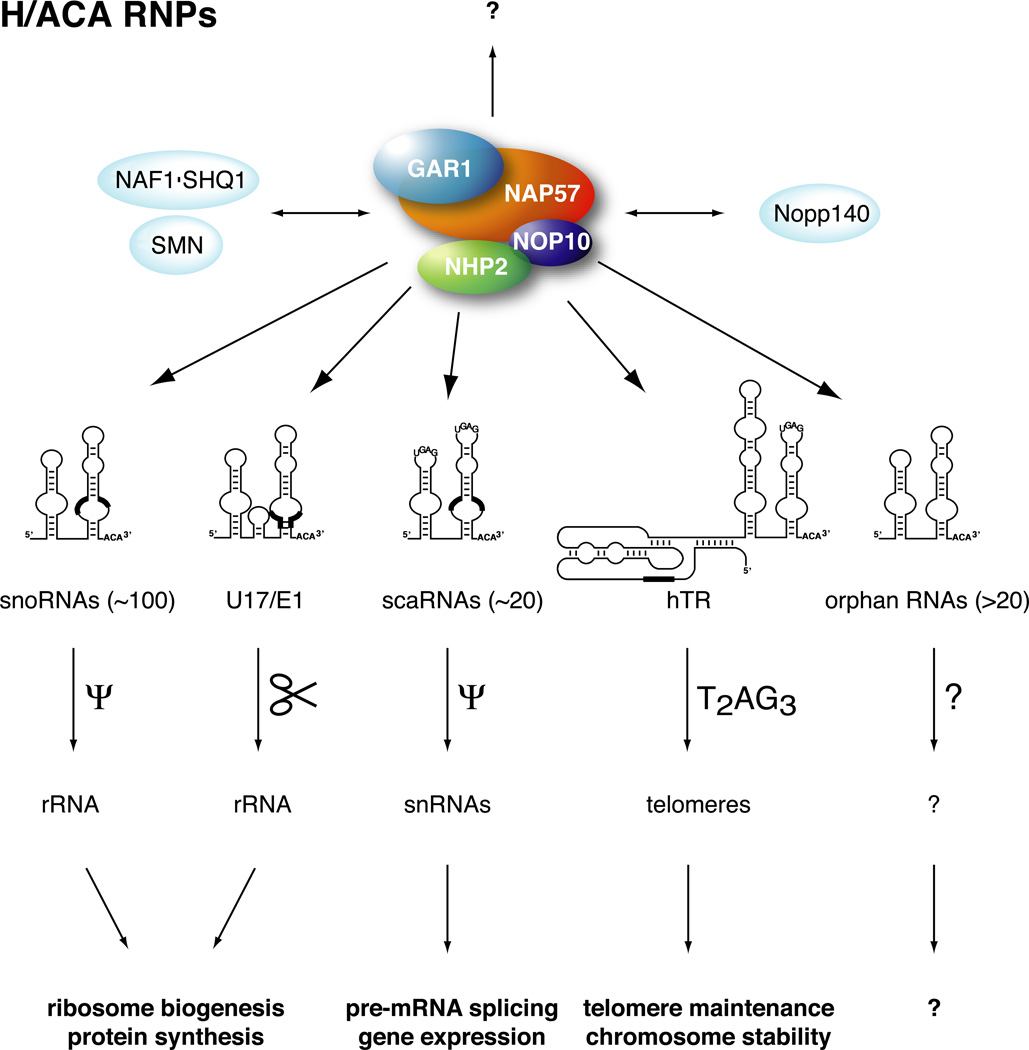
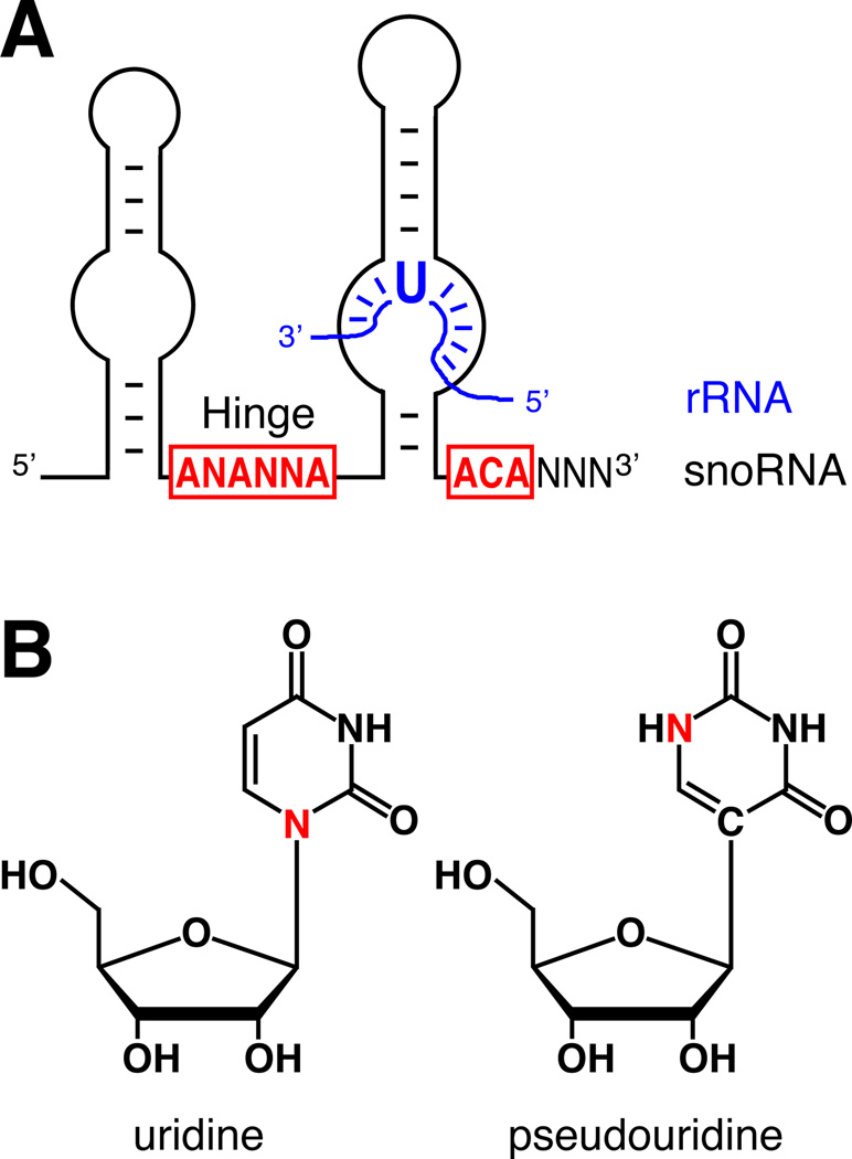
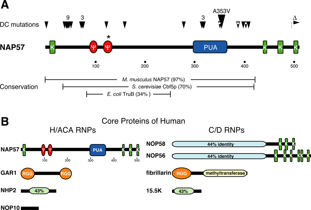
The H/ACA ribonucleoproteins (RNPs) are known as one of the two major classes of small nucleolar RNPs. They predominantly guide the site-directed pseudouridylation of target RNAs, such as ribosomal and spliceosomal small nuclear RNAs. In addition, they process ribosomal RNA and stabilize vertebrate telomerase RNA. Taken together, the function of H/ACA RNPs is essential for ribosome biogenesis, pre-mRNA splicing, and telomere maintenance. Every cell contains 100-200 different species of H/ACA RNPs, each consisting of the same four core proteins and one function-specifying H/ACA RNA. Most of these RNPs reside in nucleoli and Cajal bodies and mediate the isomerization of specific uridines to pseudouridines. Catalysis of the reaction is mediated by the putative pseudouridylase NAP57 (dyskerin, Cbf5p). Unexpectedly, mutations in this housekeeping enzyme are the major determinants of the inherited bone marrow failure syndrome dyskeratosis congenita. This review details the many diverse functions of H/ACA RNPs, some yet to be uncovered, with an emphasis on the role of the RNP proteins. The multiple functions of H/ACA RNPs appear to be reflected in the complex phenotype of dyskeratosis congenita.


Figures

Figure 1
Figure 1. Schematic overview of mammalian H/ACA RNP proteins, RNAs, targets, and functions
four H/ACA core proteins forming a protein only complex (solid colors) and interacting proteins (light blue) are depicted. Interactions between the core proteins are based on experimental evidence, i.e., GAR1 and NOP10 associate independently with NAP57 while NHP2 requires the prior association of NAP57 and NOP10. At least one set of four core proteins associates with each individual H/ACA RNA to form an RNP (arrows). RNP-independent functions of the proteins are also possible (question mark). The basic secondary structures of the five types of H/ACA RNAs and their names and numbers are shown underneath the proteins. Note hairpin sizes vary and insertions may also be present in individual RNAs. The location of antisense elements, highly conserved sequences, and the template region of hTR are indicated by thick black lines. Cajal body localization consensus elements are drawn in the stemloops (UGAG). Below the RNAs, the targets of the various RNPs, ribosomal RNA (rRNA), small nuclear RNAs (snRNAs), and telomeres, which are pseudouridylated (Ψ), processed (scissors), and extended by T2AG3 repeats, respectively, are shown. Finally, the cellular processes affected by the function of the individual RNPs are indicated at the bottom. Question marks refer to unknown targets and functions. In the case of one orphan H/ACA RNA these seem to be brain-specific (see text).
Figure 2
Figure 2. Structures of H/ACA RNA-rRNA hybrid, uridine, and pseudouridine
(A) Schematic representation of the basic two-hairpin structure of H/ACA RNAs containing the conserved sequence elements ANANNA in the hinge region (Hinge) and ACA three nucleotides from the 3’ end in red. The name of the RNAs is based on the hinge region and the ACA triplet. An rRNA hybridizing to a bulge in the 3’ hairpin with the unpaired uridine (U) to be targeted for pseudouridylation is shown (blue). Note for simplification, a second unpaired nucleotide 3’ to the target uridine is not depicted. Also, H/ACA RNAs guide pseudouridylation by the bulge in the 3’ and/or 5’ hairpin. (B) Structural formulas of uridine (left) and pseudouridine (right). Note the different location of one of the base nitrogens (red) and the nitrogen-carbon versus carbon-carbon glycosidic bond between the two isomers.
Figure 3
Figure 3. Domains of H/ACA and C/D core proteins
(A) Schematic of human NAP57 drawn to scale (amino acid positions marked under sequence) indicating various sequence elements, lysine-rich stretches (green, K) often separated by acidic serine clusters, two motifs conserved in most pseudouridylases (red, Ψ), and a domain conserved in pseudouridylases and archaeosine transglycosylases (blue, PUA). The catalytic aspartate at amino acid 125 is highlighted (asterisk). The positions of mutations identified in patients with DC are indicated above the sequence (arrowheads). The total number of tightly clustered mutations is printed over the arrowheads. The most frequent mutation, A353V, observed in ~40% of X-DC cases is enlarged. White dots in the arrowheads indicate that this residue has been mutated two different amino acids. Additionally, a carboxyterminal truncation is specified (Δ). The percent identity of human NAP57 to its mouse (M. musculus), yeast (S. cerevisiae), and bacterial (E. coli) homologs over a certain range (brackets) is given below. (B) Comparison of the domains of core proteins of H/ACA (left) and C/D RNPs (right). The sequences are drawn to scale and the domains are as in (A). In addition, glycine-arginine-rich (RGG) and conserved methyltransferase domains are indicated. Sequence identity between two proteins is given in light green or blue. Note 15.5K is also known as NHP2L1/NHPX and Snu13p in yeast.
Figure 4
Figure 4. Double immunofluorescence of NAP57 and nucleolin in HeLa cells
Indirect immunolocalization of NAP57 (A) and nucleolin (B) in fixed and permeabilized HeLa cells. (C) Merged immunofluorescence of (A) and (B) combined with DNA stain (blue). (D) Phase contrast image of the same two cells shown in the other panels. Note the granular staining of NAP57 reflecting its localization to the dense fibrillar component of nucleoli as compared to the more uniform staining of nucleolin, which is present in all parts of nucleoli. Note how the green of the nucleolin label extends beyond that of the partially colocalizing NAP57 (yellow in the merged image C). In addition and unlike nucleolin, NAP57 also concentrates in Cajal bodies (extra nucleolar red dots, particularly well visible in the merged image C).

References

    1. Aravind L, Koonin EV. Novel predicted RNA-binding domains associated with the translation machinery. J. Mol. Evol. 1999;48:291–302. - PubMed
    1. Arnez JG, Steitz TA. Crystal structure of unmodified tRNA(Gln) complexed with glutaminyl-tRNA synthetase and ATP suggests a possible role for pseudo-uridines in stabilization of RNA structure. Biochemistry. 1994;33:7560–7567. - PubMed
    1. Atzorn V, Fragapane P, Kiss T. U17/snR30 is a ubiquitous snoRNA with two conserved sequence motifs essential for 18S rRNA production. Mol. Cell. Biol. 2004;24:1769–1778. - PMC - PubMed
    1. Bachellerie JP, Cavaille J, Huttenhofer A. The expanding snoRNA world. Biochimie. 2002;84:775–790. - PubMed
    1. Badis G, Fromont-Racine M, Jacquier A. A snoRNA that guides the two most conserved pseudouridine modifications within rRNA confers a growth advantage in yeast. RNA. 2003;9:771–779. - PMC - PubMed

Publication types

MeSH terms

Substances