. 2005 Mar;96(3):157-63.
doi: 10.1111/j.1349-7006.2005.00024.x.

Inducible nitric oxide synthase-dependent DNA damage in mouse model of inflammatory bowel disease

Affiliations

Inducible nitric oxide synthase-dependent DNA damage in mouse model of inflammatory bowel disease

Xiaohui Ding et al. Cancer Sci. 2005 Mar.

Abstract

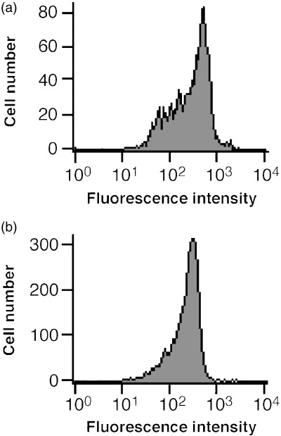
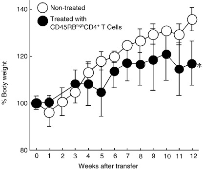
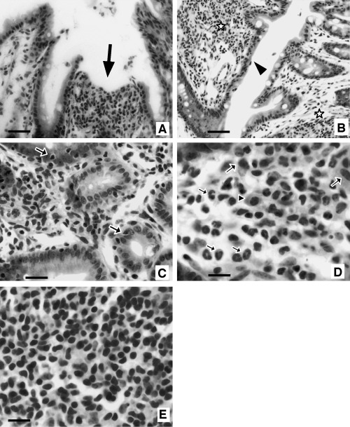
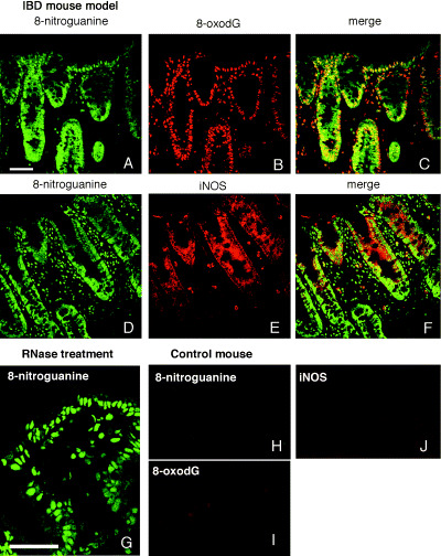
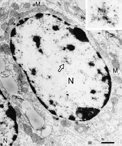
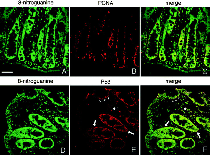
Increased cancer risk occurs in inflammatory bowel disease (IBD) undergoing long-term chronic inflammation. To evaluate whether inducible nitric oxide synthase (iNOS)-dependent DNA damage plays a role in the carcinogenic process triggered by IBD, we prepared a mouse model of IBD induced by transfer of CD45RBhighCD4+ T cells lacking regulatory T cells to female severe combined immunodeficiency (SCID) mice. CD45RBhighCD4+ T cells were isolated from mouse spleen after staining with fluorescein isothiocyanate (FITC)-conjugated anti-CD45RB monoclonal antibody, followed by anti-FITC-conjugated microbeads. This IBD mouse model showed that the bodyweight increased with aging to a lesser extent than non-treated controls, and that the intestine was shortened. Pathological findings of this mouse model, which showed severe inflammation in colon tissues, were similar to IBD patients. Double immunofluorescence technique revealed that both 8-nitroguanine and 8-oxo-7,8-dihydro-2'-deoxyguanosine (8-oxodG) were formed mainly in epithelial cells of the IBD mouse model. 8-Nitroguanine was formed in most of 8-oxodG-immunoreactive nuclei of epithelial cells. iNOS, proliferating cell nuclear antigen and p53 protein were also expressed in the colon epithelium. These results indicate that nitrative DNA damage, as well as oxidative DNA damage, is induced in colon epithelial cells of the IBD mouse model followed by proliferation of these cells, which may contribute to colon carcinogenesis.


Figures

Figure 1
Figure 1
Flow cytometric analysis of CD45RB on total CD4+ T cells and isolated CD45RBhighCD4+ T cells. CD45RBhighCD4+ T cells were positively selected from CD4+ T cells obtained from BALB/c mice spleen after staining with fluorescein isothiocyanate (FITC)‐conjugated anti‐CD45RB monoclonal antibody, followed by anti‐FITC‐conjugated microbeads. (a) CD45RB on CD4+ T cells (before isolation), and (b) isolated CD45RBhighCD4+ T cells. The horizontal axis shows the fluorescence intensity, and the vertical axis shows the cell number.
Figure 2
Figure 2
Change in bodyweight of the mice in the experiment duration. Bodyweight of the mice transferred with CD45RBhighCD4+ T cells (•) was significantly lower than that of non‐treated controls (○) at 12 weeks (n = 4, *P < 0.05).
Figure 3
Figure 3
Pathological findings in the colon of CD45RBhighCD4+ T cell‐transferred mice (hematoxylin–eosin staining). (a,b) Low power view of colon mucosa. (a) Open ulcer (arrow), which is characterized by sloughing of epithelial cells and leukocyte infiltration in lamina propria is observed. (b) Regenerated epithelial cells with mild dysplasia and goblet cell depression are prominent (arrowhead). Marked or moderate inflammatory cell infiltration and fibrosis are apparent in subepithelial tissue (asterisk). (c) Inflammatory cell infiltration in epithelial cell layer (arrow). (d,e) High power view of infiltrated inflammatory cells in lamina propria. (d) Many neutrophils (small arrow), plasma cells (arrowhead), macrophages (large arrow), and (e) lymphocytes permeate in lamina propria layer. Bars: b, 50 µm; a,c, 25 µm; d,e, 10 µm.
Figure 4
Figure 4
Formation of 8‐oxo‐7,8‐dihydro‐2′‐deoxyguanosine (8‐oxodG), 8‐nitroguanine and inducible nitric oxide synthase (iNOS) in the mouse model of inflammatory bowel disease. (a,d) 8‐Nitroguanine is accumulated in the nuclei and the cytoplasm of epithelial cells. 8‐Nitroguanine is also present in infiltrated cells. (b) 8‐oxodG formation is observed in the nuclei of epithelial cells and the infiltrated cells in lamina propria, (c) 8‐oxodG and 8‐nitroguanine co‐localized in the nuclei of most epithelial cells and infiltrated cells, (e) iNOS is expressed in the cytoplasm of epithelial cells and infiltrated cells in lamina propria, and (f) iNOS and 8‐nitroguanine colocalized in the cytoplasm of many epithelial cells and some infiltrated cells. The colon tissue sections were pretreated with ribonuclease and then immunohistochemical analysis was performed. (g) 8‐Nitroguanine immunoreactivity is more clearly observed in the nuclei of epithelial cells. (h) Little or no immunoreactivity of 8‐nitroguanine, (i) 8‐oxodG, and (j) iNOS was observed in control group mice. Scale bars: 50 µm.
Figure 5
Figure 5
Immunoelectron micrograph of 8‐nitroguanine immunoreactivity in colon epithelial cell of the mouse model of inflammatory bowel disease. 8‐Nitroguanine is accumulated mainly in the nucleus (N) of the colon epithelial cells. Immunogold particles were spread in the cytoplasm, and weak immunoreactivity was observed in mitochondria (M). Inset, the magnification of the area indicated by an arrow. Scale bar: 1 µm.
Figure 6
Figure 6
Proliferating cell nuclear antigen (PCNA) and p53 expression in the mouse model of inflammatory bowel disease. (a–c) Double immunostaining for 8‐nitroguanine and PCNA. (a,d) 8‐Nitroguanine localizes widely in the mucosa, (b) intense PCNA is expressed in the nuclei of many glandular epithelial cells and infiltrated cells in lamina propria, (c) PCNA is expressed partially in the nuclei of 8‐nitroguanine‐immunoreactive epithelial cells, (d–f) double immunostaining for 8‐nitroguanine and p53, (e) significant p53 expression is observed in the nuclei of regenerated epithelial cells (arrowhead) and some infiltrated cells (small arrow). p53 is also induced in the cytoplasm of many epithelial cells in lower portions of the gland (large arrow). (f) p53 is expressed in most of 8‐nitroguanine‐immunoreactive epithelial cells. Scale bar: 50 µm.

Similar articles

Cited by

References

    1. Podolsky DK. Inflammatory bowel disease. N Engl J Med 2002; 347: 417–29. - PubMed
    1. Bouma G, Strober W. The immunological and genetic basis of inflammatory bowel disease. Nat Rev Immunol 2003; 3: 521–33. - PubMed
    1. Coussens LM, Werb Z. Inflammation and cancer. Nature 2002; 420: 860–7. - PMC - PubMed
    1. Ekbom A, Helmick C, Zack M, Adami HO. Increased risk of large‐bowel cancer in Crohn's disease with colonic involvement. Lancet 1990; 336: 357–9. - PubMed
    1. Langholz E, Munkholm P, Davidsen M, Binder V. Colorectal cancer risk and mortality in patients with ulcerative colitis. Gastroenterology 1992; 103: 1444–51. - PubMed

Publication types

Substances