. 2005 Mar;96(3):183-90.
doi: 10.1111/j.1349-7006.2005.00026.x.

Regulation of YKL-40 expression during genotoxic or microenvironmental stress in human glioblastoma cells

Affiliations

Regulation of YKL-40 expression during genotoxic or microenvironmental stress in human glioblastoma cells

Nanna Junker et al. Cancer Sci. 2005 Mar.

Abstract

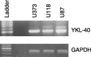
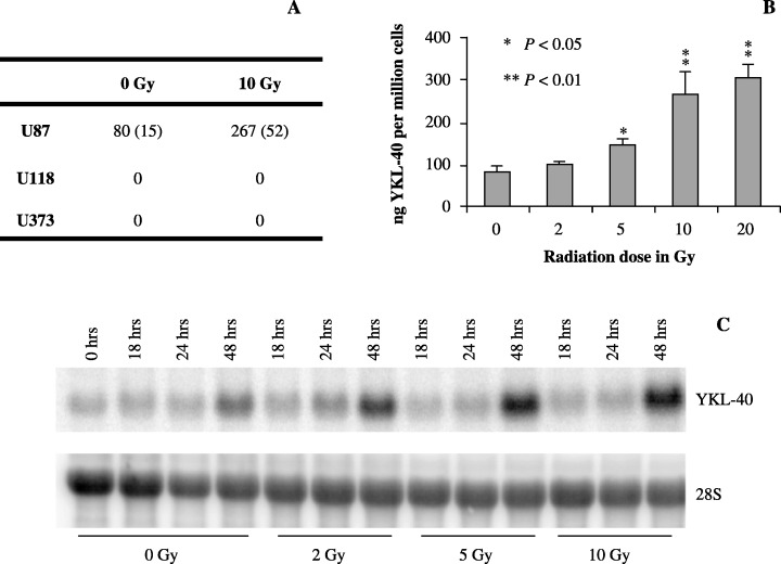
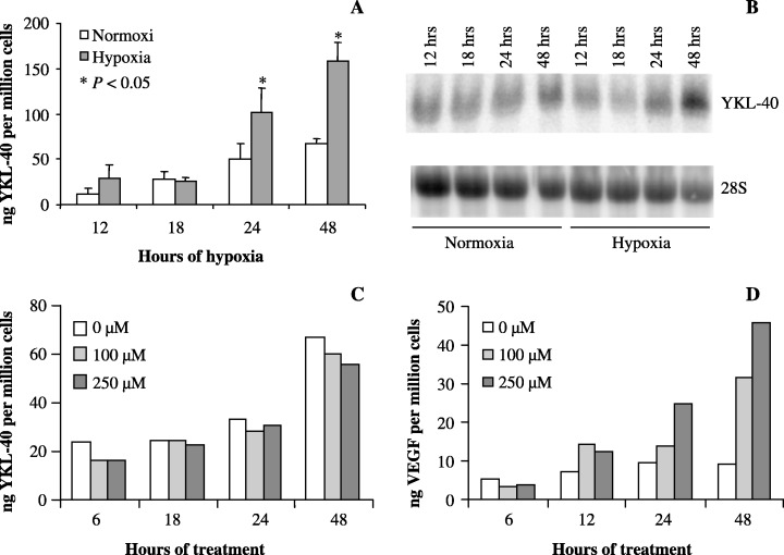
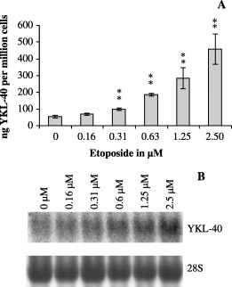
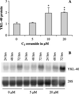
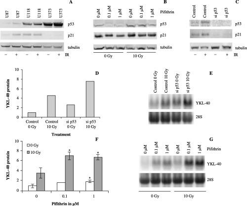
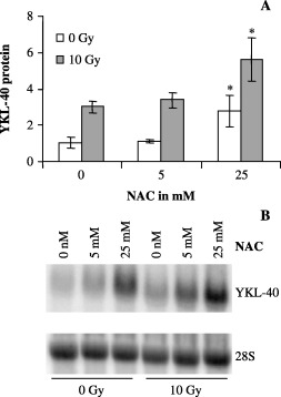
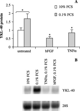
YKL-40 is a 40 kDa secreted glycoprotein belonging to the family of 'mammalian chitinase-like proteins', but without chitinase activity. YKL-40 has a proliferative effect on fibroblasts, chondrocytes and synoviocytes, and chemotactic effect on endothelium and vascular smooth muscle cells. Elevated YKL-40 levels are found in serum of patients with diseases characterized by inflammation, fibrosis and tissue remodeling. Several studies have reported that high serum YKL-40 levels in patients with cancer are associated with poor prognosis. YKL-40 expression is strongly elevated in serum and biopsy material from glioblastomas patients. We investigated the expression of YKL-40 in three human malignant glioma cell lines exposed to different types of stress. Whereas a polymerase chain reaction transcript was detectable in all three cell lines, only U87 produced measurable amounts of YKL-40 protein. In U87, hypoxia and ionizing radiation induced a significant increase in YKL-40 after 24-48 h. The hypoxic induction of YKL-40 was independent of HIF1. Etoposide, ceramide, serum depletion and confluence all led to elevated YKL-40. Inhibition of p53 augmented the YKL-40 expression indicating that YKL-40 is attenuated by p53. In contrast, both basic fibroblast growth factor and tumor necrosing factor-alpha repressed YKL-40. These are the first data on regulation of YKL-40 in cancer cells. Diverse types of stress resulted in YKL-40 elevation, which strongly supports an involvement of YKL-40 in the malignant phenotype as a cellular survival factor in an adverse microenvironment.


Figures

Figure 1
Figure 1
Reverse transcriptase‐polymerase chain reaction (RT‐PCR) of cDNA from U87, U118 and U373 cells with primers specific for YKL‐40. The PCR products were separated on a 1.2% agarose gel and visualized by ethidium bromide staining. A ladder was included as a size control and a separate RT‐PCR with primers for the housekeeping gene glyceraldehyde‐3‐phosphate dehydrogenase was performed to verify the quality of the cDNA.
Figure 2
Figure 2
(a) U87, U118 and U373 cells were irradiated with 10 Gy. After 48 h, conditioned cell culture media was collected and YKL‐40 protein levels were determined by enzyme‐linked immunosorbent assay and divided by the cell number. Experiments were performed in triplicates. Mean (ng/million cells) and standard deviation (SD) are shown. (b) U87 cells were irradiated with 0, 2, 5, 10 and 20 Gy and harvested as in (a). Mean and SD are shown. (c) In a parallel experiment total ribonucleic acid was extracted at different time points after 0, 2, 5 and 10 Gy and analyzed by Northern blotting with a YKL‐40 probe. The stripped membrane was probed for 28S to verify equal loading.
Figure 3
Figure 3
(a,b) U87 cells were exposed to hypoxia (0.1%) or normoxia. At different time points conditioned cell culture media and cells were collected for (a) enzyme‐linked immunosorbent assay (ELISA), and (b) total ribonucleic acid (RNA) for Northern blot. (a) The YKL‐40 protein level was correlated to cell number. Experiments were performed in triplicates. Mean and standard deviation (SD) are shown. (b) YKL‐40 messenger RNA was analyzed by Northern blotting with a YKL‐40 probe. The stripped membrane was probed for 28S to verify equal loading. (c,d) Cells were treated with cobalt chloride at 100 µM and 250 µM to induce ‘chemical hypoxia’. At different time points, conditioned cell culture media was collected for determination of YKL‐40 protein levels by ELISA (c), and vascular endothelial growth factor protein levels by ELISA (d).
Figure 4
Figure 4
U87 cells were treated with etoposide (0.16, 0.31, 0.63, 1.25 and 2.5 µM, or vehicle only [0.1% DMSO]) for 48 h. (a) YKL‐40 was measured in the conditioned media by enzyme‐linked immunosorbent assay and divided by the cell number. Experiments were performed in triplicates. Mean and standard deviation are shown. (b) In a parallel experiment total ribonucleic acid was extracted and analyzed by Northern blotting with a YKL‐40 probe. The stripped membrane was probed for 28S to verify equal loading.
Figure 5
Figure 5
U87 cells were treated with C2 ceramide (5 µM or 20 µM, or vehicle only [0.1% dimethyl sulfoxide]) for 24, 48 and 72 h. (a) YKL‐40 was measured in the conditioned media by enzyme‐linked immunosorbent assay after 72 h of treatment and normalized to 0 µM. Experiments were performed in triplicates. Mean and standard deviation are shown. (b) Cells were collected after 24, 48 and 72 h of treatment, and YKL‐40 messenger ribonucleic acid was analyzed by Northern blotting with an YKL‐40 probe. The stripped membrane was probed for 28S to verify equal loading.
Figure 6
Figure 6
(a) U87, U118 and U373 cells were irradiated (10 Gy). Protein from irradiated and control cells was used for Western blotting with p53 and p21 antibodies. Equal loading was confirmed by tubulin antibody. (b) Confirmation of siRNA treatment against p53. Protein from siRNA treated U87 cells and controls with and without irradiation (10 Gy) was used for Western blotting with p53 and p21 antibodies. Equal loading was confirmed by tubulin antibody. (c) Similar to (b) with protein from cyclic pifithrin‐α treated U87 cells. (d) U87 cells were treated with siRNA against p53 (si p53) and ionizing radiation (10 Gy). Controls were treated with siRNA buffer only. YKL‐40 was measured in conditioned media by enzyme‐linked immunosorbent assay (ELISA) 48 h after ionizing radiation. The experiment was performed twice and the mean is shown. YKL‐40 levels were normalized to control (0 Gy). (e) YKL‐40 mRNA from the same experiment was analyzed by Northern blotting with an YKL‐40 probe. The stripped membrane was probed for 28S to verify equal loading. (f) U87 cells were treated with the small molecule p53 inhibitor cyclic pifithrin‐α, 1 h prior to ionizing radiation (10 Gy). Control cells were treated with vehicle (0.1% dimethyl sulfoxide) only. Media was harvested 48 h after irradiation for YKL‐40 protein measurement by ELISA. YKL‐40 levels are normalized to control (0 Gy). Experiments were performed in triplicates. Mean and standard deviation are shown. (g) The increases in YKL‐40 levels were confirmed on RNA levels by Northern blotting with YKL‐40 and 28S probes.
Figure 7
Figure 7
U87 cells were exposed to N‐acetyl cysteine (NAC) (5 µM and 25 µM) and ionizing radiation (10 Gy). Cells were treated with NAC 24 h prior to irradiation. Conditioned cell culture media and cells were collected for (a) enzyme‐linked immunosorbent assay and (b) Northern blot 48 h after irradiation. (a) Experiments were performed in triplicates and YKL‐40 levels were normalized to control, 0 Gy. Mean and standard deviation are shown. (b) YKL‐40 messenger ribonucleic acid was analyzed by Northern blotting with an YKL‐40 probe. The stripped membrane was probed for 28S to verify equal loading.
Figure 8
Figure 8
U87 cells were treated with 0.1% and 10% fetal bovine serum (FBS), as well as with tumor necrosing factor‐α (10 ng/mL) and basic fibroblast growth factor (25 ng/mL) in 0.1% FBS media for 72 h. Conditioned cell culture media and cells were collected for (a) enzyme‐linked immunosorbent assay and (b) Northern blot. (a) Experiments were performed in triplicates and YKL‐40 levels were normalized to untreated controls. Mean and standard deviation are shown. (b) YKL‐40 messenger ribonucleic acid was analyzed by Northern blotting with an YKL‐40 probe. The stripped membrane was probed for 28S to verify equal loading.

References

    1. Johansen JS, Williamson MK, Rice JS, Price PA. Identification of proteins secreted by human osteoblastic cells in culture. J Bone Miner Res 1992; 7: 501–12. - PubMed
    1. Hakala BE, White C, Recklies AD. Human cartilage gp‐39, a major secretory product of articular chondrocytes and synovial cells, is a mammalian member of a chitinase protein family. J Biol Chem 1993; 268: 25803–10. - PubMed
    1. Millis AJ, Hoyle M, Reich E, Mann DM. Isolation and characterization of a Mr = 38,000 protein from differentiating smooth muscle cells. J Biol Chem 1985; 260: 3754–61. - PubMed
    1. Shackelton LM, Mann DM, Millis AJ. Identification of a 38‐kDa heparin‐binding glycoprotein (gp38k) in differentiating vascular smooth muscle cells as a member of a group of proteins associated with tissue remodeling. J Biol Chem 1995; 270: 13076–83. - PubMed
    1. Malinda KM, Ponce L, Kleinman HK, Shackelton LM, Millis AJ. Gp38k, a protein synthesized by vascular smooth muscle cells, stimulates directional migration of human umbilical vein endothelial cells. Exp Cell Res 1999; 250: 168–73. - PubMed

Publication types