. 2005 May;16(5):2433-42.
doi: 10.1091/mbc.e04-11-1004. Epub 2005 Mar 16.

Disruption of the MAP1B-related protein FUTSCH leads to changes in the neuronal cytoskeleton, axonal transport defects, and progressive neurodegeneration in Drosophila

Affiliations

Disruption of the MAP1B-related protein FUTSCH leads to changes in the neuronal cytoskeleton, axonal transport defects, and progressive neurodegeneration in Drosophila

Alexandre Bettencourt da Cruz et al. Mol Biol Cell. 2005 May.

Abstract

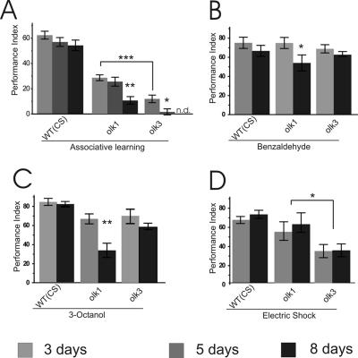
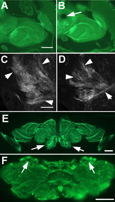
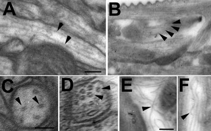
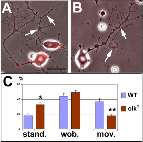
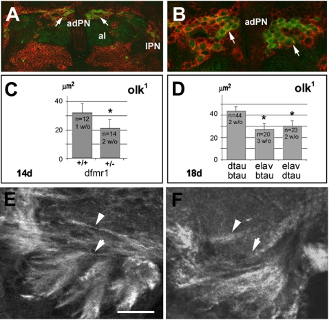
The elaboration of neuronal axons and dendrites is dependent on a functional cytoskeleton. Cytoskeletal components have been shown to play a major role in the maintenance of the nervous system through adulthood, and changes in neurofilaments and microtubule-associated proteins (MAPs) have been linked to a variety of neurodegenerative diseases. Here we show that Futsch, the fly homolog of MAP1B, is involved in progressive neurodegeneration. Although Futsch is widely expressed throughout the CNS, degeneration in futsch(olk) primarily occurs in the olfactory system and mushroom bodies. Consistent with the predicted function of Futsch, we find abnormalities in the microtubule network and defects in axonal transport. Degeneration in the adult brain is preceded by learning deficits, revealing a neuronal dysfunction before detectable levels of cell death. Futsch is negatively regulated by the Drosophila Fragile X mental retardation gene, and a mutation in this gene delays the onset of neurodegeneration in futsch(olk). A similar effect is obtained by expression of either fly or bovine tau, suggesting a certain degree of functional redundancy of MAPs. The futsch(olk) mutants exhibit several characteristics of human neurodegenerative diseases, providing an opportunity to study the role of MAPs in progressive neurodegeneration within an experimentally accessible, in vivo model system.


Figures

Figure 2.
Figure 2.
Fiber structure defects and progressive degeneration in olk1. (A) Silver staining reveals fibers and axon bundles in a wild-type head section. (B) In olk1, the fibers are much less prominent, revealing a defect in fiber structure within the mutant. (C) The mean size of vacuoles in the mechanosensory neuropil of olk1 increases with age (8 d, p < 0.1; 14 d, p < 0.01), whereas the number of flies that do not show vacuoles in this region (w/o) decreases with age. Histograms indicate mean ± SEM, and the number flies measured per condition is indicated. (D) Western blot using the anti-futsch antibody 22C10. The amount of Futsch protein (arrowhead) is significantly reduced in olk2 and olk3 mutants compared with wild-type controls. In olk1 flies, we could not detect any protein. Scale bar, (A and B) 50 μm.
Figure 1.
Figure 1.
Progressive degeneration in olk1. (A and D) Sections from the brain of a 1-d-old olk fly show no abnormalities compared with wild-type controls (unpublished data). (B and F) At 10 d into adult life, vacuoles have formed in the antennal lobes (al, arrows in F) and the mechanosensory neuropil of the lateral protocerebrum (arrow in B). (C) The lesion in the mechanosensory neuropil (arrow) enlarges dramatically with further aging (20 d). (E) The first vacuoles can be found in the calyx (c) of a 6-d-old mutant (arrows). (G) In 20-d-old olk flies, vacuoles can also be found in the region of the lateral horn (lh) of the protocerebrum (arrows). Sections are horizontal 1-μm plastic sections stained with toluidine blue. Scale bar, (A–C) 50 μm; (D and E) 10 μm; (F) 20 μm; (G) 25 μm.
Figure 3.
Figure 3.
Degeneration of projection neurons in olk1 mutants (visualized by GFP expression). (A) Wild-type, 10 d. The PNs send their dendrites into the antennal lobe (AL) and their axons into the calyx via the inner antennocerebral tract (iACT) and into the lateral horn (LH) via the medial antennocerebral tract (mACT). (B) In 1-d-old mutants, the PNs appear normal. (C) In a 10-d-old mutant, the staining of the dendritic field in the AL was noticeably weaker, and the iACT appeared both thinner and more weakly stained (arrowhead). The staining of the mACT (arrow) and its termination field and of the cell bodies of all three types of PNs was not noticeably altered at this age. (D) Further aging (23 d) resulted in severely reduced staining in all of these regions, suggesting that most or all of the PNs had undergone cell death. Images show whole-mounts using GH146-GAL4 and UAS-GFP. adPN, vPN, and lPN = anterodorsal, ventral, and lateral projection neurons, respectively. Scale bar, 50 μm.
Figure 4.
Figure 4.
Progressive loss of olfactory learning ability in olk mutants. Associative and nonassociative olfactory behavior was tested at the age of 3, 5, and 8 d. (A) Performance of olfactory short-term memory progressively declines in olk mutant flies. Already by day 3, performance was significantly impaired (p < 0.001 for both olk1 and olk3 compared with wild-type Canton-S). Performance indices (PIs) were near zero by day 5 in olk3 (p < 0.05) and by day 8 in olk1 (p < 0.01). Day 8 was not measured in olk3 mutants (n.d.). (B–D) Response to the task-relevant stimuli was measured for the associated odorants (B and C) and the behavioral reinforcer electric shock (D). Olfactory acuity was also affected in an age-dependent manner in olk1 mutant flies (p < 0.05 for benzaldehyde and p < 0.01 for 3-octanol, respectively), whereas there was no progressive effect on acuity of response to electric shock. However, overall sensitivity was significantly reduced in olk3 mutants (p < 0.05). Histograms show means ± SEM of six experiments with 100 flies each.
Figure 5.
Figure 5.
Impact of the olk mutation on tubulin organization and Futsch expression. (A) Tubulin staining in a paraffin section of a 14-d-old wild-type fly. (B) Overall tubulin staining appeared stronger in an age-matched olk1 mutant animal. (C and D) Confocal microscopic images taken from the calyx of these sections from (C) wild-type and (D) olk1 animals reveal a more densely localized pattern of staining in the mutant (arrowheads). (E and F) Immunohistochemistry of wild-type brain sections using 22C10 (anti-Futsch). Futsch is widely distributed in the brain, with strong expression in the antennal lobes (arrows in E) and calyx (arrows in F). Scale bar, (A and B) 50 μm; (C and D) 10 μm; (E and F) 50 μm.
Figure 6.
Figure 6.
Disorganized microtubule in the antennal lobes of olk1. (A) Microtubules (arrowheads) in a 14-d-old wild-type antennal lobe are straight and evenly spaced. (B) In an age-matched olk1 mutant, a noticeable increase in the number of microtubules was apparent (arrowheads), leading to reduced spacing between microtubules. This difference in density could also be seen in cross sections from wild-type (C) and olk1 mutant flies (D). (E and F) In addition, microtubules in the mutant were seen to bend, touch, or cross each other (arrowheads), a phenotype not observed in wild-type brains. Scale bar, 0.05 μm.
Figure 7.
Figure 7.
olk mutations affect mitochondrial transport. Neurons cultured for 24 h from (A) wild-type and (B) olk1 pupae show similar patterns of outgrowth and branching. (C) To analyze axonal transport, we labeled mitochondria (red, arrows in A and B) and observed their movement for 98 s with pictures taken every 2 s. During this interval, significantly more mitochondria in olk1 did not change their position (stand.) and fewer mitochondria showed active movement within the neurites (mov.). The number of mitochondria exhibiting small back-and-forth movements (<2 μm; wob.) was not significantly changed in olk1 neurons (movies can be seen in Supplementary Figure 3). Histograms show means ± SEMs. (stand. Phosphate-buffered saline < 0.05, mov. Phosphate-buffered saline < 0.01). Scale bar, (A and B) 10 μm.
Figure 8.
Figure 8.
Interaction of olk with dfmr1 and tau. (A) Double staining using an anti-DFMR1 antibody (red) and GFP staining of PNs (green). (B) A higher magnification view shows that the anterodorsal PNs (adPN; arrows) express DFMR1. Al, antennal lobes; lPN, lateral projection neurons. (C) The average size of vacuoles in the mechanosensory neuropil of the lateral protocerebrum was significantly reduced in day 14 olk1 flies carrying the dfmr1 deletion (+/-), compared with age-matched olk1 flies from the same cross but carrying a balancer chromosome (+/+). Average vacuole size was reduced by approximately one-third compared with control olk flies (p < 0.01). (D) Eighteen-day-old olk1 flies that also expressed either fly tau (dtau) or bovine tau (btau) in all neurons (driven by the ELAV promoter) had significantly fewer vacuoles than in olk1 flies (p < 0.05). Histograms show means ± SEM, and the number of flies measured in each strain are indicated. (E and F) Anti-tubulin immunostaining of brain sections from 14-d-old olk1 flies without the dfmr1 deficiency (E) and with the dfmr1 deficiency (F). Removing one copy of dfmr1 restored the overall pattern of tubulin staining pattern toward the more diffuse pattern seen in wild-type flies (compare with Figure 5). Scale bar, (A) 15 μm; (B) 6 μm; (E and F) 10 μm.

Similar articles

Cited by

References

    1. Blest, A. D. (1961). Some modifications of Holme's silver method for insect central nervous systems. Q. J. Microsc. Sci. 102, 413.
    1. Bouquet, C., Soares, S., von Boxberg, Y., Ravaille-Veron, M., Propst, F., and Nothias, F. (2004). Microtubule-associated protein 1B controls directionality of growth cone migration and axonal branching in regeneration of adult dorsal root ganglia neurons. J. Neurosci. 24, 7204-7213. - PMC - PubMed
    1. Brandt, R. (2001). Cytoskeletal mechanisms of neuronal degeneration. Cell Tissue Res. 305, 255-265. - PubMed
    1. Buchner, S., Buchner, E., and Hofbauer, A. (1989). Immunohistochemistry of the brain. In: Drosophila: A Laboratory Manual. ed. M. Ashburner, Cold Spring Harbor, NY: Cold Spring Harbor Press, 271-273.
    1. Ding, J., Liu, J. J., Kowal, A. S., Nardine, T., Bhattacharya, P., Lee, A., and Yang, Y. (2002). Microtubule-associated protein 1B: a neuronal binding partner for gigaxonin. J. Cell Biol. 158, 427-433. - PMC - PubMed

Publication types

MeSH terms

LinkOut - more resources