. 2005 May;16(5):2458-69.
doi: 10.1091/mbc.e03-12-0917. Epub 2005 Mar 16.

The unique N-terminus of R-ras is required for Rac activation and precise regulation of cell migration

Affiliations

The unique N-terminus of R-ras is required for Rac activation and precise regulation of cell migration

Stephen P Holly et al. Mol Biol Cell. 2005 May.

Abstract

The Ras family GTPase, R-Ras, elicits important integrin-dependent cellular behaviors such as adhesion, spreading and migration. While oncogenic Ras GTPases and R-Ras share extensive sequence homology, R-Ras induces a distinct set of cellular behaviors. To explore the structural basis for these differences, we asked whether the unique N-terminal 26 amino acid extension of R-Ras was responsible for R-Ras-specific signaling events. Using a 32D mouse myeloid cell line, we show that full-length R-Ras activates Rac and induces Rac-dependent cell spreading. In contrast, truncated R-Ras lacking its first 26 amino acids fails to activate Rac, resulting in reduced cell spreading. Truncated R-Ras also stimulates more beta3 integrin-dependent cell migration than full-length R-Ras, suggesting that the N-terminus may negatively regulate cell movement. However, neither the subcellular localization of R-Ras nor its effects on cell adhesion are affected by the presence or absence of the N-terminus. These results indicate that the N-terminus of R-Ras positively regulates specific R-Ras functions such as Rac activation and cell spreading but negatively regulates R-Ras-mediated cell migration.


Figures

Figure 8.
Figure 8.
Full-length R-Ras produces a Rac-like, spread morphology but truncated R-Ras fails to induce maximal spreading. (A) Cells were transfected as indicated and treated as in Figure 7, except that cells were plated on 0.5 μg/ml vitronectin (CA, constitutively active). An anti-HA antibody, 12CA5, was used to detect R-Ras and a polyclonal c-myc antibody was used to detect myc-tagged Rac. (B) Rac-like phenotypes of cells shown in A were scored by microscopically counting the percentage of randomly chosen transfected cells having the Rac phenotype defined as a round, cell area greater than 400 μm2. Phase micrographs were used for control cells not expressing epitope-tagged proteins. Each bar represents 190 cells for CA-Rac, 271 cells for pCGN, 307 cells for NΔ87L, 275 cells for NΔ87L+DN-Rac, 384 cells for Q87L, 374 cells for Q87L+DN-Rac, and 364 cells for DN-Rac alone. (C) The total cell area of empty vector control cells (n = 31), NΔ87L R-Ras–expressing cells (n = 25, *p < 0.00003 vs. Q87L), Q87L R-Ras–expressing cells (n = 15), and Q87L R-Ras + DN-Rac coexpressing cells (n = 8) was examined using ImagePro software.
Figure 1.
Figure 1.
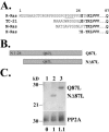
R-Ras contains a unique N-terminus that does not alter protein expression. (A) Shown is a primary amino acid sequence alignment of Ras family members most closely related to R-Ras. The initiator methionine of R-Ras is labeled as 1. Conserved regions are bolded, including the invariant glutamine at position 87. Amino acids that define a putative SH3-binding domain in the N-terminus of R-Ras are underlined. (B) A schematic representation of the full-length versus the truncation mutant of R-Ras lacking its first 26 amino acids is depicted. (C) Whole-cell lysates were prepared from mouse 32D cells transiently transfected (see Materials and Methods) with empty pCMV vector (lane 1), NΔ87L R-Ras (lane 2), or Q87L R-Ras (lane 3). Myc-tagged R-Ras proteins were detected with the polyclonal anti-R-Ras antibody, C19. Bottom numbers indicate ratios of R-Ras expression normalized to protein phosphatase 2A levels, which served as a loading control. Myc-tagged R-Ras proteins were also detected in 32D and human K562 cell lysates with the 9E10 anti-myc monoclonal antibody (unpublished data).
Figure 2.
Figure 2.
The N-terminus of R-Ras is not critical for integrin-dependent cell adhesion to vitronectin. (A) Mouse 32D cells were transiently cotransfected with empty pCMV vector, pCMV NΔ87L R-Ras, or pCMV Q87L R-Ras plus pGL-3, a luciferase reporter plasmid. Cells (50,000) were loaded into a 96-well plate coated with the indicated concentrations of vitronectin. Cells were incubated for 40 min, washed, and assayed for luciferase activity (see Materials and Methods). Cells not used for adhesion were lysed to determine expression levels of truncated and full-length R-Ras (Figure 2A, inset). (B) 32D cells were cotransfected with pCMV Q87L R-Ras and pGL-3 and pretreated with the indicated integrin blocking antibodies at the indicated concentrations for 15 min at RT. Cells were loaded onto wells coated with 0.5 μg/ml vitronectin in the presence of antibodies and assayed for adhesion as in A. Means of triplicate samples ± SEM from a representative experiment are shown for both A (n >5 experiments) and B (two experiments).
Figure 3.
Figure 3.
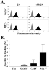
Integrin αIIbβ3 is present on 32D cells but R-Ras does not increase β3 integrin affinity for ligand. (A) Mouse 32D cells were cotransfected with empty pCGN control vector or pCGN Q87L R-Ras plus pGL-3. Cells were analyzed by flow cytometry using control hamster IgG (gray histograms), the anti-β3 antibody, 2C9.G2 (black histograms) or the anti-αIIbβ3 antibody, 1B5 (black histograms). (B) As a positive control, mouse 32D cells were treated with Mn2+ to activate integrins or transfected with the negative control GFP-tagged dynamin 2 (Con), GFP-NΔ38V R-Ras (NΔ38V) or GFP-G38V R-Ras (G38V). Alexa 546–conjugated fibrinogen binding to GFP-transfected cells was assessed by flow cytometry. Specific binding was determined by subtracting nonspecific fibrinogen binding occurring in the presence of an inhibitory β3 integrin antibody, 2C9.G2, from total fibrinogen binding. Results were expressed as the mean percentage ± SEM of transfected cells specifically binding fibrinogen from four experiments.
Figure 4.
Figure 4.
Inhibition of actin polymerization and phosphatase activity block R-Ras–dependent cell adhesion. Mouse 32D cells were cotransfected with empty pCGN control vector, pCGN NΔ87L R-Ras, pCGN Q87L R-Ras, or pCGN S43N R-Ras plus pGL-3 and assayed for cell adhesion as described in Figure 2A. Cells were pretreated with the indicated concentrations of latrunculin B (LB), cytochalasin D (CD), calyculin A (CA), or 0.1% DMSO control for 30 min at 37°C before adhesion to vitronectin. Data represent triplicate means of a representative experiment from four independent trials.
Figure 5.
Figure 5.
Activated R-Ras colocalizes with actin in filopodia and membrane ruffles. Mouse 32D cells were transfected with pCGN vector alone (one cell shown in a–d), pCGN Q87L R-Ras (one cell shown in e–h), or pCGN NΔ87L R-Ras (one cell shown in i–l). An anti-HA antibody, 12CA5, was used to detect R-Ras (e and i) and Alexa 568-phalloidin was used to detect F-actin (b, f, and j). “Merge” signifies an overlay of two single focal plane images ∼0.15-μm-thick collected by z-series scanning, whereas “smash” refers to an overlay of all slices in the z-series and thus reveals additional data. Images of single focal planes depict dorsal sections of the cells. Arrowheads in panel g and k point to membrane ruffles at the cell periphery. The arrow in panel h indicates an actin “rib” or filopod associated with a lamellipod.
Figure 6.
Figure 6.
The absence of the N-terminus of R-Ras produces a Rac-dependent dendritic morphology. Mouse 32D cells were transfected as in Figure 2A (without pGL-3) and adhered to coverslips. (A) Myc-tagged R-Ras was visualized with the 9E10 monoclonal antibody (mAb) and filamentous actin was stained with Alexa 568-phalloidin. Shown are a representative nonspreading control cell (a–c) and a representative cell expressing Q87L R-Ras (d–f). The bottom six panels depict two cells expressing NΔ87L R-Ras (first cell, g–i; second cell, j–l). The merged image is an overlay of two single focal planes near the ventral surface of the cells. (B) The filopodia-like morphology of the 32D cells having two or more protrusions (A, cells g–l) was scored microscopically by an observer who was unaware of sample identity. Data represent means from three independent experiments (*p = 0.007). (C) The percentage of cells containing four or more protrusions was scored in cells transfected with empty vector (n = 347), NΔ87L R-Ras (n = 959) or Q87L R-Ras (n = 1002), NΔ87L R-Ras plus DN-Rac (n = 442) or DN-Rac alone (n = 255). Data represent means from three independent experiments (*p < 0.03 for NΔ87L vs. Q87L and p < 0.02 for NΔ87L vs. NΔ87L+DN Rac).
Figure 7.
Figure 7.
Full-length but not truncated R-Ras stimulates Rac activation in a PI3K-dependent manner in 32D cells. Mouse 32D cells were transfected as in Figure 6, washed four times, and adhered to 60-mm dishes coated with 10 μg/ml vitronectin for 1 h. Adherent cells were lysed and recombinant GST-Pak1 PBD was used to precipitate GTP-bound Rac. R-Ras expression levels were determined in whole-cell lysates by reprobing the same blot using an anti-HA mAb (bottom panel). (A) Lane 1, empty vector control; lane 2, NΔ87L R-Ras; and lane 3, Q87L R-Ras. (B) Relative Rac activation was calculated by dividing Rac-GTP by total Rac present in whole cell lysates (*p < 0.002 for Q87L vs. NΔ87L). Shown is the mean ± SEM of three independent pull-down experiments (control n = 2). (C) Cells were transfected as in A and treated with 0.12% DMSO or 25 μM LY294002 for 30 min at 37°C before plating for 1 h as above. An example of four independent experiments is shown. Lane 1, vector control + DMSO; lane 2, NΔ87L R-Ras + DMSO; lane 3, NΔ87L R-Ras + LY; lane 4, Q87L R-Ras + DMSO; and lane 5, Q87L R-Ras + LY. (D) Inhibition of Rac activation by LY294002 was calculated as in B. Data represent the mean ± SEM of four independent experiments (*p < 0.03 for Q87L vs. Q87L + LY).
Figure 9.
Figure 9.
NΔ87L R-Ras and Q87L R-Ras activate PI3K in 3T3 cells. NIH 3T3 cells were transfected with empty pCMV vector (lanes 1 and 2), pCGN NΔ87L R-Ras (lanes 3 and 4) or pCGN Q87L (lanes 5 and 6). Cells were treated with 0.37% DMSO (odd lanes) or 25 μM LY294002 (even lanes) for 30 min and lysed. Whole cell lysates were subjected to SDS-PAGE and phosphorylation of Akt on serine 473 was detected with a polyclonal antibody. Phosphorylated Akt was used as a reporter of PI3K activation. The phospho-Akt blot was stripped and reprobed for HA-tagged R-Ras using the 12CA5 mAb. Total Akt was detected with a distinct polyclonal antibody to assess protein loading.
Figure 10.
Figure 10.
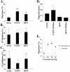
Removal of the R-Ras N-terminus increases cell migration in response to serum. (A) Mouse 32D cells were transfected as in Figure 2A. The underside of a 96-well plate embedded with 3-μm pores was coated with 1 μg/ml vitronectin. Serum-containing media was placed in the bottom chamber and 1 × 10⁵ cells were loaded on top of the chamber. Migration was expressed as the percentage of migrated luciferase-expressing cells (**p = 0.001 for NΔ87L R-Ras vs. vector control and *p < 0.006 for Q87L R-Ras vs. vector control; p < 0.04 for NΔ87L R-Ras vs. Q87L R-Ras). Data represent triplicate means from five experiments ± SEM (B) Cells were transfected as above but were pretreated with an anti-integrin β3 antibody (clone 2C9.G2) or control hamster IgG for 30 min at 37°C. Percent inhibition of migration was calculated according to the formula: (migrationIgG - migrationβ3)/migrationIgG× 100 and represents means from three independent experiments ± SEM. (C) Cells were transfected as above and treated with 0.37% DMSO or 25 μM LY294002 for 30 min at 37°C. Cells were allowed to migrate in the presence of these reagents. Percent inhibition was calculated according to the formula: (migrationDMSO - migrationLY)/migrationDMSO× 100. Data represent means from three independent experiments ± SEM (*p = 0.02 for DMSO vs. LY294002). (D) Cells were transfected with pCGN R-Ras plasmid alone or pCGN R-Ras plus pCMV DN-Rac as indicated such that the amount of DNA totaled 12 μg for each condition. Cell migration was performed as above, except that a migration index was calculated by normalizing migration values to R-Ras levels determined by densitometry of anti-HA immunoblots. Control cells are not shown because the data cannot be normalized to R-Ras expression levels. Data represent means ± SEM from two separate experiments. (E) Checkerboard analysis was performed to determine if migration to serum was chemotactic. Mouse 32D cells were transfected as in Figure 6 and resuspended in 0, 0.5, 10, or 20% serum-containing media (abscissa) and allowed to migrate toward 0.5% serum-containing media in the bottom chamber. Shown is a representative experiment from two independent trials.

Similar articles

Cited by

References

    1. Berrier, A. L., Mastrangelo, A. M., Downward, J., Ginsberg, M., and LaFlamme, S. E. (2000). Activated R-ras, Rac1, PI 3-kinase and PKCepsilon can each restore cell spreading inhibited by isolated integrin beta1 cytoplasmic domains. J. Cell Biol. 151, 1549-1560. - PMC - PubMed
    1. Bondeva, T., Balla, A., Varnai, P., and Balla, T. (2002). Structural determinants of Ras-Raf interaction analyzed in live cells. Mol. Biol. Cell 13, 2323-2333. - PMC - PubMed
    1. Borisy, G. G., and Svitkina, T. M. (2000). Actin machinery: pushing the envelope. Curr. Opin. Cell Biol. 12, 104-112. - PubMed
    1. Calderwood, D. A. (2004). Integrin activation. J. Cell Sci. 117, 657-666. - PubMed
    1. Caron, E., Self, A. J., and Hall, A. (2000). The GTPase Rap1 controls functional activation of macrophage integrin alphaMbeta2 by LPS and other inflammatory mediators. Curr. Biol. 10, 974-978. - PubMed

Publication types

MeSH terms

LinkOut - more resources