

Comparative Study
doi: 10.1073/pnas.0500508102.
Epub 2005 Mar 16.
Human alpha-defensins neutralize anthrax lethal toxin and protect against its fatal consequences
Affiliations
- PMID: 15772169
- PMCID: PMC555714
- DOI: 10.1073/pnas.0500508102
Item in Clipboard
Comparative Study
Human alpha-defensins neutralize anthrax lethal toxin and protect against its fatal consequences
Proc Natl Acad Sci U S A.
.
Abstract
Anthrax caused by Bacillus anthracis represents a major bioterroristic threat. B. anthracis produces lethal toxin (LeTx), a combination of lethal factor (LF) and protective antigen that plays a major role in anthrax pathogenesis. We demonstrate that human neutrophil alpha-defensins are potent inhibitors of LF. The inhibition of LF by human neutrophil protein (HNP-1) was noncompetitive. HNP-1 inhibited cleavage of a mitogen-activated protein kinase kinase and restored impaired mitogen-activated protein kinase signaling in LeTx-treated macrophages. HNP-1 rescued murine macrophages from B. anthracis-induced cytotoxicity, and in vivo treatment with HNP-1-3 protected mice against the fatal consequences of LeTx.
Figures
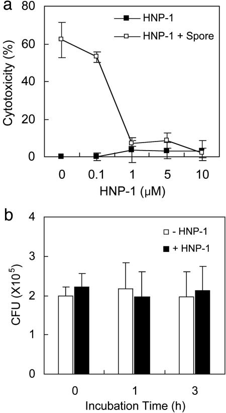
HNP-1 protects macrophages against B. anthracis-induced cell death. (a) RAW 264.7 cells were infected with B. anthracis spores and then treated with the indicated amounts of HNP-1. Cytotoxicity was determined by measuring released lactate dehydrogenase levels. (b)An in vitro killing assay was performed against spores in the presence or absence of 1 μM HNP-1. After the indicated incubation times, colony-forming units (CFU) were determined.
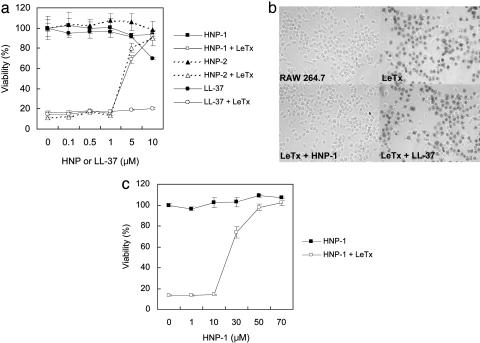
Human α-defensins protect macrophages against cytolysis by anthrax LeTx. (a) RAW 264.7 cells were treated with LeTx (400 ng/ml LF and 1,600 ng/ml PA) in the presence of the indicated amounts of HNP-1, HNP-2, or LL-37. Cell viability was determined by methyl thiazole tetrazolium (MTT) assay. (b) RAW 264.7 cells were treated with LeTx (400 ng/ml LF and 1,600 ng/ml PA) in the presence of 7 μM HNP-1 or LL-37. Five hours after treatment, cells were stained with trypan blue. (c) Viability of RAW 264.7 cells was determined by MTT assay after treatment with LeTx (400 ng/ml LF and 1,600 ng/ml PA) and various concentrations of HNP-1. This assay was performed in RPMI medium 1640 supplemented with 5% FCS.
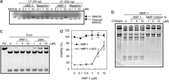
HNP-1 is an inhibitor of LF. (a) In vitro translated MKK3b was incubated for 1 h with the indicated amounts of LF and either HNP-1 or magainin I. Cleavage of MKK3b was analyzed by SDS/PAGE and autoradiography. (b) Collagen was incubated with MMP-1 in the presence of the indicated amounts of HNP-1 or MMP inhibitor. (c) PA was incubated with furin in the presence of HNP-1 or hexa-d -arginine (D6R). (d) RAW 264.7 cells were incubated with HNP-1 at 37°C. After 1 h, the medium was removed and replaced with fresh medium containing LeTx (400 ng/ml LF and 1,600 ng/ml PA). Cells were incubated further at 37°C for 5 h. Viability was determined by MTT assay.
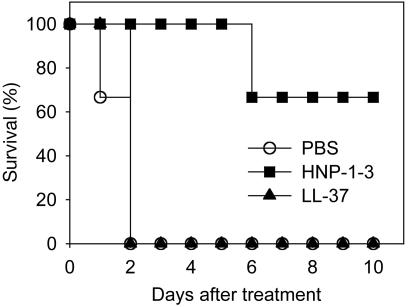
HNP-1-3 protects BALB/c mice from LeTx intoxication. Mice were injected with 50 μg of LF plus 50 μg of PA per mouse through one tail vein and then were immediately injected with PBS, 500 μg of purified HNP-1-3, or 500 μg of LL-37 through the other tail vein. Three animals per group were used for this experiment. In another set of experiments, administration of 500 μg of HNP-1-3 achieved 100% protection (data not shown).
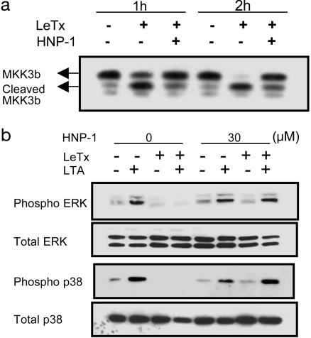
HNP-1 inhibits proteolysis of MKK in LeTx-treated macrophages. (a) LeTx was added to RAW 264.7 macrophages with (+) or without (-) HNP-1. At the indicated time points, cell lysates were prepared and assessed by Western blotting with an anti-MKK3 antibody. (b) RAW 264.7 cells were treated (+) with LeTx (200 ng/ml LF and 1,600 ng/ml PA) and HNP-1 (30 μM). Two hours after treatment, cells were stimulated with 10 μg/ml B. subtilis lipoteichoic acid (LTA) for 30 min, and the lysates were assessed by immunoblotting with antibodies against MAPKs (Total) and their phosphorylated forms (Phospho). ERK, extracellular signal-regulated kinase.
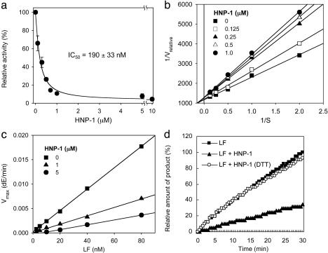
Characterization of LF inhibition by HNP-1. (a) HNP-1 inhibited 50% of LF activity at a concentration of 190 ± 33 nM. (b) Lineweaver-Burk plot indicates noncompetitive inhibition mode. (c) A plotting of Vmax versus concentrations of LF confirms that HNP-1 is a reversible noncompetitive inhibitor. dE, release of p-Nitroaniline. (d) DTT-treated HNP-1 did not show any significant effect on LF.
Similar articles
-
Human alpha-defensins inhibit Clostridium difficile toxin B.Gastroenterology. 2008 Jun;134(7):2049-58. doi: 10.1053/j.gastro.2008.03.008. Epub 2008 Mar 10. Gastroenterology. 2008. PMID: 18435932
-
Cisplatin inhibition of anthrax lethal toxin.Antimicrob Agents Chemother. 2006 Aug;50(8):2658-65. doi: 10.1128/AAC.01412-05. Antimicrob Agents Chemother. 2006. PMID: 16870755 Free PMC article.
-
Proteomic analyses of murine macrophages treated with Bacillus anthracis lethal toxin.Microb Pathog. 2006 Oct-Nov;41(4-5):157-67. doi: 10.1016/j.micpath.2006.07.002. Epub 2006 Sep 1. Microb Pathog. 2006. PMID: 16950595
-
Retrocyclins kill bacilli and germinating spores of Bacillus anthracis and inactivate anthrax lethal toxin.J Biol Chem. 2006 Oct 27;281(43):32755-64. doi: 10.1074/jbc.M603614200. Epub 2006 Jun 21. J Biol Chem. 2006. PMID: 16790431 Free PMC article.
-
Potent inhibition of tumor angiogenesis by the matrix metalloproteinase-activated anthrax lethal toxin: implications for broad anti-tumor efficacy.Cell Cycle. 2008 Mar 15;7(6):745-9. doi: 10.4161/cc.7.6.5627. Epub 2008 Jan 18. Cell Cycle. 2008. PMID: 18245947 Review.
Cited by
-
Human monoclonal antibodies against anthrax lethal factor and protective antigen act independently to protect against Bacillus anthracis infection and enhance endogenous immunity to anthrax.Infect Immun. 2007 Nov;75(11):5425-33. doi: 10.1128/IAI.00261-07. Epub 2007 Jul 23. Infect Immun. 2007. PMID: 17646360 Free PMC article.
-
Capsular antigen fraction 1 and Pla modulate the susceptibility of Yersinia pestis to pulmonary antimicrobial peptides such as cathelicidin.Infect Immun. 2008 Apr;76(4):1456-64. doi: 10.1128/IAI.01197-07. Epub 2008 Jan 28. Infect Immun. 2008. PMID: 18227173 Free PMC article.
-
Molecular determinants for a cardiovascular collapse in anthrax.Front Biosci (Elite Ed). 2014 Jan 1;6(1):139-47. doi: 10.2741/e697. Front Biosci (Elite Ed). 2014. PMID: 24389148 Free PMC article. Review.
-
Sometimes it takes two to tango: contributions of dimerization to functions of human α-defensin HNP1 peptide.J Biol Chem. 2012 Mar 16;287(12):8944-53. doi: 10.1074/jbc.M111.332205. Epub 2012 Jan 23. J Biol Chem. 2012. PMID: 22270360 Free PMC article.
-
Cooperative Function of LL-37 and HNP1 Protects Mammalian Cell Membranes from Lysis.Biophys J. 2020 Dec 15;119(12):2440-2450. doi: 10.1016/j.bpj.2020.10.031. Epub 2020 Nov 4. Biophys J. 2020. PMID: 33157121 Free PMC article.
References
-
- Turnbull, P. C. (1999) J. Appl. Microbiol. 87, 237-240. - PubMed
-
- Duesbery, N. S., Webb, C. P., Leppla, S. H., Gordon, V. M., Klimpel, K. R., Copeland, T. D., Ahn, N. G., Oskarsson, M. K., Fukasawa, K., Paull, K. D. & Vande Woude, G. F. (1998) Science 280, 734-737. - PubMed
-
- Bradley, K. A., Mogridge, J., Mourez, M., Collier, R. J. & Young, J. A. (2001) Nature 414, 225-229. - PubMed
-
- Collier, R. J. & Young, J. A. (2003) Annu. Rev. Cell Dev. Biol. 19, 45-70. - PubMed
Publication types
MeSH terms
Substances
LinkOut - more resources
Full Text Sources
Other Literature Sources
Molecular Biology Databases
