. 2005 Apr;17(4):1233-51.
doi: 10.1105/tpc.104.030452. Epub 2005 Mar 16.

Two P-type ATPases are required for copper delivery in Arabidopsis thaliana chloroplasts

Affiliations

Two P-type ATPases are required for copper delivery in Arabidopsis thaliana chloroplasts

Salah E Abdel-Ghany et al. Plant Cell. 2005 Apr.

Abstract

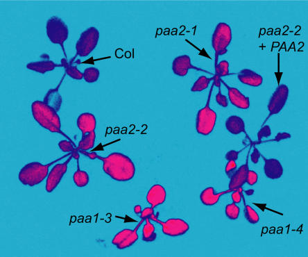
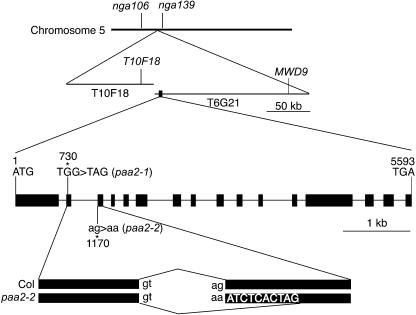
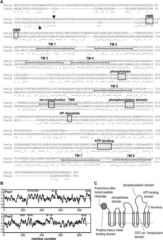
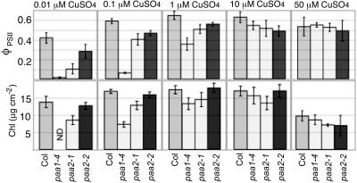
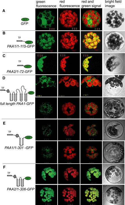
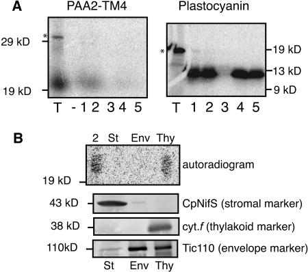
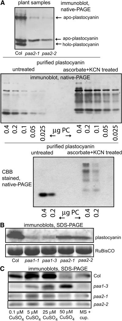
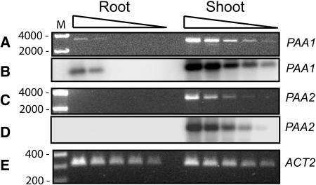
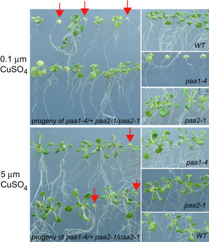
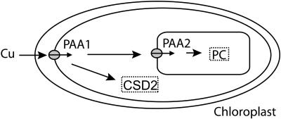
Copper delivery to the thylakoid lumen protein plastocyanin and the stromal enzyme Cu/Zn superoxide dismutase in chloroplasts is required for photosynthesis and oxidative stress protection. The copper delivery system in chloroplasts was characterized by analyzing the function of copper transporter genes in Arabidopsis thaliana. Two mutant alleles were identified of a previously uncharacterized gene, PAA2 (for P-type ATPase of Arabidopsis), which is required for efficient photosynthetic electron transport. PAA2 encodes a copper-transporting P-type ATPase with sequence similarity to PAA1, which functions in copper transport in chloroplasts. Both proteins localized to the chloroplast, as indicated by fusions to green fluorescent protein. The PAA1 fusions were found in the chloroplast periphery, whereas PAA2 fusions were localized in thylakoid membranes. The phenotypes of paa1 and paa2 mutants indicated that the two transporters have distinct functions: whereas both transporters are required for copper delivery to plastocyanin, copper delivery to the stroma is inhibited only in paa1 but not in paa2. The effects of paa1 and paa2 on superoxide dismutase isoform expression levels suggest that stromal copper levels regulate expression of the nuclear genes IRON SUPEROXIDE DISMUTASE1 and COPPER/ZINC SUPEROXIDE DISMUTASE2. A paa1 paa2 double mutant was seedling-lethal, underscoring the importance of copper to photosynthesis. We propose that PAA1 and PAA2 function sequentially in copper transport over the envelope and thylakoid membrane, respectively.


Figures

Figure 1.
Figure 1.
High Chlorophyll Fluorescence Phenotype of paa2 Mutants. Wild-type, paa1, and paa2 seedlings were grown in soil for 4 weeks. A chlorophyll fluorescence image was captured after 1 min of illumination with actinic light (300 μmol·m−2·s−1). The fluorescence level is indicated by false coloring in this order: red > pink > blue. Col, Columbia gl1 wild type; paa2-2 + PAA2, paa2-2 transformed with the genomic PAA2 sequence.
Figure 2.
Figure 2.
Positional Cloning of the PAA2 Gene Encoding a Cu-Transporting P-Type ATPase. The paa2 mutation was mapped to the 140-kb region spanning two BAC clones (T10F18 and T6G21) on chromosome 5. The small box on T6G21 represents the position of PAA2. Exons (boxes) and introns (lines) were predicted from the cDNA sequence. The positions of the paa2 mutations are indicated. The paa2-2 mutation causes incorrect splicing of exons 2 and 3.
Figure 3.
Figure 3.
Sequence Comparison of the PAA1 and PAA2 Proteins. (A) Sequence alignment of PAA1 and PAA2. Identical residues are indicated by stars, and similar residues are indicated by dots. The predicted transit sequence cleavage sites are indicated with vertical arrows. Functional regions of Cu-pumping P-type ATPases are indicated by boxes. HMB, putative heavy metal binding domain; TM 1 to TM 8, possible transmembrane regions. The GenBank accession numbers for PAA1 and PAA2 are BAA23769 and AY297817, respectively. (B) Hydrophobicity profiles of PAA1 and PAA2. The scale of Kyte and Doolittle (1982) was used with a window of 15 residues. Hydrophobic regions are indicated by positive values. Gray boxes above each sequence indicate the putative transmembrane regions. (C) Topology model of PAA2.
Figure 4.
Figure 4.
Light Intensity Dependence of Electron Transport Rate. The relative electron transport rate was calculated from the product of ΦPSII and light intensity (μmol·m−2·s−1). ΦPSII is a chlorophyll fluorescence parameter representing the efficiency of PSII photochemistry (Genty et al., 1989). Each point represents the mean ± sd. The number of replicates is five for each line except for paa2-1 + PAA2. paa2-1 + PAA2 represents paa2-1 transformed by the wild-type PAA2 sequence. Ten independent kanamycin-resistant seedlings were tested.
Figure 5.
Figure 5.
Response of paa1 and paa2 Plants to CuSO4. Both alleles of paa2, paa1-4, and the wild type (Col) were cultured on MS medium containing various concentrations of CuSO4. The standard MS medium contains 0.1 μM CuSO4. The response to Cu was evaluated by the chlorophyll fluorescence parameter ΦPSII (efficiency of PSII photochemistry) at a light intensity of 200 μmol·m−2·s−1 and chlorophyll (Chl) content. paa1-4 seedlings were too small to determine the chlorophyll content. Data are averages of five independent replicates. ND, not determined.
Figure 6.
Figure 6.
Subcellular Localization of PAA1 and PAA2. Arabidopsis protoplasts were transformed with plasmids that express the indicated gene constructs under the control of the constitutive 35S Cauliflower mosaic virus promoter. The unfused GFP coding sequence served as a control. PAA1/1-115-GFP encodes the fusion of the PAA1 transit peptide to the N terminus of GFP; PAA2/1-72-GFP is the fusion of the PAA2 transit peptide to the N terminus of GFP. Full-length PAA1-GFP contains the full coding sequence of the PAA1 precursor fused to GFP. PAA1/1-301-GFP is the fusion of the N-terminal part of the PAA1 precursor, including the first two transmembrane domains, to GFP; PAA2/1-306-GFP is the fusion of the N-terminal part of the PAA2 precursor, including the first four transmembrane domains, to GFP. After 16 h of expression, cells were observed using a confocal laser scanning microscope. Green fluorescence signals, chlorophyll red autofluorescence, an overlay of green and red signals, and bright-field images are shown.
Figure 7.
Figure 7.
In Vitro Chloroplast Import and Fractionation. (A) In vitro chloroplast import. PAA2-TM4 contains residues 1 to 333, including the first four transmembrane domains of PAA2. The precursor of plastocyanin was used as a control for import and fractionation. The radiolabeled precursor proteins in the translation mixture (T) were produced by in vitro translation in the presence of 35S-Met and incubated with isolated pea chloroplasts. The translation mixture samples represent 10% of the precursor added to lanes 1 to 5. Precursor proteins of the expected sizes are indicated by asterisks. Chloroplasts were reisolated and proteins were either analyzed directly (lane 1) or after treatment with protease (lane 2). Protease-treated chloroplasts were lysed and further fractionated into a soluble fraction (lane 3) and a membrane fraction before (lane 4) and after (lane 5) protease treatment. Proteins were separated by SDS-PAGE, and the radiolabeled protein bands were visualized using a PhosphorImager. (B) Sucrose gradient fractionation. Pea chloroplasts, after import and protease treatment (lane 2), were lysed in hypotonic buffer and fractionated into stroma (St), envelope (Env), and thylakoid (Thy) fractions by sucrose gradient centrifugation. Proteins were separated by SDS-PAGE and visualized using a PhosphorImager (autoradiogram) or blotted onto a nitrocellulose membrane and probed with the indicated antibodies.
Figure 8.
Figure 8.
Analysis of Plastocyanin. (A) Separation of apoplastocyanin and holoplastocyanin on native gels. The top gel shows soluble lumen proteins extracted from thylakoids equivalent to 3 μg of chlorophyll of the wild type (Col) and PAA2 mutants separated on a 15% nondenaturing gel, blotted, and immunodetected with plastocyanin antibody. The bands were identified as either holoplastocyanin or apoplastocyanin by comparison with control native gels (middle and bottom gels) on which purified plastocyanin (PC) was present in either the holo form (untreated) or the apo form (ascorbate + KCN treated). Bands on the middle gel were immunodetected, whereas protein on the bottom gel was visualized by staining with Coomassie Brilliant Blue (CBB). (B) Analysis of total plastocyanin polypeptides. Proteins (20 μg) in total leaf homogenate of the wild type (Col) and the indicated mutants were separated by SDS-PAGE (15% gel), blotted, and immunodetected with either a plastocyanin antibody or the large subunit of ribulose-1,5-bis-phosphate carboxylase/oxygenase (RuBisCO) antibody. (C) Effect of Cu supplementation on plastocyanin levels in the wild type (Col) and paa1 and paa2 mutants. Arabidopsis seedlings were grown in MS medium supplemented with the indicated CuSO4 concentrations or with the Cu chelator cuprizone (10 μM) for 3 weeks. Proteins (20 μg) in the total homogenate were fractionated by SDS-PAGE, blotted, and probed with plastocyanin antibody. Data shown are representative of at least two independent replicates.
Figure 9.
Figure 9.
Analysis of SOD Isozyme Activity and Expression. Wild-type (Col) and mutant seedlings were grown in MS medium supplemented with the indicated CuSO4 concentrations or with the Cu chelator cuprizone (10μM) for 3 weeks. (A) SOD isozyme activities in total shoot homogenate. Total soluble proteins (30 μg) were separated on nondenaturing 12% acrylamide gels, and the gels were stained for SOD activity. (B) Immunodetection of total SOD isoform polypeptides. Shoot proteins were fractionated by SDS-PAGE, and each SOD isoform was detected by immunoblotting using a specific antibody. (C) SOD transcript levels. Ten micrograms of total RNA was separated by electrophoresis, transferred to Hybond N+ membranes, and probed with 32P-labeled gene-specific probes. (D) Analysis of SOD activity in stromal fractions. Intact chloroplasts were isolated from Arabidopsis seedlings grown in MS medium supplemented with the indicated concentrations of CuSO4. Stromal proteins (15 μg) were separated on 15% nondenaturing gels and stained for SOD activity. Gels were stained without inhibitor (no inhib.) or preincubated with KCN, which inhibits Cu/ZnSOD, to distinguish the activities of FeSOD and Cu/ZnSOD. (E) Controls for chloroplast intactness and purity. Protein fractions of the total leaf homogenate of plants grown on MS medium and chloroplasts (CP) from plants grown on MS medium or MS medium with the indicated Cu concentrations were separated by SDS-PAGE and analyzed by immunoblotting. Fractions equivalent to 3 μg of chlorophyll were loaded. Immunoblots were probed with Arabidopsis CpNifS antibody as a stromal marker or with P28 antibody as a cytosolic marker. Data shown are representative of at least two independent replicates.
Figure 10.
Figure 10.
mRNA Expression of PAA1 and PAA2. One microgram of DNase-treated total RNA from either root or shoot was reverse-transcribed into cDNA. Serial dilutions from these cDNAs (left to right bands in each group: undiluted, 1:4, 1:16, 1:64, and 1:256) were used as templates for PCR using gene-specific primers for PAA1, PAA2, or the constitutively expressed actin (ACT2) gene as an internal control. Gels were stained with ethidium bromide ([A], [C], and [E]). PCR products were blotted and probed with gene-specific 32P-labeled probes for PAA1 (B) and PAA2 (D). The sizes of markers (M) are shown in base pairs.
Figure 11.
Figure 11.
Seedling-Lethal Phenotype of paa1 paa2. Progeny of paa1-4/+ paa2-1/paa2-1 as well as the wild type (WT) and single mutants were cultured on MS medium containing 0.1 or 5 μM CuSO4. paa1-4 paa2-1 double mutants are indicated by red arrows. The genotype of the double mutants was confirmed by genome analysis using PCR.
Figure 12.
Figure 12.
The Cu Delivery Systems in Plant Chloroplasts. CSD2, stromal Cu/ZnSOD; PAA1 and PAA2, Cu-transporting P-type ATPases of Arabidopsis; PC, plastocyanin.

References

    1. Asada, K. (1999). The water-water cycle in chloroplasts: Scavenging of active oxygen and dissipation of excess photons. Annu. Rev. Plant Physiol. Plant Mol. Biol. 50, 601–639. - PubMed
    1. Asakura, Y., Hirohashi, T., Kikuchi, S., Belcher, S., Osborne, E., Yano, S., Terashima, I., Barkan, A., and Nakai, M. (2004). Maize mutants lacking chloroplast FtsY exhibit pleiotropic defects in the biogenesis of thylakoid membranes. Plant Cell 16, 201–214. - PMC - PubMed
    1. Askwith, C., Eide, D., Van Ho, A., Bernard, P.S., Li, L., Davis-Kaplan, S., Sipe, D.M., and Kaplan, J. (1994). The FET3 gene of S. cerevisiae encodes a multicopper oxidase required for ferrous iron uptake. Cell 76, 403–410. - PubMed
    1. Axelsen, K.B., and Palmgren, M.G. (2001). Inventory of the superfamily of P-type ion pumps in Arabidopsis. Plant Physiol. 126, 696–706. - PMC - PubMed
    1. Baxter, I., Tchieu, J., Sussman, M.R., Boutry, M., Palmgren, M.G., Gribskov, M., Harper, J.F., and Axelsen, K.B. (2003). Genomic comparison of P-type ATPase ion pumps in Arabidopsis and rice. Plant Physiol. 132, 618–628. - PMC - PubMed

Publication types

MeSH terms

Associated data