. 2005 Apr;17(4):1090-104.
doi: 10.1105/tpc.104.029272. Epub 2005 Mar 16.

Sites and regulation of auxin biosynthesis in Arabidopsis roots

Affiliations

Sites and regulation of auxin biosynthesis in Arabidopsis roots

Karin Ljung et al. Plant Cell. 2005 Apr.

Abstract

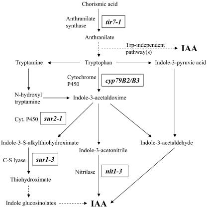
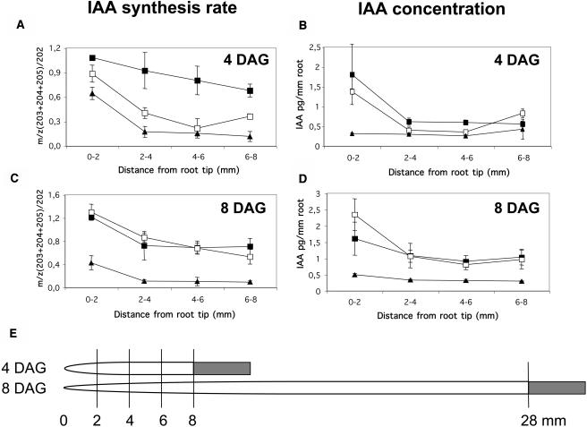
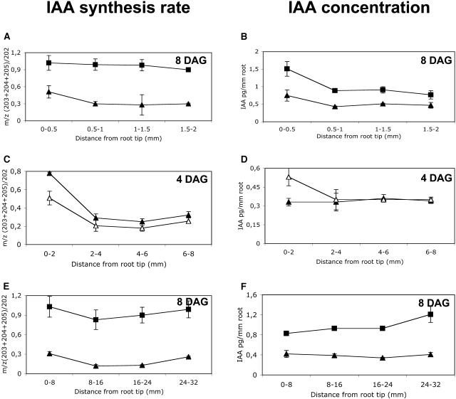
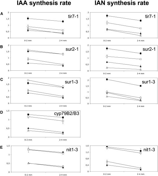
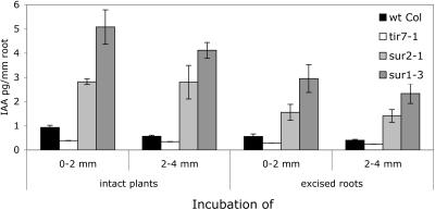
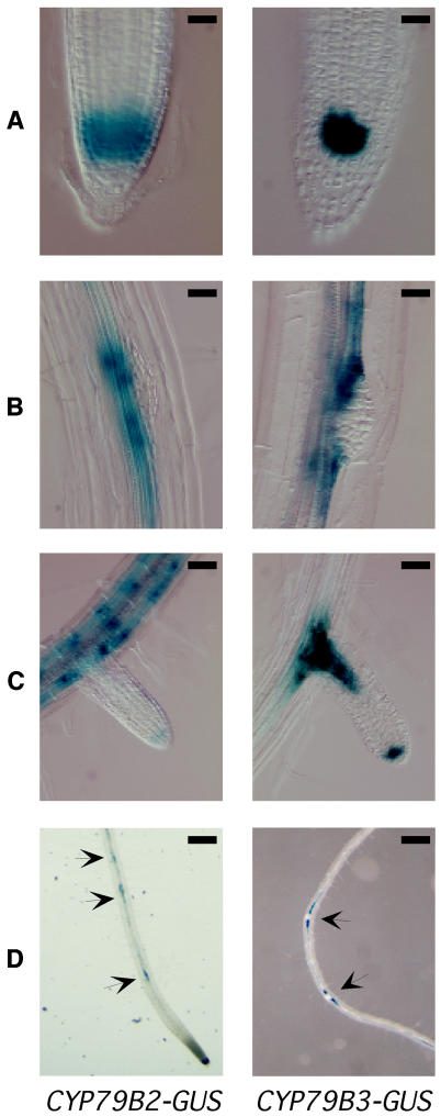
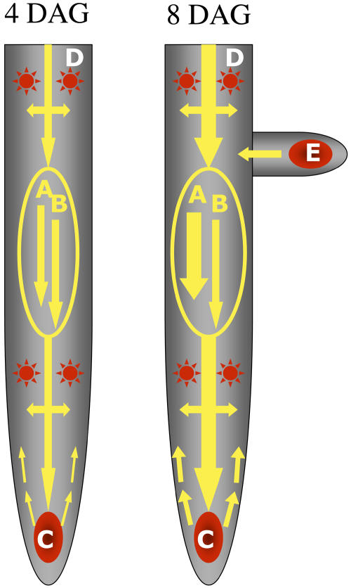
Auxin has been shown to be important for many aspects of root development, including initiation and emergence of lateral roots, patterning of the root apical meristem, gravitropism, and root elongation. Auxin biosynthesis occurs in both aerial portions of the plant and in roots; thus, the auxin required for root development could come from either source, or both. To monitor putative internal sites of auxin synthesis in the root, a method for measuring indole-3-acetic acid (IAA) biosynthesis with tissue resolution was developed. We monitored IAA synthesis in 0.5- to 2-mm sections of Arabidopsis thaliana roots and were able to identify an important auxin source in the meristematic region of the primary root tip as well as in the tips of emerged lateral roots. Lower but significant synthesis capacity was observed in tissues upward from the tip, showing that the root contains multiple auxin sources. Root-localized IAA synthesis was diminished in a cyp79B2 cyp79B3 double knockout, suggesting an important role for Trp-dependent IAA synthesis pathways in the root. We present a model for how the primary root is supplied with auxin during early seedling development.


Figures

Figure 1.
Figure 1.
IAA Biosynthesis Pathways. Putative pathways for Trp-dependent IAA synthesis in Arabidopsis. De novo biosynthesis of IAA and IAN was measured in root tips from different mutants in these pathways.
Figure 2.
Figure 2.
IAA Biosynthesis Measurements. (A) Electron-impact mass spectrum and fragmentation pattern for IAA-Me-TMS. D indicates positions on the indole ring and side chain that can be labeled with deuterium. (B) Mass isotopomer profiles for the molecular ion (m/z 261) and base peak (m/z 202) of IAA-Me-TMS. Natural isotope contribution (open bars) and deuterium labeling (closed bars) are indicated. Arrows indicate the metastable transitions that are monitored to calculate deuterium incorporation after feeding with 2H2O. (C) Ten-day-old Arabidopsis seedlings were incubated for 12 and 24 h in medium containing 2H2O, and measurements of IAA biosynthesis rates were then performed on samples containing pooled root tissue from 10 seedlings. Three different experiments followed: (I) intact seedlings were incubated in medium containing 30% 2H2O; (II) intact seedlings were incubated in medium containing 30% 2H2O and 40 μM NPA; (III) excised roots were incubated in medium containing 30% 2H2O. Measurements were performed by SRM and HR-SIM-MS. Samples were measured in triplicate, and corrections were made for background and natural isotope abundances. Error bars indicate sd.
Figure 3.
Figure 3.
Distribution of IAA Biosynthesis and Concentration in the Arabidopsis Primary Root. (A) to (D) De novo IAA biosynthesis rates ([A] and [C]) were measured in 2-mm root sections from Arabidopsis seedlings incubated for 24 h in medium containing 2H2O starting at 3 and 7 DAG. Endogenous IAA levels ([B] and [D]) in 2-mm root sections at 4 and 8 DAG were calculated from the data obtained in the IAA biosynthesis measurements by combining the corrected areas of the ions from deuterium-labeled IAA (m/z 203 to 205) with that of unlabeled IAA (m/z 202) after correcting for the natural abundance of stable isotopes. Error bars indicate sd. Experiments were performed with intact seedlings incubated in medium containing 30% 2H2O (closed squares), intact seedlings incubated in medium containing 30% 2H2O and 40 μM NPA (open squares), and excised roots incubated in medium containing 30% 2H2O (closed triangles). (E) Root length and sampling positions for root tissue collected after feeding experiments performed 3 to 4 and 7 to 8 DAG.
Figure 4.
Figure 4.
IAA Biosynthesis and Levels in Different Parts of the Arabidopsis Primary Root. De novo IAA biosynthesis rate ([A], [C], and [E]) and IAA concentration ([B], [D], and [F]) were measured in different root sections after incubation of Arabidopsis seedlings in medium containing 2H2O. Error bars indicate sd. (A) and (B) Intact seedlings (closed squares) and excised roots (closed triangles) were incubated in medium containing 30% 2H2O for 24 h starting at 7 DAG. Sections (0.5 mm) of the root tip were collected for analysis. (C) and (D) Excised roots were incubated in medium containing 30% 2H2O (closed triangles) or 30% 2H2O and 40 μM NPA (open triangles) starting at 3 DAG. Sections (2 mm) of the root were collected for analysis. (E) and (F) Intact seedlings (closed squares) and excised roots (closed triangles) were incubated in medium containing 30% 2H2O for 24 h starting at 7 DAG. Sections (8 mm) of the root were collected for analysis.
Figure 5.
Figure 5.
IAA and IAN Synthesis in Root Tips of IAA Biosynthesis Mutants. IAA and IAN synthesis rates were measured in root tips from wild-type Columbia and the mutant lines tir7-1 (A), sur2-1 (B), sur1-3 (C), cyp79B2 cyp79B3 (D), and nit1-3 (E). Intact seedlings of the wild type (closed squares) and mutants (open squares) and excised roots of the wild type (closed triangles) and mutants (open triangles) were incubated in medium containing 30% 2H2O for 24 h starting at 7 DAG. The most apical 4-mm part of the primary root was collected in 2-mm sections. Error bars indicate sd.
Figure 6.
Figure 6.
IAA Concentration in Root Tips of IAA Biosynthesis Mutants. Endogenous IAA levels in 2-mm root sections were calculated from the data obtained in the IAA biosynthesis measurements (Figure 5) of different IAA synthesis mutants by combining the corrected areas of the ions from deuterium-labeled IAA (m/z 203 to 205) with that of unlabeled IAA (m/z 202). Error bars indicate sd. wt Col, wild-type Columbia.
Figure 7.
Figure 7.
CYP79B2-GUS and CYP79B3-GUS Reporter Expression in Arabidopsis Roots. CYP79B2-GUS transgenic plants are shown at left, and CYP79B3-GUS transgenic plants are shown at right. CYP79B2-GUS plants were stained with X-Gluc for 1 h, and CYP79B3-GUS plants were stained for 18 h. (A) to (C) Expression patterns of CYP79B2 and CYP79B3 in the primary root tip (A), sites of lateral root formation (B), and developing lateral roots (C) of 7-DAG seedlings. (D) The primary root after treatment with 1 μM IAA for 24 h at 7 DAG, showing expression at all sites of lateral root formation (arrows) but not in the rest of the primary root. Bars = 25 μm in (A) and (B), 50 μm in (C), and 250 μm in (D).
Figure 8.
Figure 8.
Model Showing How Different Sources Supply the Root with Auxin during Early Seedling Development. Sources (red) and transport (yellow) of de novo synthesized IAA in the root system of Arabidopsis. At 4 DAG, auxin produced in the aerial part of the seedling is transported to the root via two routes of equal importance: phloem-mediated transport (A) and polar transport (B). At 8 DAG, the phloem-mediated transport is relatively more important than the polar transport. Already early in root development, two internal sources are established, one specifically localized to the outermost 0.5 mm of the root (C), and one representing all tissues upward from the root tip with low but important synthesis capacity (D). In the later stages of development, all emerging lateral roots gain synthesis capacity (E) and will be important contributors to the auxin pool in the root system.

References

    1. Åstot, C., Dolezal, K., Moritz, T., and Sandberg, G. (2000). Deuterium in vivo labelling of cytokinins in Arabidopsis thaliana analysed by capillary liquid chromatography/frit-fast atom bombardment mass spectrometry. J. Mass Spectrom. 35, 13–22. - PubMed
    1. Bak, S., Tax, F.E., Feldmann, K.A., Galbraith, D.W., and Feyereisen, R. (2001). CYP83B1, a cytochrome P450 at the metabolic branch point in auxin and indole glucosinolate biosynthesis in Arabidopsis. Plant Cell 13, 101–111. - PMC - PubMed
    1. Barlier, I., Kowalczyk, M., Marchant, A., Ljung, K., Bhalerao, R., Bennett, M., Sandberg, G., and Bellini, C. (2000). The SUR2 gene of Arabidopsis thaliana encodes the cytochrome P450 CYP83B1, a modulator of auxin homeostasis. Proc. Natl. Acad. Sci. USA 97, 14819–14824. - PMC - PubMed
    1. Bartel, B., and Fink, G.R. (1994). Differential regulation of an auxin-producing nitrilase gene family in Arabidopsis thaliana. Proc. Natl. Acad. Sci. USA 91, 6649–6653. - PMC - PubMed
    1. Bartel, B., LeClere, S., Magidin, M., and Zolman, B.K. (2001). Inputs to the active indole-3-acetic acid pool: De novo synthesis, conjugate hydrolysis, and indole-3-butyric acid β-oxidation. J. Plant Growth Regul. 20, 198–216.

Publication types

MeSH terms

Substances

Associated data