Comparative Study
. 2005 Mar 16;25(11):2793-802.
doi: 10.1523/JNEUROSCI.4090-04.2005.

The disulfide isomerase Grp58 is a protective factor against prion neurotoxicity

Affiliations
Comparative Study

The disulfide isomerase Grp58 is a protective factor against prion neurotoxicity

Claudio Hetz et al. J Neurosci. .

Abstract

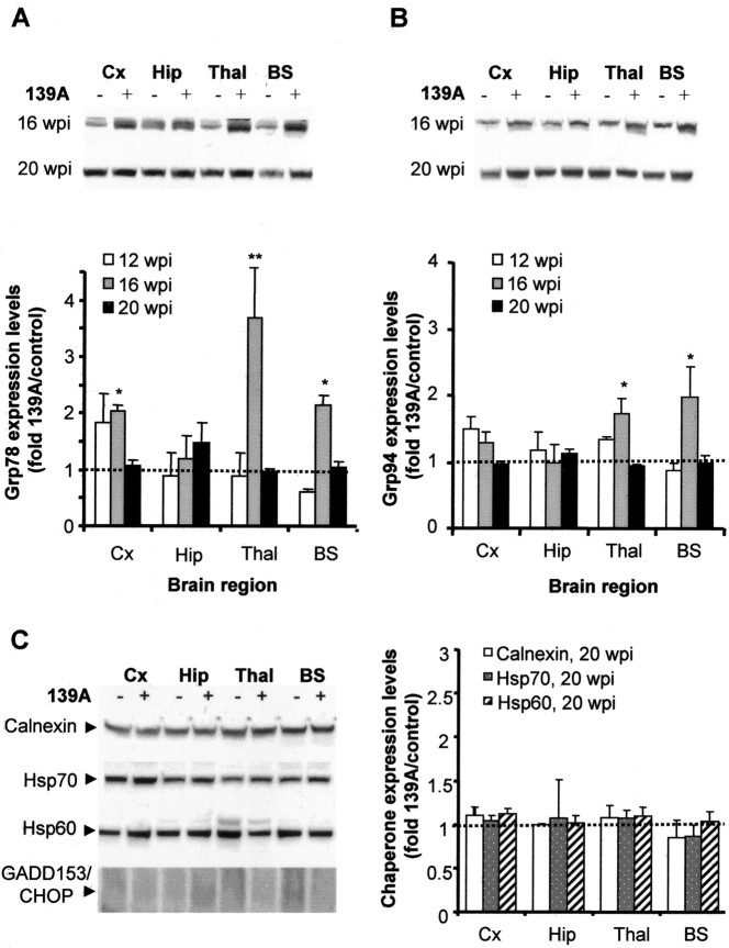
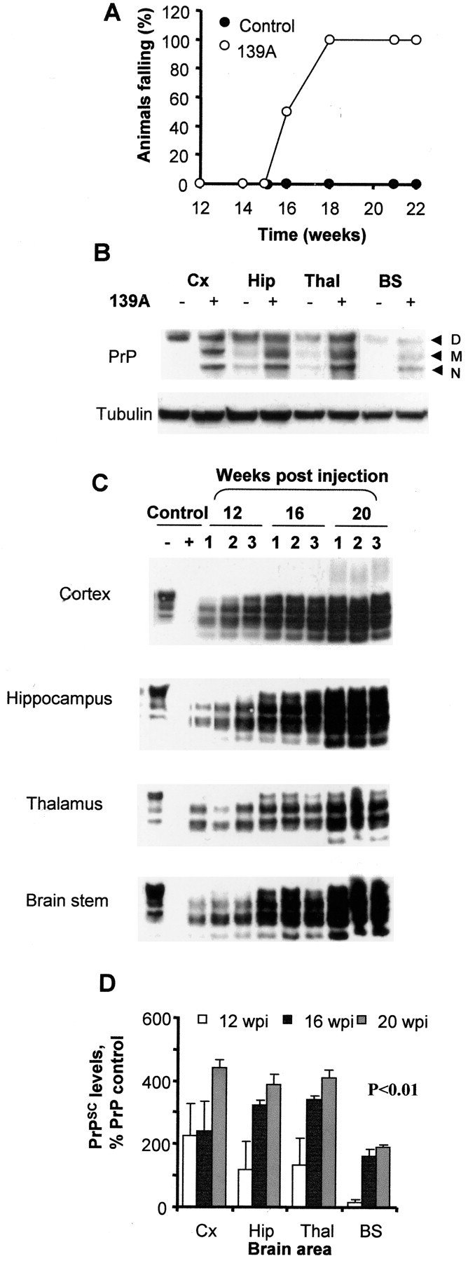
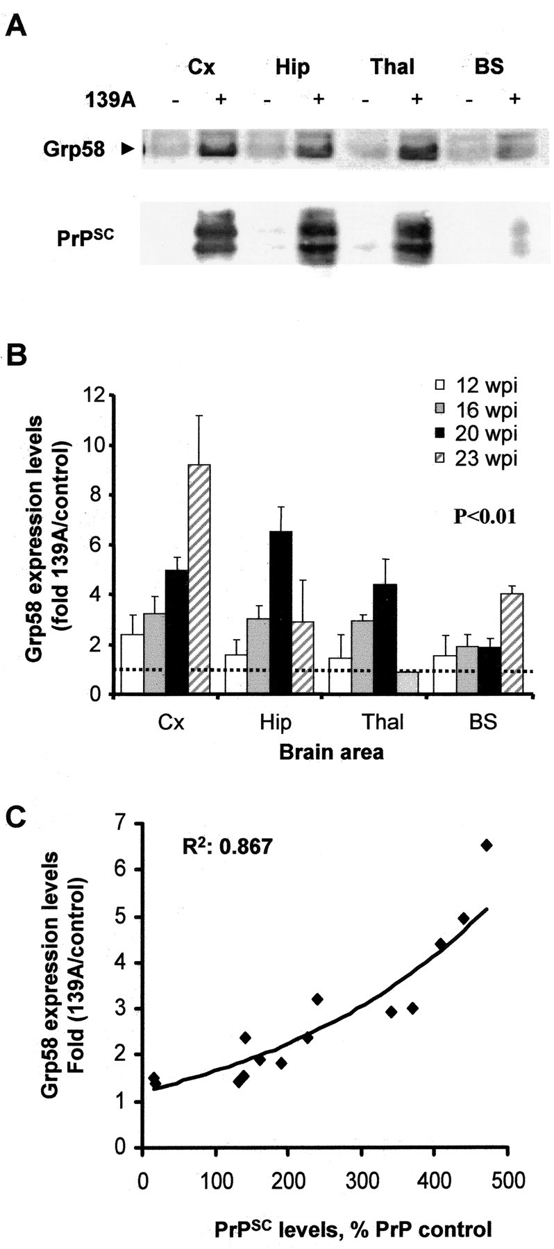
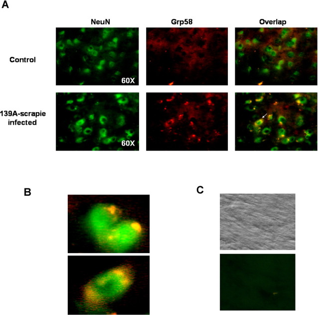
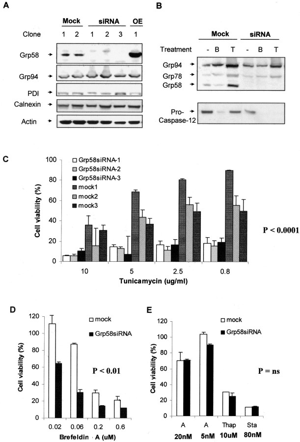
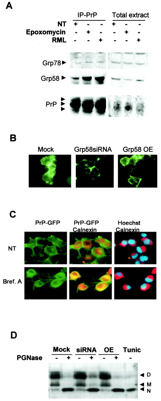
Prion diseases are transmissible neurodegenerative disorders characterized by extensive neuronal apoptosis and accumulation of misfolded prion protein (PrP(SC)). Recent reports indicate that PrP(SC) induces neuronal apoptosis via activation of the endoplasmic reticulum (ER) stress pathway and activation of the ER resident caspase-12. Here, we investigate the relationship between prion replication and induction of ER stress during different stages of the disease in a murine scrapie model. The first alteration observed consists of the upregulation of the ER chaperone of the glucose-regulated protein Grp58, which was detected during the presymptomatic phase and followed closely the formation of PrP(SC). An increase in Grp58 expression correlated with PrP(SC) accumulation at all stages of the disease in different brain areas, suggesting that this chaperone may play an important role in the cellular response to prion infection. Indeed, in vitro studies using N2a neuroblastoma cells demonstrated that inhibition of Grp58 expression with small interfering RNA led to a significant enhancement of PrP(SC) toxicity. Conversely, overexpression of Grp58 protected cells against PrP(SC) toxicity and decreased the rate of caspase-12 activation. Grp58 and PrP were shown to interact by coimmunoprecipitation, observing a higher interaction in cells infected with scrapie prions. Our data indicate that expression of Grp58 is an early cellular response to prion replication, acting as a neuroprotective factor against prion neurotoxicity. Our findings suggest that targeting Grp58 interaction may have applications for developing novel strategies for treatment and early diagnosis of prion diseases.


Figures

Figure 4.
Figure 4.
Grp78 and Grp94 are transiently induced by 139A scrapie infection. A, B, Grp78 (A) and Grp94 (B) expression levels at 12, 16, and 20 wpi were determined by Western blotting, and the band intensities from three different infected animals were quantified by densitometry. Top, A representative Western blot analysis at 16 and 20 wpi. Bottom, Averaged intensities were compared with the corresponding values from three control animals for both Grp78 and Grp94. Analysis by two-way ANOVA, using brain regions and weeks postinjection as the variables, showed that Grp78 and Grp94 expression levels were not significantly different. t test analysis of individual values showed some statistically significant differences (*p < 0.05; **p < 0.01). Cx, Cortex; Hip, hippocampus; Thal, thalamus; BS, brainstem. C, Calnexin, Hasp70, Hsp60, and GADD153/CHOP levels were analyzed in different brain areas at 20 wpi by Western blotting (left) and quantified as described in A and B (right).
Figure 6.
Figure 6.
Grp58 protects N2a cells against PrPSC toxicity. A, N2a clones transfected with Grp58siRNA, mock siRNA, or full-length Grp58 were treated with different concentrations of purified PrPSC, and cell viability was determined after 48 h by MTS analysis. Data were analyzed by Student's t test to compare the differences between mock-transfected cells and those overexpressing Grp58 or transfected with Grp58siRNA. *p < 0.05; **p < 0.01. B, In parallel, cells were treated with PrPSC, and caspase-12 activation was measured by Western blot analysis. Actin levels are shown as an internal control of protein loading. C, N2a clones transfected with Grp58siRNA, mock siRNA, or full-length Grp58 (OE) were treated with 50 nm PrPSC or 800 ng/ml tunicamycin for 48 h, and GADD153/CHOP and actin levels were determined by Western blot analysis.
Figure 1.
Figure 1.
Disease progression and prion propagation in a 139A murine scrapie model. A, Eight animals per group were injected in the hippocampus with 1 μl of 10% brain homogenate from normal (controls) or 139A scrapie-infected mice. Muscle strength was determined by measuring the time during which animals were able to cling to an inverted grill. Values represent the percentage of animals in each group that fell within 1 min after inversion. B, Western blot analysis of total PrP levels in control and infected brains in different brain areas after 16 wpi. Tubulin levels were used as an internal control for protein loading. Cx, Cortex; Hip, hippocampus; Thal, thalamus; BS, brainstem. D, M, and N represent the diglycosylated, monoglycosylated, and nonglycosylated forms of PrP, respectively. C, Western blot analysis of PrPSC in PK-digested brain homogenates from infected animals at 12, 16, and 20 wpi. Results from three different animals per group are shown. As controls, normal brain homogenates were treated with PK or left untreated. D, Quantification of PrPSC levels in different brain areas of scrapie-infected animals as a percentage of total PrPC in controls animals. Differences in PrPSC accumulation were statistically significant (p < 0.001), as analyzed by two-way ANOVA using brain regions and weeks postinjection as the variables.
Figure 2.
Figure 2.
Grp58 is upregulated in the brain of 139A-infected animals during early stages of the disease and correlates with PrPSC levels. A, Grp58 and PrPSC levels were analyzed by Western blot in different brain areas at 16 wpi. B, Grp58 expression levels were determined by quantitative Western blot analysis of brain homogenates taken at different stages of the disease, and the results from three animals were averaged. The ratio of the Grp58 signal from infected and normal animals was determined for different brain areas at various times postinjection. Results represent the average ± SD of three different animals. Differences in Grp58 expression levels were statistically significant (p < 0.01), as analyzed by two-way ANOVA using brain regions and weeks postinjection as the variables. Cx, Cortex; Hip, hippocampus; Thal, thalamus; BS, brainstem. C, The levels of PrPSC and Grp58 were compared from all brain homogenates described in B and Figure 1 E.
Figure 3.
Figure 3.
Neuronal expression of Grp58. A, Thalamus brain section from control and scrapie-infected animals were coimmunostained with anti-Grp58 (red fluorescence) and anti-NeuN (green fluorescence). Right panels indicate superposition of NeuN and Grp58 staining. B, Magnification of two pictures obtained from scrapie-infected animals, as described in A. C, Control experiments performed in the absence of primary antibodies. Phase-contrast picture is shown as control.
Figure 5.
Figure 5.
Blocking the expression of Grp58 increases the susceptibility of N2a cells to some forms of ER stress. A, Different N2a subclones were obtained, which were stably transfected with a vector-based siRNA against Grp58 RNA (RNAi), an irrelevant siRNA (mock), or a Grp58 expression plasmid (OE). The expression levels of Grp58, Grp94, PDI, calnexin, and actin were analyzed in several different clones by Western blot. B, The induction levels of Grp58, Grp78, and Grp94 (top) were determined after treatment with tunicamycin (T) or brefeldin A (B). In parallel, activation of caspase-12 was determined in the same samples (bottom). Actin levels were measured as a control for proper protein loading. C, Three different clones transfected with Grp58siRNA or with mock siRNA were treated with different concentrations of tunicamycin, and cell viability was determined after 48 h by MTS analysis. The effect of tunicamycin on cell death of Grp58siRNA was found highly significantly different (p < 0.0001) compared with the effect on mock-transfected cells as analyzed by one-way ANOVA. D, One representative clone transfected with Grp58siRNA or mock siRNA was treated with different concentrations of brefeldin A. Analysis by one-way ANOVA found the effect significantly different (p < 0.01) compared with mock-transfected cells. E, The same clone was treated with 5 or 20 nm A23187 (A), thapsigargin (Thap; 10 μm), or staurosporine (Sta; 80 nm). In these experiments, the differences between Grp58siRNA cells were not considered significantly different from mock-transfected cells, as evaluated by Student's t tests. In all of these studies, data show the mean and SD of at least two different experiments performed in triplicate.
Figure 7.
Figure 7.
Grp58 interacts with PrP but does not alter its maturation process. A, N2a cells untreated (NT), infected with RML scrapie prions, or treated with 5 μm epoxomycin were lysed and immunoprecipitated (IP) with the anti-PrP monoclonal antibody 6H4. The levels of Grp58 and Grp78 were analyzed by Western blot. As control, the levels of these proteins were analyzed in the total extracts before immunoprecipitation of PrP. B, Prp-GFP fusion protein was transiently expressed in N2a clones transfected with Grp58siRNA, mock siRNA, or full-length Grp58 (Grp58 OE), and the GFP fluorescence distribution was analyzed. C, N2a cells transfected with PrP-GFP were treated with 50 μm brefeldin A (Bref. A) or left untreated, and PrP distribution was analyzed (green fluorescence). As an intracellular marker, stained calnexin was analyzed (red fluorescence). Nuclear was stained with Hoechst (blue fluorescence). Superposition of GFP and calnexin or calnexin and Hoechst staining are shown. D, Murine PrP was overexpressed, cell extracts were treated with PGNase F or left untreated, and the PrP pattern was analyzed by Western blot. As a positive control, wild-type cells were treated with 5 μg/ml tunicamycin (Tunic) for 12 h. D, Diglycosylated; M, monoglycosylated; N, nonglycosylated.

References

    1. Barros LF, Stutzin A, Calixto A, Catalan M, Castro J, Hetz C, Hermosilla T (2001) Nonselective cation channels as effectors of free radical-induced rat liver cell necrosis. Hepatology 33: 114-122. - PubMed
    1. Beranger F, Mange A, Goud B, Lehmann S (2002) Stimulation of PrP(C) retrograde transport toward the endoplasmic reticulum increases accumulation of PrP(Sc) in prion-infected cells. J Biol Chem 277: 38972-38977. - PubMed
    1. Breckenridge DG, Germain M, Mathai JP, Nguyen M, Shore GC (2003) Regulation of apoptosis by endoplasmic reticulum pathways. Oncogene 22: 8608-8618. - PubMed
    1. Butler DA, Scott MR, Bockman JM, Borchelt DR, Taraboulos A, Hsiao KK, Kingsbury DT, Prusiner SB (1988) Scrapie-infected murine neuroblastoma cells produce protease-resistant prion proteins. J Virol 62: 1558-1564. - PMC - PubMed
    1. Capellari S, Zaidi SI, Urig CB, Perry G, Smith MA, Petersen RB (1999) Prion protein glycosylation is sensitive to redox change. J Biol Chem 274: 34846-34850. - PubMed

Publication types

MeSH terms