Review
. 2005;6(3):213.
doi: 10.1186/gb-2005-6-3-213. Epub 2005 Mar 1.

The opsins

Affiliations
Review

The opsins

Akihisa Terakita. Genome Biol. 2005.

Abstract

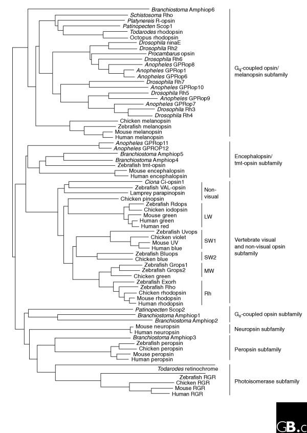
The photosensitive molecule rhodopsin and its relatives consist of a protein moiety - an opsin - and a non-protein moiety - the chromophore retinal. Opsins, which are G-protein-coupled receptors (GPCRs), are found in animals, and more than a thousand have been identified so far. Detailed molecular phylogenetic analyses show that the opsin family is divided into seven subfamilies, which correspond well to functional classifications within the family: the vertebrate visual (transducin-coupled) and non-visual opsin subfamily, the encephalopsin/tmt-opsin subfamily, the Gq-coupled opsin/melanopsin subfamily, the Go-coupled opsin subfamily, the neuropsin subfamily, the peropsin subfamily and the retinal photoisomerase subfamily. The subfamilies diversified before the deuterostomes (including vertebrates) split from the protostomes (most invertebrates), suggesting that a common animal ancestor had multiple opsin genes. Opsins have a seven-transmembrane structure similar to that of other GPCRs, but are distinguished by a lysine residue that is a retinal-binding site in the seventh helix. Accumulated evidence suggests that most opsins act as pigments that activate G proteins in a light-dependent manner in both visual and non-visual systems, whereas a few serve as retinal photoisomerases, generating the chromophore used by other opsins, and some opsins have unknown functions.


Figures

Figure 1
Figure 1
A molecular phylogenetic tree of the opsin family. The tree was inferred by the neighbor-joining method [81]. It shows that members of opsin family are divided into seven subfamilies, whose names are given on the right of the tree. Common names of species shown: Anopheles, mosquito; Branchiostoma, amphioxus; Ciona, ascidian; Drosophila, fruit fly; Patinopecten, scallop; Platynereis, polychaete annelid worm; Procambarus, crayfish; Schistosoma, blood fluke; Todarodes, squid. Abbreviations: LW, long-wavelength-sensitive opsin; SW, short-wavelength-sensitive opsin; MW, middle-wavelength-sensitive opsin; Rh, rhodopsin; RGR, retinal G-protein-coupled receptor. Other abbreviations are protein names; where only a color is given for a protein name, it refers to a cone opsin that detects that color.
Figure 2
Figure 2
Structures of opsins and of the chromophore retinal. (a) A model of the secondary structure of bovine rhodopsin. Amino-acid residues that are highly conserved in the whole opsin family are shown with a gray background. The retinal-binding site (K296) and the counterion position (E113) are marked with bold circles, as is E181, the counterion in opsins other than the vertebrate visual and non-visual ones. C110 and C187 form a disulfide bond. (b) The chemical structures of the 11-cis and all-trans forms of retinal. (c) The crystal structure of bovine rhodopsin (Protein DataBank ID: 1U19 [PDB:1U19]). The chromophore 11-cis-retinal, K296 and E113 are shown in stick representation in the ringed area. (d) The structure of the Schiff base linkage formed by retinal within the bovine opsin, together with the counterion that stabilizes it.

References

    1. Shichida Y, Imai H. Visual pigment: G-protein-coupled receptor for light signals. Cell Mol Life Sci. 1998;54:1299–1315. doi: 10.1007/s000180050256. A review of the properties and functions of the various visual opsins. - DOI - PMC - PubMed
    1. Gartner W. Invertebrate visual pigments. In: Stavenga DG, DeGrip WJ, Pugh Jr EN, editor. Handbook of Biological Physics. Vol. 3. Amsterdam: Elsevier Science; 2000. pp. 297–388. A detailed review of invertebrate visual and non-visual opsins.
    1. Takahashi Y, Ebrey TG. Photobiology of retinal proteins. In: Coohill T, Valenzeno D, editor. Photobiology for the 21th Century. Overland Park, Kansas: Valdenmar; 2001. pp. 101–133. A detailed review of vertebrate visual opsins with amino-acid sequence comparisons.
    1. Sakmar TP, Menon ST, Marin EP, Awad ES. Rhodopsin: insights from recent structural studies. Annu Rev Biophys Biomol Struct. 2002;31:443–484. doi: 10.1146/annurev.biophys.31.082901.134348. A recent structure-based review of vertebrate rhodopsins. - DOI - PubMed
    1. Spudich JL, Yang CS, Jung KH, Spudich EN. Retinylidene proteins: structures and functions from archaea to humans. Annu Rev Cell Dev Biol. 2000;16:365–392. doi: 10.1146/annurev.cellbio.16.1.365. A review of various non-animal opsins, comparing them with animal opsins. - DOI - PubMed

Publication types

LinkOut - more resources